A Tumor-Penetrating Peptide Modification Enhances the
Antitumor Activity of Thymosin Alpha 1
Xingzhen Lao., Meng Liu., Jiao Chen, Heng Zheng*
Department of Life Science and Technology, China Pharmaceutical University, Nanjing, Jiang Su Province, P.R. China
Abstract
A serious limitation of numerous antitumor drugs is the incapacity to penetrate solid tumors. However, addition of an RGD fragment to peptide drugs might solve this problem. In this study, we explored whether the introduction of a permeability-enhancing sequence, such as iRGD (CRGDK/RGPD/EC) fragments, would enhance the activity of thymosin alpha 1 (Ta1). The modified Ta1 (Ta1-iRGD) was successfully expressed and purified, and the in vitro assay showed that Ta1-iRGD presented a similar activity as Ta1 in promoting proliferation of mouse splenocytes. Meanwhile, cell adhesion analysis revealed that Ta 1-iRGD exhibited more specific and greater binding with tumor cells compared with Ta1. Furthermore, the iRGD fragment evidently enhanced the basal ability of Ta1 to inhibit proliferation of cancer cells in vitro, particularly of mouse melanoma cell line B16F10 and human lung cancer cell line H460. Our findings indicated that the addition of an iRGD fragment increased the anti-proliferative activity of Ta1 against cancer cells by improving the ability of Ta1 to penetrate the tumor cells. This study highlighted the important roles of an iRGD sequence in the therapeutic strategy of Ta1-iRGD. Thus, Ta 1-iRGD could be a novel drug candidate for cancer treatment.
Citation:Lao X, Liu M, Chen J, Zheng H (2013) A Tumor-Penetrating Peptide Modification Enhances the Antitumor Activity of Thymosin Alpha 1. PLoS ONE 8(8): e72242. doi:10.1371/journal.pone.0072242
Editor:Maxim Antopolsky, University of Helsinki, Finland
ReceivedMay 7, 2013;AcceptedJuly 10, 2013;PublishedAugust 19, 2013
Copyright:ß2013 Lao et al. This is an open-access article distributed under the terms of the Creative Commons Attribution License, which permits unrestricted use, distribution, and reproduction in any medium, provided the original author and source are credited.
Funding:This work was supported by a grant from the National High Technology Research and Development Program of China (863) (number 2012AA020304) and the Fundamental Research Funds for the Central Universities of China (number JKQ2011044). The funders had no role in study design, data collection and analysis, decision to publish, or preparation of the manuscript.
Competing Interests:The authors have declared that no competing interests exist. * E-mail: zhengh18@hotmail.com
.These authors contributed equally to this work.
Introduction
Thymosin alpha 1 (Ta1; generic drug name: thymalfasin; trade name: Zadaxin) is a 28-amino acid peptide that was first isolated from the thymus gland in mammals by Goldestein in 1977 [1]. Ta1 has already been considered as a potential agent for treating immune deficiencies and cancers for several years. Numerous experiments have confirmed the anti-proliferative activity of Ta1 against melanoma, lung cancer [2], breast cancer [3], and glioblastoma [4]. Furthermore, Ta1 activity has been evaluated in a phase II trial of patients with metastatic melanoma [5]. However, the activity of Ta1 is pleiotropic. Thus, we should find an effective way of delivering Ta1 to the target tumor cells to exert its full adjustive effect on tumor cells. During the past decades, integrins, particularly the integrinavb3, were proven to recognize the RGD (Arg-Gly-Asp) fragment, and avb3 is specifically associated with the upregulated expression of tumor vessels and certain tumor cells on the endothelium [6]. Therefore, the RGD fragment was considered a useful tool for targeting drugs to tumor [7–10].
The more powerful tumor-penetrating peptide iRGD (CRGDK/RGPD/EC) was identified recently based on the mechanism of RGD [11,12]. The iRGD achieved tumor homing by initially binding to integrinavb3 and then being proteolytically cleaved to CRGDK/R, which can penetrate into tumor cells and tissues by binding with neuropilin-1. Therefore, we introduced iRGD to Ta1 by conjugating iRGD to the C-terminus of Ta1 with an ordinary GGGG linker. We then investigated the activity
changes of the modified Ta1-iRGD. The results showed that the modified Ta1-iRGD obviously exhibited the similar or improved anti-proliferative activity against various tumor cell lines, thereby making make it a better candidate drug for specific treatment of cancers.
Materials and Methods
Materials
B16F10 mouse melanoma cell, human gastric cancer cell line BGC-823, human lung cancer cell line H460, and human colon cancer cells line HT-29 were available in our institute. Our institute purchased the cell lines from American Type Cell Culture (ATCC, Shanghai, China). Concanavalin A (ConA) was pur-chased from sigma-aldrich Company (USA). Paclitaxel (Taxol, was provided by JiangsuYew Pharmaceutical Company Limited (Wuxi, Jiangsu province of China). Enterokinase was obtained from New England Biolabs, USA. Nickel-chelating column was purchased from GE Healthcare Company. All the restriction enzymes were purchased from Takara Biotechnology (Dalian) Co., Ltd., and T4DNA ligase was purchased from Promega (USA).
Methods
positive clones were selected based on ampicillin resistance, which was further confirmed by DNA sequencing.Escherichia coli BL21
(DE3) harboring positive recombinant plasmid was grown at 37uC on Luria-Bertani medium containing 100mg/ml of ampicillin at
37uC. Lactose (5 mM) was added to the medium for 4 h to induce protein expression, when the absorbance at 600 nm reached 0.6. Then, the cells were harvested by centrifugation (10,000 g, 10 min, and 4uC), suspended in ice-cold 10 mM Tris and 1 mM EDTA at pH 7.6, and disrupted by sonication. The supernatant of the cell lysate was further purified by a nickel-chelating affinity resin according to the manufacturer’s instruction, and the eluted fraction was analyzed by 15% sodium dodecyl sulfate-polyacryl-amide gel electrophoresis (SDS-PAGE). The fusion proteins were digested by enterokinase at 23uC for 16 h. The target peptides were obtained by loading the nickel-chelating affinity column with equilibration buffer. The eluted peptides were collected, desali-nated by gel filtration chromatography, and confirmed by mass spectroscopy.
Spleen lymphocyte proliferation assay. All experimental procedures using animals used in the our study were performed in strict accordance with the recommendations from the Guide for the Care and Use of Laboratory Animals, which is promulgated by the United States National Institute of Health, and was approved by Jiangsu Provincial Experimental Animal Management Com-mittee under Contract 2012(su)-0035. Spleen lymphocyte prolif-eration experiments were performed as previously described [13]. Suspensions of single spleen cells were prepared from ICR mice (SPF, Comparative Medicine Center of Yangzhou University, China). Single cell suspension (26106/mL) was prepared in RPMI 1640 containing 10% FBS (HyClone, USA) after teasing through a sterilized autoclaved mesh. The adjusted cells were placed in a 96-well microtiter plate and added with 200mL aliquots per well. The
samples were incubated with or without recombinant Ta1 or Ta1-iRGD at 37uC in an atmosphere containing 5% CO2and 95%
humidity. ConA (5mg/mL) and RPMI 1640 were set as positive
and negative control, respectively. After 68 h, cells were counted by the 3-(4, 5-dimethylthiazol-2-yl)-2, 5-diphenyltetrazolium bro-mide (MTT) assay [14,15]. MTT was dissolved in 5 mg/mL of PBS and filtered for sterilization. The MTT solution (20mL) was
added to each well, and the samples were cultured for another 4 h. After all the medium were removed from the wells, DMSO (150mL) was used to solubilize the formazan crystals. The plates
were placed on a plate shaker for 10 min, and then read at 570 nm using a microplate reader. The results were expressed as the relative spleen lymphocyte proliferation (%), which is calculated by the equation (AT 2 AN)/AN6100% (where AT
refers to the absorbance of the treatment group, and ANrefers to
the absorbance of the negative control group).
B16F10 melanoma cell attachment assay. Cell attachment experiments were conducted as previously described [16]. Various concentrations of recombinant Ta1 or Ta1-iRGD were coated in a 96-well ELISA plate (Costar, USA) at 4uC overnight. Then, the plates were blocked with 2% bovine serum albumin in RPMI 1640 medium at 37uC for 2 h. B16F10 melanoma cells (3 6104/ 100mL) were added to the pre-coated plate. After 1h of incubation at 37uC, the plates were washed twice with isotonic buffer saline to remove unbound cells. Cells bound to the wells were fixed, stained by 0.5% crystal violet staining buffer, and photographed. Finally, 10% acetic acid (100mL per well) was added to each well to extract the crystal violet. The remaining adherent B16F10 melanoma Cells were determined by a microplate spectropho-tometer at 595 nm. The relative cell adhesion (%) was calculated using the equation (AT2AC)/AC6100% (where ATrefers to the
absorbance of the treatment group, and AC refers to the
absorbance of the negative control group).
Tumor cell proliferation-inhibition study
in vitro. B16F10 melanoma cell in the logarithmic growth phase were dispersed in 0.25% trypsin with RPMI 1640 complete
Figure 1. Production of Ta1 and Ta1-iRGD peptides.A. Restriction sites are indicated in underline, the translated amino acid and Trx-His6 6-(Asp)4Lys- tag are shown in the box. B. and C., pET32a/Ta1 and pET32a/Ta1-iRGD clones digested with KpnI and HindIII and analyzed by gel electrophoresis analysis (1.2%). MW = molecular weight markers, bp; D. and E. Trx-Ta1 and Trx-Ta1-iRGD purified from BL21 (DE3) lysate, Lane 1, Trx-Ta1 or Trx-Ta1-iRGD induced by lactose inE. coliBL21 (DE3); Lanes 2 and 3, nonbinding protein of crude extract that were subjected to nickel-chelating affinity resin; Lane 4, purified Trx-Ta1 or Trx-Ta1-iRGD.
doi:10.1371/journal.pone.0072242.g001
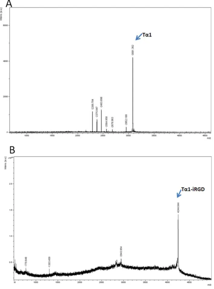
Figure 2. Mass spectroscopy result of recombinant Ta1 or Ta1-iRGD.A. Ta1; B. Ta1-iRGD. doi:10.1371/journal.pone.0072242.g002
medium. Cell suspension (50 000 cells/mL) was plated into a 96-well culture plate (0.1 mL/96-well), which was inoculated for 8 h in 37uC. The cells were incubated with different concentrations of recombinant Ta1 or Ta1-iRGD at 37uC. Paclitaxel (0.0125mmol/mL) was set as a positive control, whereas RPMI
1640 was set as a negative control. After 36 h, cells were counted by MTT assay [14,15], which was described in the spleen lymphocyte proliferation assay. The results were expressed as the relative inhibition of cell proliferation (%), which is calculated by the equation (AN 2 AT)/AN6100% (where AT refers to the
absorbance of the treatment group, and AN refers to the
absorbance of the negative control group). Other cells such as human gastric cancer cell line BGC-823, human lung cancer cell line H460, and human colon cancer cell line HT-29, were treated with the same method.
Three-Dimensional modeling. The structure of Ta1-iRGD was modeled using the DS|Built homology model (Accelrys Inc., USA) based on template (PDB file 2L9I) [17]. Then, the initial model was optimized for 2000 steps by the steepest descent minimizer and for 2000 steps by the conjugate gradient minimizer under the CHARMmH force field. The final Ta1-iRGD model was used as a ligand and docked with integrin avb3 (PDB file 1L5G) [18] by DS|ZDOCK [19] to determine whether Ta1-iRGD binds to avb3 or not. ZDOCK is the protein-protein docking software. The most probable predictions can be selected or filtered by residue conservation of the interaction sites and pose ranks.
Results
Cloning, Expression, and Purification of Ta1 and Ta 1-iRGD
Ta1 and Ta1-iRGD genes were cloned in the frame between KpnI and HindIII restriction sites of the pET32a plasmid, which produced fusion proteins partnered with Trx-A separately (Figures 1A to 1C). Most of the fusion proteins were detected as
soluble proteins. The Trx fusion Ta1 and Ta1-iRGD protein was successfully induced by lactose inE.Coli BL21 (DE3), the fusion
proteins were purified by a nickel-chelating affinity resin respectively, and then confirmed by SDS-PAGE (Figures 1D and 1E). Then, the fusion proteins were digested with enterokinase to release Ta1 and Ta1-iRGD. The molecular weight of Ta1 was detected as 3081.362 by using MALDI mass spectrometry, which is consistent with the theoretical value (Figure 2A). Meanwhile, the molecular weight of Ta1-iRGD was 4244.644, which is in agreement with the expected value as well (Figure 2B). These results confirm the successful expression of soluble Ta1 and Ta1-iRGD.
Ta1-iRGD Showed Similar Spleen Lymphocyte Proliferation Activity with Ta1 in vitro
Different concentrations of recombinant Ta1 or Ta1-iRGD were added to spleen lymphocyte to test their proliferation activity. As shown in Figure 3, recombinant Ta1 and Ta1-iRGD shared extremely similar activities in promoting the proliferation of mouse spleen cells. In addition, a significant dose-dependent proliferation of spleen lymphocyte can be observed in Figure 3. The spleen lymphocyte proliferation assays confirmed that similar to Ta1, Ta1-iRGD has the capability to stimulate the proliferation of spleen lymphocyte, indicating that the conjugation of iRGD to the C- terminus of Ta1 does not change its spleen cell proliferation activity.
Ta1-iRGD Increased B16F10 Melanoma Cell Attachment in vitro
The cell attachment of Ta1-iRGD and Ta1 to a melanoma cell line was further evaluated to determine whether the addition of the iRGD motif to Ta1 enhances its binding to the tumor cell or not. PBS wells were used as negative control. As shown in Figures 4A and 4B, both Ta1-iRGD and Ta1 showed a significant dose-dependent attachment to melanoma cells in this assay. However, Ta1-iRGD specifically enhanced the attachment effect to cancer
Figure 3. Ta1-iRGD and Ta1 showed similar spleen lymphocyte proliferation activity.Spleen lymphocytes were treated with Ta1-iRGD and Ta1. ConA was used as positive control. All data are expressed as the mean6standard deviation, n = 4. Statistical analyses were performed with Student’s t-test, n.s., not significant; *p,0.05; **p,0.01; ***p,0.001.
doi:10.1371/journal.pone.0072242.g003
cells, particularly at a concentration of 1.0mmol/mL or at an even
more low concentration of 0.04mmol/mL. For instance, at
concentrations that ranged from 0.008mmol/mL to 1.0mmol/
mL, Ta1-iRGD exhibited significantly higher activity than Ta1 (p value ,0.01) in three out of the four activities compared, particularly at 0.04 and 1.0mmol/mL, This finding indicates that
Ta1-iRGD can attach to B16F10 melanoma cell line more effectively (p value,0.01).
Ta1-iRGD Increased the Inhibition of Tumor Cell Proliferation in vitro
Tumor cell proliferation was analyzed by MTT assay to determine whether the modification of Ta1 affected its biological activity. Figure 5 indicates that the addition of iRGD to the C-terminus of Ta1 generally enhanced its basal antiproliferative activity in general. As shown in Figure 5A,cell growth was inhibited in a dose-dependent manner when the B16F10 melanoma cells were treated with various doses of Ta1 or
Ta1-iRGD (0.03125mmol/mL to 0.5000mmol/mL). However,
Ta1-iRGD exhibited significantly higher antiproliferative activity than Ta1 at concentrations of 0.03125, 0.0625 and 0.2500mmol/mL.
These results indicated that the conjugation of iRGD to the C-terminus of Ta1 might increase its basal antiproliferative activity against the melanoma cell line B16F10.
When human gastric cancer cell line BGC-823 was treated with Ta1 or Ta1-iRGD at doses that ranged from 0.03125mmol/mL to 0.5000mmol/mL (Figure 5B), Ta1 exhibited no
antiprolifera-tive activity even at high concentrations (beyond 0.25mmol/mL), By contrast, Ta1-iRGD exhibited a significant proliferative activity at high concentrations (from 0.125mmol/mL and above).-This finding indicated that the addition of the iRGD motif can improve the antiproliferative activity of Ta1 in the human gastric cancer cell line BGC-823.
Furthermore, when human lung cancer cell line H460 was treated with Ta1 or Ta1-iRGD at doses that ranged from 0.03125mmol/mL to 0.5000mmol/mL (Figure 5C), Ta1-iRGD Figure 4. Enhanced cell attachment effect by adding iRGD to the C-terminus of Ta1.A. First, cells bound to the wells were fixed, stained by 0.5% crystal violet staining buffer and photographed. B. Second, crystal violet was extracted and the remaining adherent B16F10 Melanoma Cells were recorded by a microplate spectrophotometer at 595 nm wavelengths. All data are expressed as the mean6standard deviation, n = 3. Statistical analyses were performed with Student’s t-test, n.s., not significant; *p,0.05; **p,0.01; ***p,0.001.
doi:10.1371/journal.pone.0072242.g004
exhibited significantly higher antiproliferative activity than Ta1 (p value,0.001). Ta1 only exhibited a slight cell growth inhibition activity at high concentrations (from 0.2500mmol/mL to
0.5000mmol/mL). By contrast, Ta1-iRGD exhibited significant
antiproliferative activity even at very low concentrations. For instance, Ta1-iRGD inhibited human lung cancer cell line H460 proliferation by 16.8% at 0.03125mmol/mL, whereas Ta1 had no
antiproliferative activity at the same concentration. At 0.125mmol/mL, Ta1-iRGD inhibited human lung cancer cell
line H460 proliferation by 24.0%, whereas Ta1 only had 0.64% inhibition.
Moreover, when human colon cancer cell line HT-29 was treated with Ta1 or Ta1-iRGD at doses that ranged from 0.03125mmol/mL to 0.5000mmol/mL (Figure 5D), Ta1-iRGD
also exhibited significantly higher antiproliferative activity than Ta1. For instance, Ta1 exhibited cell growth inhibition activity at 0.5000mmol/mL, whereas Ta1-iRGD exhibited a significant
antiproliferative activity even at low concentrations. At 0.1250mmol/mL, Ta1-iRGD inhibited human colon cancer cell
line HT-29 proliferation by 14.3%, whereas Ta1 had no antiproliferative activity. These results indicated that the addition of the iRGD fragment increased the antiproliferative activity of Ta1 against cancer cells.
Figure 5. Inhibition of cancer cell proliferation by Ta1 or Ta1-iRGD.Ta1 or Ta1-iRGD was added to the cell suspension and incubated for 36 h and counted by MTT assay. Paclitaxel (0.0125mmol/mL) was set as a positive control. RPMI 1640 was set as a negative control. All data were
expressed as the mean6standard deviation, n = 4. Statistical analyses were performed with Student’s t-test, n.s., not significant; *p,0.05; **p,0.01; ***p,0.001.
doi:10.1371/journal.pone.0072242.g005
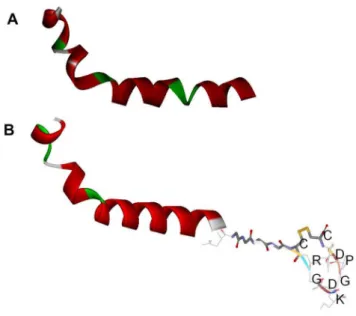
Figure 6. Modeling of the structures of the composed peptides by DS|Built homology model.A. Ta1 B. Ta1-iRGD.
doi:10.1371/journal.pone.0072242.g006
Three-Dimensional Modeling
The 3D structures of Ta1 and Ta1-iRGD were compared using computational modeling to determine how the addition of the iRGD motif improves the efficacy of Ta1 to arrest tumor cell growth. However, no obvious differences can be observed from the modeling (Figure 6A and 6B). Although the modeling method might not totally reflect the subtle changes of Ta1 structure due to computational ability, this result gave an implication that the iRGD might not directly affect the structure significantly. Based on previous studies on iRGD [11,12], we assumed that the main reason of the better antiproliferative activity against tumor cells of Ta1-iRGD than Ta1 is attributed to the ability of iRGD to facilitate the more effective targeting and attachment of Ta1 to tumor cells by recognizing integrins. Thus, we further investigated the interaction between Ta1-iRGD and integrin avb3 using computational modeling approach. The 3D structure of integrin avb3 (PDB file 1L5G) was used as the receptor model in our dock research. The 1L5G describes a crystal structure of the outside membrane segment of integrin avb3 binding with a cyclic pentapeptide ligand that contains the Arg-Gly-Asp sequence. The structure revealed that the cyclic pentapeptide binds at the head interface between theav and theb3 subunits, and the main contact area with the integrin involves Asp150, Gln180, and Asp218 in theav subunit; and Ser121, Ser123, Asn215, Asp217, and Ala 218 in theb3 subunit. The iRGD part of Ta1 -iRGD was found capable of inserting into a groove betweenav andb3 on the integrin head in our dock model (Figure 7), sharing a similar interaction manner with the structure ofavb3 of an RGD ligand. The contacts between the iRGD part and theav subunit primarily involve Asp150, whereas the contacts between the iRGD part and the b3 subunit primarily involve Ser121, Ser123, and Ala 218.
Here, the computational model may have limitations to reflect real protein-ligand interaction in detail. However, it is important to illustrate the phenomenon observed from experiments and understand the potential mechanism of the iRGD motif. Therefore, our model revealed that the adding iRGD to the Ta1 might have enabled the new function of binding to avb3, resulting in tumor-homing. However, both Ta1 and the Ta1 part in Ta1-iRGD shared the same helical structure in our models. Thus, the similarity between the structures of Ta1 and Ta1-iRGD might also explain why Ta1 and Ta1-iRGD exhibited similar spleen lymphocyte proliferation capability.
Discussion
Ta1 has already been approved for the treatment of Hepatitis B and C in several countries [20,21].Clinically, Ta1 has been clinically proven to exert an immune modulatory activity on maturation of T cell [22,23] and the natural killing cell and activation of dendritic cells [24] and the up-regulation of high affinity interleukin-2 receptors [25]. However, the effect of Ta1 is pleiotropic. Garaci E et al. recently reported that Ta1 is capable of increasing the expression of major histocompatibility complex class I surface molecules and tumor antigens in murine and human tumor cell lines [26]. These effects represent the potential factors for increasing the antitumor activity of Ta1. The therapeutic use of the Ta1 in human melanoma is currently on phase II trial. Although the antitumor activity exhibited by Ta1 has numerous benefits, the pleiotropic effect of Ta1 decreases the specificity of the treatments. Therefore, Ta1 also has relatively adverse effects on healthy cells similar to other anti-cancer drugs. Furthermore, the therapeutic efficacy and specificity of Ta1 is expected to increase by improving its capability to penetrate tumor cells. Given the fact that the expression or elevation of integrinavb3 and neuropilin-1 are only restricted to tumors [9,27,28], we propose a strategy to improve the targeted delivery of Ta1 to tumor cells by adding the iRGD fragment to the C-terminus of Ta1. The motif of iRGD is a tumor-homing peptide that has a high affinity partnering with integrinavb3, which has been proven to remarkably enhance the activity of antitumor drugs [12].
In this study, both Ta1 and Ta1-iRGD were successfully expressed and purified by bioengineering methods inE. coli. Cell
attachment assay, spleen cell proliferation experiments and tumor cell proliferation experiments revealed that Ta1-iRGD has higher antitumor activity than Ta1. In the melanoma cell attachment assay, we found that Ta1-iRGD has higher attachment activity than Ta1. In the cancer cell inhibition assay, we also found that Ta1-iRGD has a better anti-proliferation activity in several cancer cell lines than Ta1, particularly in mouse melanoma cell line B16F10 and human lung cancer cell line H460. With the aid of the iRGD, Ta1-iRGD directly inhibited the proliferation of human lung cancer cell line H460 and mouse melanoma cells line B16F10 at very low concentrations in contrast to the slight inhibition exhibited by Ta1.
Previous publications have reported the effectiveness of Ta1 in clinical trials of non-small cell lung cancer [29,30] and advanced metastatic melanoma patients [31,32]. Our experiment showed that Ta1-iRGD can inhibit the growth of mouse melanoma cell line B16F10 and human lung cancer cell line H460 more effectively. Thus, Ta1-iRGD can be an attractive strategy for a novel therapeutics against neoplastic diseases. The positive result of cell attachment assay might be able to explain the better anti-proliferation activity in cancer cell lines of Ta1-iRGD than Ta1. These results can be further explained by our computational model. Although the computational model may have limitations to
Figure 7. Structure of integrinavb3 complexed with Ta1-iRGD. The structure of av and b3 subunits, respectively, in a ribbon-like structure. The Ta1 part of Ta1-iRGD is shown in red, the iRGD part is shown as a spherical structure, and the linker between them is shown in yellow. Theavb3-Ta1-iRGD structure reveals that the iRGD of Ta1-iRGD inserts into a crevice betweenav andb3 on the integrin head, sharing a similar interaction manner with the structure ofavb3 in complex with a cyclic RGD ligand (PDB file code:1L5G).
doi:10.1371/journal.pone.0072242.g007
represent the real protein structure and protein-ligand interactions in detail since it merely basing on the integrations of desolvation, shape complementarity and electrostatics [19]. However, it is very helpful to understand the potential mechanism of how iRGD motif affects the function of Ta1. In our model, the iRGD part of Ta1-iRGD can form a stable complex with integrinavb3 in our model resulting in the tumor cell homing. In summary, the addition of iRGD significantly improved the antitumor effect of Ta1. However, the immune modulatory activity of Ta1-iRGD is almost the same with that of Ta1.
Conclusion
In this study, we tried to connect Ta1 and tumor-homing. We expressed and evaluated the effectiveness of Ta1- iRGD in vitro for the first time. The research on Ta1- iRGD fusion peptide provided a new effective way of maximizing the effectiveness of
Ta1 in treating solid tumors. The iRGD Ta1 fusion peptide could be an attractive strategy for designing novel therapeutics against tumors. We are currently studying the activity of Ta1- iRGD on cancer targeting, and the pharmacokinetic and pharmacodynamic aspects of Ta1- iRGD will be further investigated to guarantee better clinical application.
Acknowledgments
The authors would like to thank Dr. Jing Li for the discussions and helpful suggestions during this investigation.
Author Contributions
Conceived and designed the experiments: XL ML HZ. Performed the experiments: XL ML. Analyzed the data: XL ML. Wrote the paper: XL. Computational model: XL JC.
References
1. Goldstein AL, Low TL, McAdoo M, McClure J, Thurman GB, et al. (1977) Thymosin alpha1: isolation and sequence analysis of an immunologically active thymic polypeptide. Proc Natl Acad Sci U S A 74: 725–729.
2. Moody TW (2007) Thymosin alpha 1 as a chemopreventive agent in lung and breast cancer. Thymosins in Health and Disease: First International Symposium 1112: 297–304.
3. Moody TW, Tuthill C, Badamchian M, Goldstein AL (2002) Thymosin alpha1 inhibits mammary carcinogenesis in Fisher rats. Peptides 23: 1011–1014. 4. Sungarian A, Cielo D, Sampath P, Bowling N, Moskal P, et al. (2009) Potential
Role of Thymosin-alpha1 Adjuvant Therapy for Glioblastoma. J Oncol 2009: 302084.
5. Danielli R, Fonsatti E, Calabro L, Di Giacomo AM, Maio M (2012) Thymosin alpha1 in melanoma: from the clinical trial setting to the daily practice and beyond. Ann N Y Acad Sci 1270: 8–12.
6. Zetter BR (1997) On target with tumor blood vessel markers. Nat Biotechnol 15: 1243–1244.
7. Arap W, Pasqualini R, Ruoslahti E (1998) Cancer treatment by targeted drug delivery to tumor vasculature in a mouse model. Science 279: 377–380. 8. Zitzmann S, Ehemann V, Schwab M (2002) Arginine-glycine-aspartic acid
(RGD)-peptide binds to both tumor and tumor-endothelial cells in vivo. Cancer Res 62: 5139–5143.
9. Ruoslahti E (2002) Specialization of tumour vasculature. Nat Rev Cancer 2: 83– 90.
10. Desgrosellier JS, Cheresh DA (2010) Integrins in cancer: biological implications and therapeutic opportunities. Nat Rev Cancer 10: 9–22.
11. Sugahara KN, Teesalu T, Karmali PP, Kotamraju VR, Agemy L, et al. (2009) Tissue-Penetrating Delivery of Compounds and Nanoparticles into Tumors. Cancer Cell 16: 510–520.
12. Sugahara KN, Teesalu T, Karmali PP, Kotamraju VR, Agemy L, et al. (2010) Coadministration of a Tumor-Penetrating Peptide Enhances the Efficacy of Cancer Drugs. Science 328: 1031–1035.
13. Wang J, Chen B, Jin N, Xia G, Chen Y, et al. (2011) The changes of T lymphocytes and cytokines in ICR mice fed with Fe3O4 magnetic nanoparticles. Int J Nanomedicine 6: 605–610.
14. Carmichael J, DeGraff WG, Gazdar AF, Minna JD, Mitchell JB (1987) Evaluation of a tetrazolium-based semiautomated colorimetric assay: assessment of radiosensitivity. Cancer Res 47: 943–946.
15. Chen CC, Liou SW, Chen CC, Chen WC, Hu FR, et al. (2011) Coenzyme Q10 reduces ethanol-induced apoptosis in corneal fibroblasts. PLoS One 6: e19111. 16. Wang P, Ballestrem C, Streuli CH (2011) The C terminus of talin links integrins
to cell cycle progression. J Cell Biol 195: 499–513.
17. Elizondo-Riojas MA, Chamow SM, Tuthill CW, Gorenstein DG, Volk DE (2011) NMR structure of human thymosin alpha-1. Biochemical and Biophysical Research Communications 416: 356–361.
18. Xiong JP, Stehle T, Zhang R, Joachimiak A, Frech M, et al. (2002) Crystal structure of the extracellular segment of integrin alpha Vbeta3 in complex with an Arg-Gly-Asp ligand. Science 296: 151–155.
19. Chen R, Li L, Weng Z (2003) ZDOCK: an initial-stage protein-docking algorithm. Proteins 52: 80–87.
20. Garaci E, Favalli C, Pica F, Sinibaldi Vallebona P, Palamara AT, et al. (2007) Thymosin alpha 1: from bench to bedside. Ann N Y Acad Sci 1112: 225–234. 21. Goldstein AL, Goldstein AL (2009) From lab to bedside: emerging clinical
applications of thymosin alpha 1. Expert Opin Biol Ther 9: 593–608. 22. Peng Y, Chen Z, Yu W, Zhou Q, Xu L, et al. (2008) Effects of thymic
polypeptides on the thymopoiesis of mouse embryonic stem cells. Cell Biol Int 32: 1265–1271.
23. Yao W, Zhu Q, Yuan Y, Qiao M, Zhang Y, et al. (2007) Thymosin alpha 1 improves severe acute pancreatitis in rats via regulation of peripheral T cell number and cytokine serum level. J Gastroenterol Hepatol 22: 1866–1871. 24. Romani L, Bistoni F, Gaziano R, Bozza S, Montagnoli C, et al. (2004)
Thymosin alpha 1 activates dendritic cells for antifungal Th1 resistance through toll-like receptor signaling. Blood 103: 4232–4239.
25. Leichtling KD, Serrate SA, Sztein MB (1990) Thymosin alpha 1 modulates the expression of high affinity interleukin-2 receptors on normal human lympho-cytes. Int J Immunopharmacol 12: 19–29.
26. Garaci E, Pica F, Serafino A, Balestrieri E, Matteucci C, et al. (2012) Thymosin alpha1 and cancer: action on immune effector and tumor target cells. Ann N Y Acad Sci 1269: 26–33.
27. Eliceiri BP, Cheresh DA (2001) Adhesion events in angiogenesis. Curr Opin Cell Biol 13: 563–568.
28. Pellet-Many C, Frankel P, Jia H, Zachary I (2008) Neuropilins: structure, function and role in disease. Biochem J 411: 211–226.
29. Garaci E, Lopez M, Bonsignore G, Della Giulia M, D’Aprile M, et al. (1995) Sequential chemoimmunotherapy for advanced non-small cell lung cancer using cisplatin, etoposide, thymosin-alpha 1 and interferon-alpha 2a. Eur J Cancer 31A: 2403–2405.
30. Salvati F, Rasi G, Portalone L, Antilli A, Garaci E (1996) Combined treatment with thymosin-alpha1 and low-dose interferon-alpha after ifosfamide in non-small cell lung cancer: a phase-II controlled trial. Anticancer Res 16: 1001–1004. 31. Lopez M, Carpano S, Cavaliere R, Dilauro L, Ameglio F, et al. (1994) Biochemotherapy with Thymosin-Alpha-1, Interleukin-2 and Dacarbazine in Patients with Metastatic Melanoma - Clinical and Immunological Effects. Annals of Oncology 5: 741–746.
32. Maio M, Mackiewicz A, Testori A, Trefzer U, Ferraresi V, et al. (2010) Large randomized study of thymosin alpha 1, interferon alfa, or both in combination with dacarbazine in patients with metastatic melanoma. J Clin Oncol 28: 1780– 1787.