Contents lists available atScienceDirect
Phytomedicine
journal homepage:www.elsevier.com/locate/phymed
Red propolis ameliorates ischemic-reperfusion acute kidney injury
Marcus Felipe Bezerra da Costa
a, Alexandre Braga Libório
b,∗, Flávio Teles
c,
Conceição da Silva Martins
d, Pedro Marcos Gomes Soares
d, Gdayllon C. Meneses
e,
Francisco Adelvane de Paulo Rodrigues
a, Luzia Kalyne Almeida Moreira Leal
e, Diogo Miron
e,
Aline Holanda Silva
e, Alice Maria Costa Martins
eaDepartment of Physiology and Pharmacology, Faculty of Medicine, Federal University of Ceará, Fortaleza, Ceará, Brazil bDepartment of Clinical Medicine, Faculty of Medicine, Federal University of Ceará, Fortaleza, Ceará, Brazil
cFederal University of Alagoas, Tabuleiro dos Martins, Maceió, AL 57072-900, Brazil
dDepartment of Morphology, Faculty of Medicine, Federal University of Ceará, Fortaleza, Ceará, Brazil
eDepartment of Clinical and Toxicological Analysis, Faculty of Pharmacy, Federal University of Ceará, Fortaleza, Ceará, Brazil
a r t i c l e
i n f o
Article history:
Received 11 November 2013 Revised 2 March 2015 Accepted 19 March 2015
Keywords: Acute kidney injury Red propolis Heme-oxygenase
a b s t r a c t
Introduction: Acute kidney injury (AKI) remains a great problem in clinical practice. Renal
is-chemia/reperfusion (I/R) injury is a complex pathophysiological process. Propolis is a natural polyphenol-rich resinous substance collected by honeybees from a variety of plant sources that has anti-inflammatory and anti-oxidative properties. Red propolis (RP) protection in renal I/R injury was investigated.
Methods:Male Wistar rats underwent unilateral nephrectomy and contralateral renal I/R (60 min). Rats were
divided into four groups: (1) sham group, (2) RP group (sham-operated rats treated with RP), 3) IR group (rats submitted to ischemia) and (4) IR-RP (rats treated with RP before ischemia). At 48 h after reperfusion, renal function was assessed and kidneys were removed for analysis.
Results:I/R increased plasma levels of creatinine and reduced creatinine clearance (CrCl), and RP provided
pro-tection against this renal injury. Red propolis significantly improves oxidative stress parameters when com-pared with the IR group. Semiquantitative assessment of the histological lesions showed marked structural damage in I/R rats compared with the IR-RP rats. RP attenuates I/R-induced endothelial nitric oxide-synthase down regulation and increased heme-oxygenase expression in renal tissue.
Conclusion:Red propolis protects kidney against acute ischemic renal failure and this protection is associated
with reduced oxidative stress and eNOS and heme-oxygenase up regulation.
© 2015 Elsevier GmbH. All rights reserved.
Introduction
Acute kidney injury (AKI) remains a great problem in clinical practice. It affects approximately 20% of hospitalized patients and half of critically-ill patients admitted to intensive care unit (Poukkanen et al. 2013; Zeng et al. 2013; Uchino et al. 2005). Despite improved strategies for supporting vital organs during AKI recovery and in renal replacement therapy (dialysis), AKI mortality rates remain quite high (Leite et al. 2013). Also, renal I/R injury is a common cause of early allograft dysfunction in renal transplanted patients and represents an additional risk factor for late renal allograft failure (Ditonno et al. 2013). The prevention of kidney lesions and their progression continue to represent a great challenge. Although renal injuries are
∗ Corresponding author at: Av. Abolição, 4043 Ap. 1203 Jangada Bairro: Mucuripe,
Fortaleza, Ceará, CEP 60165-082, Brazil. Tel.:+55 85 99987995. Fax: 99987995.
E-mail address:[email protected](A.B. Libório).
multifactorial in many patients, in the clinical scenario, animal models of renal ischemia/reperfusion (I/R) remain important to understand the pathophysiology and potential treatment options for AKI.
Renal I/R injury is a complex pathophysiological process involving oxidative and inflammatory damage, endothelium-mediated injury and apoptosis. Nitric oxide (NO) is involved in the pathophysiology of ischemic AKI. Increased expression of proinflammatory inducible nitric oxide synthase (iNOS) is considered a pivotal step in renal dam-age, whereas the reduced activity of endothelial nitric oxide synthase (eNOS) contributes to renal impairment resulting from endothelial dysfunction (Heemskerk et al. 2009).
Many molecules have intrinsic cytoprotective properties that include anti-apoptotic, anti-inflammatory and antioxidant actions. Heme-oxygenase (HO) 1 and 2 are the rate-limiting enzymes in the catabolism of heme, a reaction that yields equimolar amounts of biliverdin, Fe++and carbon monoxide. Expression of HO-1 is read-ily increased upon organ I/R injury, becoming the rate-limiting fac-tor in the generation of biliverdin, Fe+and CO. Heme-oxygenase-1
788 M.F.B. da Costa et al. / Phytomedicine 22 (2015) 787–795
provides protection against renal I/R injury through its antioxidant, anti-inflammatory and cytoprotective activities (Nath et al. 1992; Agarwal Nick 2000).
Propolis is a natural polyphenol-rich resinous substance collected by honeybees from a variety of plant sources. In recent years, propolis has gained popularity as a health drink, has been extensively used in food and beverages, and is thought to improve human health and pre-vent disease (Daleprane and Abdalla 2013). Beneficial health effects are largely attributed to its polyphenolic composition. Red propolis has been classified as a separate type based on its unique chemi-cal composition, particularly rich in isoflavonoids (Righi et al. 2013). Anti-inflammatory and antioxidant properties have been attributed to red propolis (Bueno-Silva et al. 2013; Enis Yonar et al. 2012). In the present study, we aimed to evaluate the effects of red propolis extract on renal I/R injury.
Methods
Animals and red propolis
The experimental protocol was approved by the Ethical Commit-tee on Animal Research of Federal University of Ceará (no. 39/13). Wistar rats, weighing 250–300 g, were obtained from the Pharmacol-ogy Department of Federal University of Ceara and maintained under controlled temperature (21±2°C) and humidity conditions (60±5%) with a 12:12-h light:dark cycle. A standard commercial pellet diet and water were offeredad libitum.
Chemical characterization of red propolis
Red propolis was collected in the mangrove region in Marechal Deodoro (a city in the vicinity of Maceio, capital of Alagoas State, in the northeastern Brazil (SL 09.40 and WL 35.41). Forty grams of propolis was macerated in 100 ml of 40% ethanol, for 30 days, the solution was filtered, the filtrate evaporated to dryness. The residue, 12.5 g of dry extract (drug extract ratio of 3.2:1), was dissolved in 50 ml of ethanol to stock and transportation with a concentration of 0.25 g/ml. The botanic origin wasDalbergia ecasttophyllum. The chromato-graphic analysis by high-performance liquid chromatography (HPLC) was performed. The assay was performed on Alliance – Waters 2695 (Milford, MA) chromatograph with a binary pump, auto-sampler, and photodiode-array detector (Waters-2996 PDA) at 268 nm. The sepa-rations were performed with an analytical reverse-phase column C18 (Waters, 250 mm×4.6 mm, 5
µ
m) at 40°C in a thermostatic oven. The mobile phase was made from water/acetic acid 0.1% (solvent A) and methanol (solvent B) in a gradient elution for 65 min (total run time), starting with 30% B (0–15 min), increasing to 90% B (15–60 min), held at 90% B and decreasing to 30% B (60–65 min) with a sol-vent flow rate of 1 ml/min. The solsol-vents were previously degassed under vacuum by sonication during 5 min and filtered through Phe-nomenex nylon membrane (0.45µ
m). The samples were dissolvedin the initial mobile phase and filtered through a 0.45
µ
m filter unit(Millipore, USA) before injection (20 ml). The data was processed by Empower software (Waters, USA).
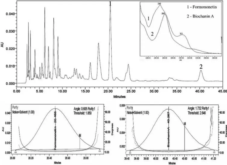
The identification of formononetin and biochanin A in RP by HPLC experiments were based on the retention time (rt) of external stan-dards. The contents of the three flavonoids were calculated using calibration curves. The ranges of calibration curves were 0.04–0.12 mg/ml for formononetin and 0.005–0.013 mg/ml for biochanin A. The linear relationship was obtained correlating the concentration of flavonoids to the correspondent peak area.
For peak purity analysis, spectra in the range of 210–400 nm were recorded at a frequency of 1 Hz. Threshold was calculated employ-ing noise and solvent angles. Reference spectra of formononetin and biochanin A standards were recorded in the Empower 2 software library for identification purposes.
The spectra search improves the identification of compounds in complex matrices since different substance can have identical reten-tion times. Formononetin and Biochanin A were identified in propolis extract chromatogram through the comparison of peak apex spec-trum against the results of reference standards solutions recorded previously in the software library. The peak height of biochanin A in propolis extract chromatogram is lower than formononetin (Fig. 1).
The peak purity analysis provided by diode array detectors is es-sential to ensure reliability and accuracy of the chromatographic mea-surements of analytes in complex matrices. In the present work, the formononetin and biochanin A peaks were found pure since the pu-rity angles were lower than the threshold angles and the threshold curves do not intersect the purity curves.
The chromatographic method shows linearity over the range eval-uated and the correlation coefficients for and formononetin and biochanin A were 0.9915 and 0.9996, respectively. The concentra-tions (mean±standard deviation forn=12) of formononetin and biochanin A in the propolis extracted were 10.25±0.21 and 0.50±
0.02
µ
g/mg, respectively. The amount of formononetin in the propolisextract is greater than 1% and was approximately fifteen times larger than biochanin A.
Surgical procedure
Animals were anesthetized with sodium pentobarbital (50 mg/kg i.p.). A midline laparotomy incision was performed, the right kidney was removed and left ischemic renal failure was induced by clamping the renal artery (with a nontraumatic clamp) for 60 min, followed by reperfusion. After 48h, animals were sacrificed to obtain blood samples for biochemical tests. Additionally, the left kidneys were col-lected for histological and immunohistochemistry evaluation.
Experimental groups
To administration, the ethanol extract was filtered and then evap-orated by using a vacuum evaporator. The gravimetric analysis was carried out in quintuplicate and disclosed a ratio of dry extract of 24.1
±0.09 (% m/m). Each 150 mg of dry extract contained approximately 6.38 mg of formononetin and 0.31 mg of biochanin A. The propo-lis samples were maintained in a dark environment, inside a deep freezer (kept at−20°C). The dried extract was administrated accord-ing animal weight (150 mg/kg of body weight). The quantity of dried extract for each animal was suspended in tap water (5 ml) just before oral administration and the total volume (5 ml) was administered by gavage.
Rats were divided into the following groups (n=8 in each group):
- Sham+tap water group (SHAM): Rats were submitted to identi-cal surgiidenti-cal procedures, except for the nephrectomy and unilateral renal occlusion shock and were kept under anesthesia for the du-ration of the experiment.
- Sham+red propolis (RP): Identical to SHAM group, receiving red propolis (150 mg/kg/day) was administered by gastric gavage 3 days before the procedure and 1 h prior to surgical procedure. - I/R+tap water group (IR): Rats were submitted to nephrectomy
and unilateral renal occlusion (60 min) followed by reperfusion. - I/R+red propolis group (IR-RP): Rats were submitted to the above
mentioned surgical procedures and red propolis (150 mg/kg/day) was administered by gastric gavage 3 days before the procedure and 1 h prior to ischemia.
Measurement of biochemical parameters
Fig. 1. Chromatogram of propolis extract and spectra and purity analysis of formononetin and biochanin A.
and urine concentrations of urea (BUN) and creatinine (Cr) were measured as indicators of impaired glomerular function. Plasma and urine concentrations of sodium (Na+) and potassium (K+) were used as indicators of renal tubular injury. BUN and Cr levels were mea-sured by means of colorimetric methods in a semi-automatic ana-lyzer (LABQUESTR
) using diagnostic kits (LabtestR
, Brazil). The de-termination of sodium and potassium levels was made by means of ion-selective electrodes (Rapid Chem 744, Bayer Diagnostics).
Determination of MDA levels
Malondialdehyde (MDA) concentration in kidney tissue was deter-mined as an indicator of lipid peroxidation, following a protocol pre-viously described byMihara et al. (1980). Briefly, the left kidney was removed and homogenized with KCl (1.15%) to make a homogenate. Then 3 ml of a solution containing phosphoric acid (1%) and 1 ml of thiobarbituric acid (0.6%) were added to 0.5 ml of the homogenate in a tube. The mixture was heated in boiling water for 45 min. Then, 4 ml of a solution ofn-butanol was added to the mixture, which was shaken vigorously. The absorbance was measured by spectrophotometry at 532 nm. The results were expressed in nmol/g tissue.
To determine the levels of MDA in urine samples, 0.4 ml of the urine sample was mixed with 0.6 ml of distilled water, 1 ml of TCA (trichloroacetic 10%) and 1 ml of thiobarbituric acid (0.6% pH 2). All these solutions were kept on ice during this stage of the process. The solutions were homogenized and then placed in a water bath at 100
°C for 20 min. After cooling and addition of 1 ml of TCA (70%), the final
mixture was centrifuged for 15 min at 3000 rpm and the absorbance was measured by spectrophotometry at 534 nm.
Determination of GSH levels
Homogenate was prepared with EDTA (0.02 M). Then, 400
µ
l of thehomogenate was removed and added to 320
µ
l of distilled water and80
µ
l of trichloroacetic acid (50%). The material was centrifuged at3000 rpm for 15 min. Then, 400
µ
l of the supernatant was collectedand 800
µ
l of Tris–HCl buffer (0.4 M pH 8.9) and 20µ
l of DTNB(0.01 M) were added. After 1 min of reaction, the absorbance was measured by spectrophotometry at 412 nm. The concentration of reduced glutathione was expressed in micrograms per gram of tissue.
Histological analyses
Renal tissue was removed and placed in a 10% solution of buffered formaldehyde. After 24 h, the tissue was transferred to an alcoholic so-lution (70%) and used for histological analysis. Kidney tissue was fixed using a 10% formalin solution and then paraffin-embedded. Slices of
5-µ
m thickness were obtained and then stained with hematoxylin and790 M.F.B. da Costa et al. / Phytomedicine 22 (2015) 787–795
Table 1
Effect of red propolis on renal function. Cr: creatinine; ClCr: creatinine clearance; FENa+: absolute
excretion of sodium; FEK+: absolute excretion of potassium.
Groups Cr Urea ClCr FENa+ FEK+
Mean+SD Mean+SD Mean+SD Mean+SD Mean+SD
SHAM 0.64±0.05 36.36±6.2 1.29±0.46 0.23±0.11 7.5±2.9
RP 0.66±0.13 40.17±0.11 0.93±0.24 0.19±0.05 10.58±6.4
IR 2.7±0.99∗ 274.3±91.81∗ 0.07±0.04∗ 1.03±0.39∗ 134.4±54.9∗
IRRP 1.82±0.5# 181.1±65.6# 0.41±0.14# 0.58±0.3# 64.7±52.4#
∗ P<0.05 vs. SHAM.
#P<0.05 vs. IRG.
Fig. 2.The effect of red propolis on tubular necrosis score after renal I/R. Values shown are mean±SE. Mean of 8 rats for each group.∗P<0.05 vs. Sham,#P<0.05 vs. IRG.
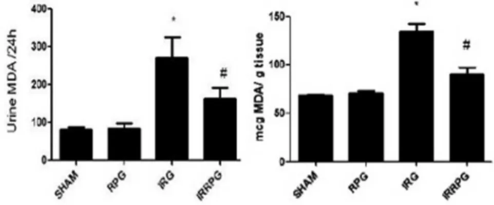
Fig. 3.Red propolis reduces malondialdehyde (MDA) levels induced by I/R injury. Values shown are mean±SE of the MDA levels. (A) MDA levels of urine samples,∗P<0.05 vs.
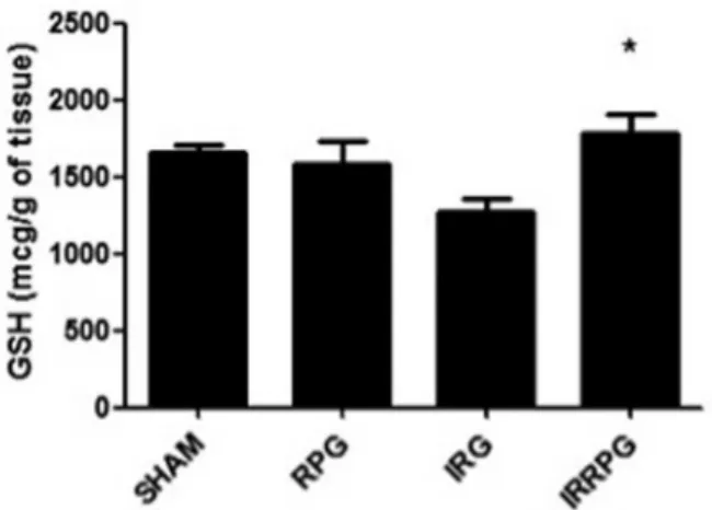
Fig. 4. Red propolis improves glutathione (GSH) levels in animals with I/R injury.
Values shown are mean±SE of the GSH levels.∗P<0.05 vs. IRG.
4 – lesions affecting 50% of kidney samples, and 5 – lesions affecting more than 75% of samples.
Immunohistochemical localization of heme-oxygenase and eNos
Immunohistochemistry for eNOS and heme-oxygenase was per-formed using the streptavidin-biotin-peroxidase method (Hsu and Raine 1981). After 48 h of reperfusion, the animals were sacrificed and the left kidney was removed and fixed in formaldehyde (10%) for 24 h, and subsequently submitted to treatment with EDTA (10%) for dem-ineralization. Subsequently, the samples were suspended in sodium sulfate (5%), and then paraffin-embedded. After this procedure, serial
4-mm sections were obtained with an appropriate microtome and placed onl -polylysine slides, suitable for immunohistochemistry
analysis. The sections were deparaffinized, hydrated in xylene and alcohol, and immersed in a citrate buffer (0.1 M; pH 6.0), by heating in a microwave oven for 15 min for antigen retrieval. After cooling to ambient temperature at 20°C, washes were performed with phos-phate buffered solution (PBS) and with an endogenous peroxidase blocking solution of H2O2(3%) for 15 min. The sections were incu-bated “overnight” (at 4°C) with primary rabbit anti-ENOS antibody diluted in PBS (1:200) and with primary goat polyclonal antibody against heme-oxygenase diluted in PBS (1:200). Then, samples were incubated with the secondary antibody for 30 min. After washing, the sections are incubated with the conjugated streptavidin peroxidase complex (ABC complex VectastainR
) for 30 min. After further wash-ing with PBS, followed by stainwash-ing with (DAB), samples were coun-terstained with Mayer’s hematoxylin. Finally, dehydration of samples was performed and they were mounted on slides. Negative controls were processed simultaneously as described above and the primary antibody was replaced by PBS–5% BSA.
When assessing immunohistochemical staining for eNOS and heme-oxygenase, each tubule interstitial grid field was graded semi-quantitatively and the mean score per kidney was calculated. Each score reflected mainly changes in the extent, rather than the inten-sity, of staining, based on the proportion of the grid field showing positive staining: 0, absent or<5%; I, 5–25%; II, 25–50%; III, 50–75%, and IV greater than 75%.
Western blot analysis
The protein expression of endothelial nitric oxide synthase (eNOS) and heme-oxygenase were assessed in the kidney samples that were
792 M.F.B. da Costa et al. / Phytomedicine 22 (2015) 787–795
individually homogenized with a Polytron in K-HEPES buffer con-taining a mixture of protease inhibitors. After incubation at 4°C for 15 min, the samples were centrifuged at 2000g. The protein con-centrations were quantified using the Bradford assay method and the 80
µ
g of protein from each sample was separated on an 8%polyacrylamide gel and transferred to a nitrocellulose membrane. Subsequently, the membranes were probed with a primary rabbit monoclonal eNOS (1:200) or a primary goat polyclonal against heme-oxygenase (1:200) primary antibody, followed by anti-rabbit (1:1000) or anti-goat (1:5000) secondary antibody, respectively. The bands were visualized using a chemiluminescence substrate and analyzed by gel documentation Alience 4.7 Uvitec (Cambridge, Cambs, UK). The relative expression of NOS and heme-oxygenase proteins in each kidney were normalized using actin antibody and the values are ex-pressed as a percentage of normal protein expression.
Statistical analyses
All continuous variables are shown as mean±standard error. One-way analyses of variance with Newman–Keuls post hoc test was used for intergroup comparisons. All statistical analyses were performed using the GraphPad Prism 5.0R
software. APvalue less than 0.05 was considered significant.
Results
Red propolis attenuates I/R-induced functional impairment
Animals submitted to I/R injury had an increment in serum Cr (2.7±0.9 vs. 0.6±0.5 mg/dl,P<0.05) and urea (274.3±91.8 vs. 39.3
±6.2 mg/dl,P<0.05), reflecting a marked reduction in CrCl (creati-nine clearance) when compared to control animals (0.07±0.04 vs. 1.29±0.46 ml/min/100 g). This reduction in CrCl was associated with a higher absolute excretion of Na+(1.03±0.39 vs. 0.23±0.01%,P <0.05). While SHAM-operated animals receiving red propolis (RP) had no alteration in these parameters in relation to control group, red propolis attenuated the functional alterations induced by I/R injury. Animals in the IR-RP group had lower serum Cr and urea when com-pared with IR group. Also, there was attenuation in CrCl reduction (0.41±0.14 vs. 0.07±0.04 ml/min/100 g in the RP-IR and IR groups, respectively,P<0.05). This improvement also was seen in the frac-tional excretion of Na+(0.58±0.30 vs. 1.03±0.39,P<0.05). Data are shown inTable 1.
Effects of red propolis on histologic injury
Light microscopy studies showed tubular necrosis, tubular dila-tion, inflammatory cell infiltration and cellular edema in the tubular
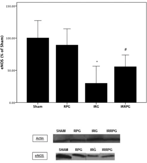
Fig. 6. eNOS expression represented graphically after actin housekeeping normalization;n=6/group. Representative image of Western blot analysis of eNOS and actin in renal
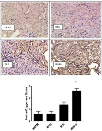
Fig. 7.Heme oxygenase-1 upregulation by red propolis. Values shown are mean±SE. Eight rats for each group.∗P=0.005 vs. Sham,#P<0.001 vs. IRG.
interstitium of the renal cortex and outer medulla from animals that were killed 48 h after renal ischemia. These lesions were less intense in rats that were treated with red propolis when compared with un-treated animals. Tubular necrosis scores can be seen inFig. 2.
Red propolis reduces I/R-induced oxidative stress
As shown inFig. 3, kidney tissue malondialdehyde (MDA) levels were found to be significantly higher in the IR group when compared with theShamgroup (133.9±23.36 vs. 68.10±3.71,P<0.05). After red propolis treatment, there was a significant decrease in MDA levels after I/R injury (90.22±20.82,P<0.05). The same results were found in urine samples, as MDA levels in the urine of animals from the IR group were significantly higher than in the urine of animals from Shamgroup (271.4±145.6 vs. 81.37±10.67,P<005). MDA levels in the urine of the animals who received red propolis treatment were significantly lower after I/R injury (161.4±81.01,P<0.05).
However, the values of glutathione (GSH) were significantly lower when compared with those of the control group (1267±229.5 vs. 1659±107.9,P>0.05), whereas the values of GSH were significantly improved by the red propolis treatment (1784±297.4 vs. 1267±
229.5,P<0.05) as shown inFig. 4.
Red propolis restores the I/R-induced downregulation of eNOS
At 48 h after the surgical procedures, the IR group rats showed markedly lower eNOS protein expression when compared with the
control rats at the semi-quantitative analysis of the area on immuno-histochemistry (0.6±0.5 vs. 1.8±0.4,P=0.003). It is noteworthy that red propolis administration attenuated the down regulation of eNOS expression (IRRPG: 2.2±0.4; IR: 0.6±0.5,P<0.001), as can be seen inFig. 5. All these results were confirmed by western-blot analysis (Fig. 6).
Heme oxygenase-1 upregulation by red propolis
Heme oxygenase-1 immunostaining was weakly present in tubules of both SHAM and RP groups (Fig. 7). Stronger positive stain-ing of HO-1 was observed in the IR group (IR: 1.4±0.5 vs. SHAM: 0.6±0.54,P=0.005). Heme oxygenase-1 immunostaining in the IR-RP group was significantly increased when compared with IR group (2.6±0.5 vs. 1.4±0.5,P<0.001). There was no difference in HO-1 between animals that received red propolis, but were not exposed to renal ischemia. Although the western-blot analysis disclosed similar HO-1 expression in SHAM and IRG groups, it confirmed the increased expression in the IRRPG group (seeFig. 8).
Discussion
794 M.F.B. da Costa et al. / Phytomedicine 22 (2015) 787–795
Fig. 8. Heme-oxygenase expression represented graphically after actin housekeeping normalization;n=6/group. Representative image of Western blot analysis of HO-1 and actin
in renal tissue according each group.∗P<0.001 vs. other groups.
Acute kidney injury is a multifaceted entity that evolves through different stages, culminating in organ failure. Its pathogenesis is com-plex and involves apoptosis, endothelial damage, ROS and inflamma-tion (Basile et al. 2012). Moreover, in the past decade it has become increasingly clear that the cytoprotective response prompted by an injury is crucial for determining the final functional outcome of an in-jured organ (Bonventre 2007). In this study, we focused on determin-ing the protective mechanisms provided by red propolis treatment in a model of renal injury, mainly through anti-oxidant system, eNOS and HO-1.
Propolis is a complex honeybee product with a resinous aspect, containing plant exudates and beeswax. In a previous study, red propolis was characterized as being especially rich in isoflavonoids (Silva et al. 2008). Several studies have demonstrated the anti-oxidative and anti-inflammatory activity of isoflavonoids (Kupeli et al. 2006; Bhandary et al. 2012), but their effects on renal I/R in-jury was never studied. In our study, animals were treated on the day before the procedure with a fixed dose of red propolis extract, re-sulting in a significant attenuation of GFR drop. Although creatinine is limited as a marker of GFR, the protective effect of red propolis was confirmed by a reduced FENa in IR-RP group, a marker of func-tional tubular viability and by lower tubular necrosis index in the histopathological analysis.
MDA and GSH were measured in renal tissue to assess oxidative stress. Reactive oxygen species can result in further renal damage by inducing apoptosis, inflammation and mitochondrial cell damage (Zou et al. 2013; Ye et al. 2010). Reduced oxidative stress induced by renal I/R in animals receiving red propolis was associated with an increase in eNOS expression. Earlier studies demonstrated that the
oxidative stress status that is enhanced during I/R is increased in NO deficiency (Küçük et al. 2012; Tajes et al. 2013). It is possible that reduced oxidative stress is at least partially mediated by increased eNOS expression.
While inducible NOS (iNOS) is fundamentally involved in the pro-cess of kidney damage, inducing inflammation and apoptosis, and the inhibition of its activity (or the absence of iNOS itself in KO-mice) improves renal I/R damagein vivo, the other NOS isoform, eNOS, has protective effects on I/R injury (de Souza et al. 2012; Kato et al. 2009). The effects of isoflavones have been demonstrated on endothe-lial production of NO. It is capable of increasing the expression of eNOS in endothelial cell culture and in the myocardium (Maulik et al. 2012; Joy et al. 2006). In the present animals, I/R injury induced a down-regulation of eNOS expression after 48 h. This down-regulation was completely prevented by previous administration of red propo-lis. In the kidney, this effect of isoflavones on eNOS has shown to be effective against diabetic nephropathy, fructose and lead-induced nephrotoxicity, but no study had evaluated I/R injury (Arya et al. 2011; Palanisamy and Venkataraman 2013;Liu et al. 2012).
(Wang et al. 2010). We investigated whether HO-1 participates in renal protection induced by red propolis. Similarly to others studies, I/R injury itself stimulated HO-1 expression (Ferenbach et al. 2010) and previous treatment with red propolis increased the renal capacity to increase I/R injury-induced HO-1. In addition to reducing oxidative stress, HO-1 can protect against I/R injury by generating CO gas as a byproduct of the breakdown of heme. Several studies have demonstrated the protective role of CO itself in limiting renal damage in ischemia-induced acute kidney injury (Hou et al. 2013).
Our study has several limitations that must be explored in future studies. First, although the main protective component of propolis is isoflavones, future studies with isolated fractions of RP are warranted. Second, other studies must be performed to investigate mechanistic pathways regarding red propolis protection in renal injury, mainly focusing on inflammatory pathway.
In conclusion, this study provides strong evidence of the benefi-cial effects of red propolis on renal I/R injury, which were evaluated for the first time. Red propolis, given at a dose of 150 mg/kg (gas-tric gavage) before the ischemic and reperfusion period, improved kidney damage. The beneficial changes in biochemical parameters, including antioxidant status, were also associated with parallel bene-ficial changes in the histopathological appearance of renal tissue and immunohistochemical evidence.
In summary, our results strongly suggest potential clinical benefits of red propolis use to protect kidneys against acute ischemic renal failure and this protection is associated with reduced oxidative stress and eNOS and heme-oxygenase up regulation.
Conflict of interest
Alexandre Braga Libório and Alice Maria Costa Martins are re-cipients of a grant from the Conselho Nacional de Desenvolvimento Científico e Tecnológico (CNPq). The funders had no role in study de-sign, data collection and analysis, decision to publish or preparation of the manuscript.
References
Agarwal, A., Nick, H.S., 2000. Renal response to tissue injury: lessons from heme oxygenase-1 GeneAblation and expression. J. Am. Soc. Nephrol. 11 (5), 965–973. Arya, A., Yadav, H.N., Sharma, P.L., 2011. Involvement of vascular endothelial nitric
oxide synthase in development of experimental diabetic nephropathy in rats. Mol. Cell. Biochem. 354 (1–2), 57–66.
Basile, D.P., Anderson, M.D., Sutton, T.A., 2012. Pathophysiology of acute kidney injury. Compr. Physiol. 2 (2), 1303–1353.
Bhandary, B., Piao, C.S., Kim, D.-S., Lee, G.-H., Chae, S.-W., Kim, H.-R., Chae, H.-J., 2012. The protective effect of rutin against ischemia/reperfusion-associated hemody-namic alteration through antioxidant activity. Arch. Pharm. Res. 35 (6), 1091–1097. Bonventre, J.V., 2007. Pathophysiology of acute kidney injury: roles of potential
in-hibitors of inflammation. Contrib. Nephrol. 156, 39–46.
Bueno-Silva, B., Alencar, S.M., Koo, H., Ikegaki, M., Silva, G.V.J., Napimoga, M.H., Rosalen, P.L., 2013. Anti-inflammatory and antimicrobial evaluation of neovesti-tol and vestineovesti-tol isolated from Brazilian red propolis. J. Agric. Food. Chem. 61 (19), 4546–4550.
Correa-Costa, M., Amano, M.T., Câmara, N.O.S., 2012. Cytoprotection behind heme oxygenase-1 in renal diseases. World J. Nephrol. 1 (1), 4–11.
Daleprane, J.B., Abdalla, D.S., 2013. Emerging roles of propolis: antioxidant, cardiopro-tective, and antiangiogenic actions. Evid. Based Complement. Alternat. Med. 2013, 175135.
Ditonno, P., Impedovo, S.V., Palazzo, S., Bettocchi, C., Gesualdo, L., Grandaliano, G., Selvaggi, F.P., Battaglia, M., 2013. Effects of ischemia-reperfusion injury in kidney transplantation: risk factors and early and long-term outcomes in a single center. Transplant. Proc. 45 (7), 2641–2644.
Enis Yonar, M., Yonar, S.M., Ural, M., Silici, S., Dü ¸sükcan, M., 2012. Protective role of propolis in chlorpyrifos-induced changes in the haematological parameters and the oxidative/antioxidative status of Cyprinus carpio carpio. Food. Chem. Toxicol. 50 (8), 2703–2708.
Ferenbach, D.A., Kluth, D.C., Hughes, J., 2010. Hemeoxygenase-1 and renal ischaemia-reperfusion injury. Nephron. Exp. Nephrol. 115 (3), e33–e37.
Heemskerk, S., Masereeuw, R., Russel, F.G.M., Pickkers, P., 2009. Selective iNOS inhibi-tion for the treatment of sepsis-induced acute kidney injury. Nat. Rev. Nephrol. 5 (11), 629–640.
Hou, J., Cai, S., Kitajima, Y., Fujino, M., Ito, H., Takahashi, K., Abe, F., Tanaka, T., Ding, Q., Li, X.-K., 2013. 5-Aminolevulinic acid combined with ferrous iron induces car-bon monoxide generation in mouse kidneys and protects from renal ischemia-reperfusion injury. Am. J. Physiol. Renal. Physiol. 305 (8), F1149–F1157. Hsu, S.M., Raine, L., 1981. Protein A, avidin, and biotin in immunohistochemistry. J.
Histochem. Cytochem. 29 (11), 1349–1353.
Joy, S., Siow, R.C.M., Rowlands, D.J., Becker, M., Wyatt, A.W., Aaronson, P.I., Coen, C.W., Kallo, I., Jacob, R., Mann, G.E., 2006. The isoflavone Equol mediates rapid vascular re-laxation: Ca2+-independent activation of endothelial nitric-oxide synthase/Hsp90
involving ERK1/2 and Akt phosphorylation in human endothelial cells. J. Biol. Chem. 281 (37), 27335–27345.
Kato, N., Abe, S., Suto, M., Hiraiwa, K., 2009. Comparison of renal dysfunction in wild-type, IL-6 KO and iNOS KO mice hind limb tourniquet-reperfusion model. Leg. Med. (Tokyo). 11 (Suppl. 1), S248–S251.
Kupeli, E., Orhan, I., Toker, G., Yesilada, E., 2006. Anti-inflammatory and antinocicep-tive potential of Maclura pomifera (Rafin.) Schneider fruit extracts and its major isoflavonoids, scandenone and auriculasin. J. Ethnopharmacol. 107 (2), 169–174. Küçük, A., Yucel, M., Erkasap, N., Tosun, M., Koken, T., Ozkurt, M., Erkasap, S., 2012. The
effects of PDE5 inhibitory drugs on renal ischemia/reperfusion injury in rats. Mol. Biol. Rep. 39 (10), 9775–9782.
Leite, T.T., Macedo, E., Pereira, S.M., Bandeira, S.R., Pontes, P.H., Garcia, A.S., Militão, F.R., Sobrinho, I.M., Assunção, L.M., Libório, A.B., 2013. Timing of renal replacement therapy initiation by AKIN classification system. Crit. Care. 17 (2), R62.
Liu, C.-M., Ma, J.-Q., Sun, Y.-Z., 2012. Puerarin protects rat kidney from lead-induced apoptosis by modulating the PI3K/Akt/eNOS pathway. Toxicol. Appl. Pharmacol. 258 (3), 330–342.
Maulik, S.K., Prabhakar, P., Dinda, A.K., Seth, S., 2012. Genistein prevents isoproterenol-induced cardiac hypertrophy in rats. Can. J. Physiol. Pharmacol. 90 (8), 1117–1125. Mihara, M., Uchiyama, M., Fukuzawa, K., 1980. Thiobarbituric acid value on fresh ho-mogenate of rat as a parameter of lipid peroxidation in aging, CCl4 intoxication, and vitamin E deficiency. Biochem. Med. 23 (3), 302–311.
Nath, K.A., Balla, G., Vercellotti, G.M., Balla, J., Jacob, H.S., Levitt, M.D., Rosenberg, M.E., 1992. Induction of heme oxygenase is a rapid, protective response in rhabdomyol-ysis in the rat. J. Clin. Invest. 90 (1), 267–270.
Palanisamy, N., Venkataraman, A.C., 2013. Beneficial effect of genistein on lowering blood pressure and kidney toxicity in fructose-fed hypertensive rats. Br. J. Nutr. 109 (10), 1806–1812.
Poukkanen, M., Vaara, S.T., Pettilä, V., Kaukonen, K.-M., Korhonen, A.-M., Hovilehto, S., Inkinen, O., Laru-Sompa, R., Kaminski, T., Reinikainen, M., Lund, V., Karlsson, S., FINNAKI study group, 2013. Acute kidney injury in patients with severe sepsis in Finnish Intensive Care Units. Acta Anaesthesiol. Scand. 57 (7), 863–872. Righi, A.A., Negri, G., Salatino, A., 2013. Comparative chemistry of propolis from eight
Brazilian localities. Evid. Based Complement. Alternat. Med. 2013, 267878. Silva, B.B., Rosalen, P.L., Cury, J.A., Ikegaki, M., Souza, V.C., Esteves, A., Alencar, S.M.,
2008. Chemical composition and botanical origin of red propolis, a new type of Brazilian propolis. Evid. Based Complement. Alternat. Med. 5 (3), 313–316. de Souza, A.C.C.P., Volpini, R.A., Shimizu, M.H., Sanches, T.R., Camara, N.O.S., Semedo, P.,
Rodrigues, C.E., Seguro, A.C., Andrade, L., 2012. Erythropoietin prevents sepsis-related acute kidney injury in rats by inhibiting NF-κB and upregulating endothelial nitric oxide synthase. Am. J. Physiol. Renal. Physiol. 302 (8), F1045–F1054. Tajes, M., Ill-Raga, G., Palomer, E., Ramos-Fernández, E., Guix, F.X., Bosch-Morató, M.,
Guivernau, B., Jiménez-Conde, J., Ois, A., Pérez-Asensio, F., Reyes-Navarro, M., Caballo, C., Galán, A.M., Alameda, F., Escolar, G., et al., 2013. Nitro-oxidative stress after neuronal ischemia induces protein nitrotyrosination and cell death. Oxid. Med. Cell. Longev. 2013, 826143.
Uchino, S., Kellum, J.A., Bellomo, R., Doig, G.S., Morimatsu, H., Morgera, S., Schetz, M., Tan, I., Bouman, C., Macedo, E., Gibney, N., Tolwani, A., Ronco, C., 2005. Beginning and ending supportive therapy for the kidney (best kidney) investigators. Acute renal failure in critically ill patients: a multinational, multicenter study. JAMA 294 (7), 813–818.
Wang, H.-Q., Sun, X.-B., Xu, Y.-X., Zhao, H., Zhu, Q.-Y., Zhu, C.-Q., 2010. Astaxanthin upregulates heme oxygenase-1 expression through ERK1/2 pathway and its pro-tective effect against beta-amyloid-induced cytotoxicity in SH-SY5Y cells. Brain Res. 1360, 159–167.
Ye, J., Li, J., Yu, Y., Wei, Q., Deng, W., Yu, L., 2010. L-carnitine attenuates oxidant injury in HK-2 cells via ROS-mitochondria pathway. Regul. Pept. 161 (1–3), 58–66. Zeng, X., McMahon, G.M., Brunelli, S.M., Bates, D.W., Waikar, S.S., 2013. Incidence,
outcomes, and comparisons across definitions of AKI in hospitalized individuals. Clin. J. Am. Soc. Nephrol. 9 (1), 12–20.