Full Length Research Paper
Quantitative evaluation of the mast cell population in
border of ulcers in America Cutaneous Leishmaniasis –
ACL
Cláudio Gleidiston Lima da Silva
Departament Medicine, Federal University of Cariri, Barbalha, Ceará, Brazil.
Author’s email: [email protected]
Accepted 07 February, 2014
Abstract
American Cutaneous Leishmaniasis (ACL) is an infectious disease with a broad spectrum of presentation, has two clinical poles, one anergic and other hyperergic. The inflammatory response to the etiologic agent is complex and involves several cell lines, especially lymphocytes, plasma cells, histiocytes, and antigen-presenting cells. Other cells are observed in inflammatory exudate in a lesser degree, such as eosinophil and mast cells – MC. Several studies have sought to investigate the role of mast cells in cutaneous Leishmaniasis. This study evaluated 64 skin biopsies of leishmanial ulcer edge. The number of mast cells and leishmania were observed in the timeline. It was found that the number of mast cells and leishmania decline with age of the lesion, there is a negative correlation p<0.05. In the late stages of injury (more than eight weeks) the number of mast cell sin the leishmanial lesion sis almost nil. The authors question whether there is really involvement of the mast cells in the chronic phase of Cutaneous Leishmaniasis.
Keywords: Mast Cell, Leishmaniasis, Cutaneous Leishmaniasis.
INTRODUCTION
American Cutaneous Leishmaniasis (ACL) is a group of infectious diseases with the clinical spectrum of presentation similar to that seen in leprosy, ranging from one pole hyperergic to another anergic (Herath et al., 2010). The inflammatory response to the etiologic agents complex and involves several cell lines, especially lymphocytes, plasma cells, histiocytes, and antigen-presenting cells (Gaafar et al., 1995; Elhassan et al., 1994). Other cells are observed in inflammatory exudate in a lesser degree, such as eosinophil and mast cells - (Katakura et al., 1993; Marovt et al., 2010; Saito et al., 1996). Knowledge about the histophysiology of mast cells and eosinophil in recent years, has gained attention from researchers. The role of these cells in the body
phase of the ACL, discussing the results based on the current literature, speculating what would be the role of this cell in the afore said morbidity.
MATERIALS AND METHODS
Ethical approval for this study was obtained from the relevant local ethics committees (Ethics committee of the Medical School of the Federal University of Ceará in Barbalha).
Tissue specimens and patients
Data relating to patients were obtained from Outpatient Tropical Medicine at the Federal University of Ceará, Campus Cariri, Ceará, Brazil. The health records of patients were retrieved and sociodemographic and clinical data were analyzed (n=64, male: female ratio =1.56; group mean age =37.46875 ±19.84381). The diagnoses of patients presenting ACL were confirmed by clinical examination, Montenegro intradermoreaction (IDRM), culture with NNN media and histopathological analysis in the Laboratory of Experimental Pathology (LAPEX) from College of Medicine at the Federal University of Ceará, Campus Cariri, Ceará, Brazil. The archived tissue blocks from surgically resected samples (n=64) at LAPEX were evaluated in this study.
Histological, histochemical staining and Immunohistochemistry for identification of mast cells and Leishmania
For the purposes of morphological and histochemical analysis, samples were fixed in formalin, embedded in paraffin, serially sectioned at 05 microns and evaluated under a conventional light microscope. Sections were stained with hematoxylin and eosin (H&E) for routine examine. In order to visualize mast cells, two sections of each sample were stained with 1% toluidine blue and counter stained with 5% methanol yellow for 5 min, following which the sections were dehydrated, cleared and mounted with synthetic balsam. The sections were stained with Giemsa for Leishmania visualization and count (Armed Forces Institute of Pathology, 1960).
For the Immunohistochemical evaluation of Mast cell, in order to compare the histochemical count, sections were mounted on organosilane-coated slides (Dako Silanized Slides Code No. S3003). The primary mouse monoclonal antibody against Tryptase Antigen (Clone AA1 - Isotype IgG1, kappa, to formalin use of the DAKO CORPORATION) was detected with the aid of an ELITE ABC KIT (product # K0690, Vector Laboratories, Inc. 30-
Cláudio. 02
Ingold Road, Burlingame, CA 94010 – USA), employing chromogen diaminobenzidine for colour development. Slides were then counter stained with Mayer’s hematoxylin and mounted. Negative controls were obtained by substituting normal whole rabbit serum (product # X0902; Dako) for the primary antibodies. A sample of the normal skin (shown previously to be strongly positive to MAST CELL) was used as a positive control.
For the Immunohistochemical evaluation of Leishmania, in order to compare the count of histochemical (Giemsa); four-micron thick histological sections were collected on previously prepared slides (Dako Silanized Slides Code No. S3003); then deparaffinated in xylene and rehydrated in ethanol and water were performed. The endogenous peroxidase activity was blocked. Antigenic retrieval was done in a 96°C water bath with citrate buffer (Dako target retrieval solution), inhibition of the specific bonds with skimmed milk (30mlTRIS + NaCl + 0.3g of bovine albumin Merck + 0.3gMolico skimmed powdered milk). After draining the milk, the primary antibodies Anti-Leishmania polyclonal antibody (diluted at 1/4000) in diluting solution with a Dako unspecific bond reducer were used, then placed in a humid chamber at room temperature for 30 minutes. The biotinylated secondary antibody from an LSAB kit DAKO was used. The development (antigen-antibody bond) was visualized with diaminobenzidine, Dako DAB+ kit, the slides counter stained with Harris’ hematoxylin, and mounted. Anti-Leishmania serum was produced at the Leishmaniasis Surveillance Laboratory of Evandro Chagas Clinical Research Institute (IPEC), FIOCRUZ, by immunization of albino rabbits inoculated with four doses of soluble protein extracts of promastigotes forms of
Leishmania (Leishmania) chagasi
(MHOM/BR/1974/PP75).
Determination of mast cells
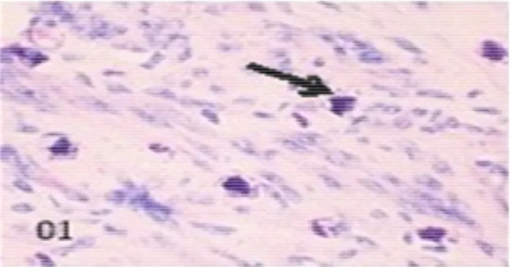
Figure 1. Mast cell stained by toluidine.
Figure 2. Mast cell by immunohistochemistry.
Figure 3. Leishmania by immunohistochemistry.
Determination of Leishmania
The Leishmania stained with Giemsa were identified as small body vacuolated with blue kinetoplast, observed within macrophages or scattered in tissue (figure 4). In the immunohistochemistry small brown point were observed (figure 3). For quantitative analysis, an Optical microscope Olympus - BH2 (model CX31, RTSF, Miami, FL, USA), with・10 ocular and・40 objective lenses, was employed, and an ocular lattice (area 0.092 mm2) with 100 points composed of 10 horizontal and 10 vertical test lines was superimposed on the test field to be measured.
A total area of 1.84 mm2 was evaluated for each of the samples, and this corresponded to 20 randomly selected high-power microscopic fields.
Statistical analysis
Cláudio. 04
Figure 4. Leishmania stained by Giemsa.
Table 1. Epidemiological variables of the population.
Epidemiological variables of the population
N(64) N(100%)
Male Female
42 22
66 34
Rural Urban
60 04
94 06
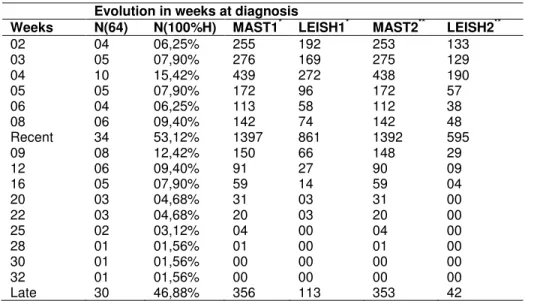
Table 2. Evolution in weeks at diagnosis.
Evolution in weeks at diagnosis
Weeks N(64) N(100%H) MAST1* LEISH1* MAST2** LEISH2**
02 04 06,25% 255 192 253 133
03 05 07,90% 276 169 275 129
04 10 15,42% 439 272 438 190
05 05 07,90% 172 96 172 57
06 04 06,25% 113 58 112 38
08 06 09,40% 142 74 142 48
Recent 34 53,12% 1397 861 1392 595
09 08 12,42% 150 66 148 29
12 06 09,40% 91 27 90 09
16 05 07,90% 59 14 59 04
20 03 04,68% 31 03 31 00
22 03 04,68% 20 03 20 00
25 02 03,12% 04 00 04 00
28 01 01,56% 01 00 01 00
30 01 01,56% 00 00 00 00
32 01 01,56% 00 00 00 00
Late 30 46,88% 356 113 353 42
*Histochemical staining. ** Immunohistochemistry.
immunohistochemistry were performed using paired sample t-test, considering ap<0.05. The comparative analysis between the results obtained for the count of mast cells and Leishmania was performed using the Pearson’s coefficient considering ap< 0,05.
RESULTS
The study population was predominantly male and living in rural areas as shown in Table 1. The youngest patient was 09 years old, the oldest 85. The average age of
cases is around 37.4 years. The study population is made up of brown (n = 43, 67.20%), blacks, (n=04, 6.20%) and whites (n=17, 26.60%). Farmers (n = 37) account for half the population (50%). The progression of the lesions was divided into recent and late. Recent injuries are considered the lesions with down to 08 week sat diagnosis, late lesions with more than 08 week sat diagnosis. The distribution of the time evolution can be observed in Table 2.
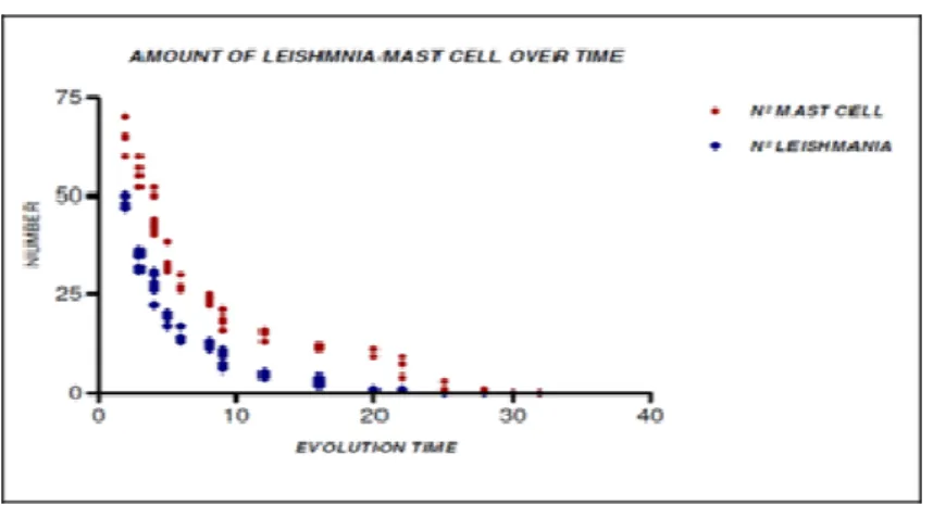
Figure 5: Amount of Leishmania/Mast cell over time
Coefficient = -0, 85061, 95% confidence interval: -0.9068 to -0.7647 with P<0.0001; Strong Negative Correlation), (Carvalho et al., 1994). A similar aspect was seen with the number of the Leishmania. The Leishmania reduce in number over time, figure 5.This reduction has a negative correlation (Pearson’s Coefficient = -0, 78745, 95% confidence interval: -0.8657 to -0.6716 with P<0.0001; Strong Negative Correlation), (Carvalho et al., 1994).
The data collected show that the number of Mast Cells and Leishmania decrease with time of infection and there’s a strong correlation between the reduction of Leishmania and the number of mast cell (Pearson’s Coefficient = 0,9831, 95% confidence interval: 0.9068 to -0.7647 with P<0.0001).
There was no statistical difference between the count of mast cells in histochemistry and immunohistochemistry. On the other hand count for Leishmania the immunohistochemistry was more sensitive than histochemistry.
DISCUSSION
The experimental model of the American Cutaneous Leishmaniasis caused by Leishmania Mexican, determines a progressive disease which rapidly spreads in the susceptible BALB/c mouse, whereas in C57BL/6 mice the disease tends to be self-healing. The first model it has been established that Th1 cytokines lead to disease resistance. On the other hand in the BALB/c mice early presence of IL-4 and IL-10 determine a Th2 response and disease progression (Sacks and Noben-Trauth, 2002). The cells responsible of producing these IL-4 and IL-10 cytokines in the early phases of the disease remain controversial, although cells of the innate immune system are a likely source. The mast cells are possible candidates that produce these cytokines (Vonstebut, 2007; Villaseñor-Cardoso et al., 2008). Besides, due to their location in the skin and mucosa, mast cells are one of the first cells to encounter invading pathogens. They are found in association with blood
vessels, as well as in tissues and surface exposed to external environment such as skin and mucosal linings, which are common portals of infection (Galli et al., 2005). This fact would have an important role in the early stages of Leishmaniasis, say some authors (Villaseñor-Cardoso et al., 2008; Romão et al., 2009). In experimental model of Visceral Leishmaniasis an interleukin-3-dependent augmentation in mast cell committed progenitors is observed in BALB/c but not in C57BL/6 mice during
Leishmania infection. The mast cell supernatants inhibit
IFNg -dependent restriction of Leishmania growth in macrophages in BALB/c mice whereas the reverse phenomenon occurs in C57BL/6 mice. Most of this data point to a possible involvement of mast cells in cutaneous infection by Leishmania, although KATAKURA, 1993, demonstrated that the evolution of the Cutaneous Leishmaniasis is independent of the Mast cell (Katakura et al., 1993).
In this work we observed the presence of mast cells in regular issue in the early stages of skin lesions. The number of mast cells decreased over time. The old lesions showed a smaller number of mast cells. Reducing the number of mast cells over time showed a direct correlation with the decrease in the number of Leishmania in the wound.
with age of the lesion. This observation suggests that even though the mast cell is a cell involved n the early stages of ACL, probably should not being the late phase of the skin lesion. Who would be responsible for the maintenance of the lesion where as mast cells and Leishmania reducer significantly in the late phase of Cutaneous Leishmaniasis?
Sakaguchi and associates (Sakaguchi et al., 1995; Asano et al., 1996) reactivated interest in the concept of T-cell-mediated suppression around the year 1990 by showing that a minor population (<10%) of CD4+ T cells, which co-expresses the interleukin-2 receptor (IL-2R) α -chain (CD25), is primordial for the control of auto reactive T cells in vivo. Subsequent in vitro studies by several groups showed that CD4+CD25+ T cells are both hyporesponsive and suppressive (Thornton and Shevach, 1998; Read et al., 1998). CD4+CD25+ T cells were discovered originally in mice, but a population with identical phenotypic and functional properties has been defined recently in humans (Levings et al., 2001; Baecher-Allen et al., 2001).Studies have related the presence of this cell as the main stay in the maintenance of some diseases, especially Leishmaniasis. Considering that it produces IL4 and especially IL10. Thus, the presence of mast cells in the maintenance of the lesion would not be essential in the late phase of the Cutaneous Leishmaniasis, corroborating the findings of this study (Campanelli et al., 2006; Holaday et al., 1993; Holaday, 1999).
In the present study also found that the use of Giemsa stain to identify Leishmaniain late lesions (more than eight weeks) is not a good method. The immunohistochemistry was more sensitive and statistically representative.
CONCLUSIONS
1. The number of mast cells in Leishmanial infection
reduces numerically overtime;
2. The number of Leishmania in skin lesion reduces
numerically with the passage of time;
3. There is a positive and statistically significant
correlation with the simultaneous reduction of mast cells and Leishmania in skin lesions leishmaniotic, whose cause is unknown;
4. The search for Leishmania in cutaneous lesions
of Leishmanias is with more than 8 weeks should be discouraged.
ACKNOWLEDGEMENTS
The patients who agreed to participate in this study. Evandro Chagas Institute for providing the polyclonalanti-Leishmania antibody. The Teachers Sousa and Moraes to challenge presented; the under graduate students for
Cláudio. 06
data collection and assistance in the routine laboratory.
REFERENCES
Armed Forces Institute of Pathology (1960). Manual of Histologic and Special Staining Technics, ed. 2. New York, The Blakiston Division McGraw-Hill Book Company, Inc.
Asano M, Toda M, Sakaguchi N, Sakaguchi S (1996). Autoimmune disease as a consequence of developmental abnormality of a T-cell subpopulation. J. Exp. Med. 184, 387–396.
Baecher-Allen C, Brown J A, Freeman G J, Hafler DA (2001). CD4+CD25+ regulatory cells in human peripheral blood.J. Immunol. 167, 1245–1253.
Campanelli AP, Roselino AM, Cavassani KA, Pereira MSF, Mortara RA, Claudia I. Brodskyn CI, Gonçalves HS, Belkaid Y, Barral-Netto M, Barral A, Silva JS (2006). CD4+CD25+ T Cells in Skin Lesions of Patients with Cutaneous Leishmaniasis Exhibit Phenotypic and Functional Characteristics of Natural Regulatory T Cells. The Journal of Infectious Diseases; 193:1313–22.
Carvalho Em, Barrala Jml, Costab Jml, Bittencourt A (1994). Clinical and immunopathological aspects of disseminated cutaneous leishmaniasis. Acta Tropica 56: 315-325.
Costa SF (2007). Introdução Ilustrada à Estatística. Editora Habra..
Duarte ML; Rochael MC (2006). Histopathological and immunohistochemical profile of the american cutaneous leishmaniasis with emphasis on fxiiia dermal dendrocytes. An Bras Dermatol. 81(6):537-4.
Elhassan AM, Gaafar A, Theander TG (1994). Antigen-presenting cells in human cutaneous Leishmaniasis due to Leishmania major. Clin. Exp. Immunol. 99:445-453.
Gaafar A, EL Kadaroay, Theander TG, Permin H, Ismail A, Kharazmi A, EL Hassan AM (1995). The Pathology of Cutaneous Leishmaniasis Due to Leishmania Major in Sudan. Am. J. Trop. Med. Hyg. 52:438-442.
Galli S, Kalesnikoff J, Grimbaldeston M, Piliponski AM, Williams CM, Tsai M (2005). Mast cells as ‘tunable’ effector and immunoregulatory cells: recent advances. Annu. Rev. Immunol.23:749-786.
Galli S, Nakae S, Tsai M (2005). Mast cells in the development of adaptive immune responses. Nat Immunol; 6: 135–142.
Gontijo B, De Carvalho MLR (2003). Leishmaniose tegumentar americana. Revista da Sociedade Brasileira de Medicina Tropical. 36(1):71-80, jan-fev.
Herath CHP, Ratnatunga NVI, Waduge R, Ratnayake P, Ratnatunga CN, Ramadasa S (2010). A histopathological study of cutaneous leishmaniasis in sri lanka. Ceylon Med. J. 55: 106-11.
Holaday B (1999). “Immunotherapy for Visceral Leishm,aniasis: Ability of Factors Produced during Anti_leishmania Responses of Skin Test Positive Adults to Inhibit Peripheral Blood Mononuclear Cell Activities Associated with Visceral Leishmaniasis”. Memorias do Instituto Oswaldo Cruz. 94: 55-66.
Holaday B, Pompeu M, Jeronimo S, Texeira M, Sousa A, Vasconcelos W, Pearson R, Abrams J, Locksley R (1993). “Potential Role for Interleukin-10 in Immunosuppression Associated with Kala Azar”. J. Clini. Inves. 92: 2626-2632.
Janssens AS, Heide R, Den Hollander JC, Mulder PGM, Tank B, Oranje AP (2005). Mast cell distribution in normal adult skin. J. Clin. Pathol. 58:285–289.
Jeziorska M, Salamonsen LA, Woolley DE (1995). Mast Cell and Eosinophil Distribution and Activation in Human Endometrium throughout the Menstrual Cycle. Biology of Reproduction53, 312-320. Katakura K, Saito S, Hamada A, Matsuda H, Watanabe N (1993). Cutaneous Leishmaniasis in Mast Cell-Deficient W/Wv Mice. Infection and immunity May, p. 2242-2244.
Levings MK, Sangregorio R, Roncarolo MG (2001). Human CD25+CD4+ T cells suppress naïve and memory T-cell proliferation and can be expanded in vitro without loss of suppressor function. J. Exp. Med. 193, 1295–1302.
responses. Trends in Immunology. Vol.28 No. 5. 234-241.
Oliani SM, Gil CD (2006). Proteína anti-inflamatória anexina 1- mecanismos celulares e relevância clínica.ArqCiênc Saúde out/dez;13(4):186-191.
Oliveira MP, Lima MCR, Calheiros AS, Martins MA, Antas PRZ, De Luca PM, Pirmez C (2004). Leishmania (Viannia) braziliensis: human mast cell line activation induced by logarithmic and stationary promastigote derived-lysates. Clin. Exp. Immunol; 137:19–23. Raposo G, Tenza D, Mecheri S, Peronet R, Bonnerot C, Desaymard C
(1997). Accumulation of major histocompatibility complex class ii molecules in mast cell secretary granules and their release upon degranulation. Molecular Biology of the Cell.Vol. 8, 2631–2645, Read S, Mauze S, Asseman C, Bean A, Coffman R, Powrie F (1998).
CD38+ CD45RBlow CD4+ T cells: a population of T cells with immune regulatory activities in vitro. Eur. J. Immunol. 28: 3435– 3447.
Romão PR, Da Costa Santiago H, Ramos CD, De Oliveira CF, Monteiro MC, De Queiroz Cunha F, Vieira LQ (2009). Mast cell degranulation contributes to susceptibility to Leishmania major. Parasite Immunol Mar; 31(3):140-6.
Sacks D, Noben-Trauth N (2002). The immunology of susceptibility and resistance to Leishmania major in mice. Nat Rev Immunol. 2: 845– 858.
Saha B, Tonkal A, Croft S, Roy S (2004). Mast cells at the host– pathogen interface: host-protection versus immuneevasion in Leishmaniasis. ClinExp Immunol; 137:19–23.
Saito S, Hamada A, Watanabe N, Obata T, Katakura K, Ohtomo H (1996). Eosinophil chemotactic activity in Leishmania amazonensis promastigotas. Parasitology Research. Volume 82, Number 6.
Sakaguchi S, Sakaguchi N, Asano M, Itoh M, Toda, M (1995). Immunologic self-tolerance maintained by activated T cells expressing IL-2 receptor α-chains. J. Immunol. 155, 1151–1164.
Silva JRL (2008). Inflamação crônica na asma brônquica. Pulmao RJ; Supl1:S2-S7.
Theoharides TC, Cochrane DE (2004). Critical role of mast cells in inflammatory diseases and the effect of acute stress. J. Neuroimmunol. 146 1–12.
Thornton AM, Shevach EM (1998). CD4+CD25+ immunoregulatory T cells suppress polyclonal T-cell activation in vitro by inhibiting interleukin-2 production. J. Exp. Med. 188, 287–296
Villaseñor-Cardoso MI, Salaiza N, Delgado J, Gutiérrez-Kobeh L, Pérez-Torres A, Becker I (2008). Mast cells are activated by Leishmania mexicana LPG and
regulate the disease outcome depending on the genetic background of the host. Parasite Immunology; 30; 425–434.
Vonstebut E (2007). Immunology of cutaneous leishmaniasis: the role of mast cells, phagocytes and dendritic cells for protective immunity. Eur. J. Immunol. 17: 115–122.
Walls AF, Jones DB, Williams JH, Church MK, Holgate ST (1990). Immunohistochemical identification of mast cells in formaldehyde-fixed tissue using monoclonal antibodies specific for tryptase. J. Pathol. Oct; 162(2):119-26.