Developing Gonads
Davood Sabour1, Xueping Xu2, Arthur C. K. Chung2,3, Damien Le Menuet2,4, Kinarm Ko5,6, Natalia Tapia1, Marcos J. Arau´zo-Bravo1,7, Luca Gentile1, Boris Greber1, Karin Hu¨bner1, Vittorio Sebastiano8,
Guangming Wu1, Hans R. Scho¨ler1,9*, Austin J. Cooney2*
1Department of Cell and Developmental Biology, Max Planck Institute for Molecular Biomedicine, Mu¨nster, Germany,2Department of Molecular and Cellular Biology, Baylor College of Medicine, Houston, Texas, United States of America,3Centre for Inflammatory Diseases and Molecular Therapies, The University of Hong Kong, Pokfulam, Hong Kong,4INSERM, U693, Faculte´ de Me´decine Paris-Sud, Paris, France,5Center for Stem Cell Research, Institute of Biomedical Sciences and Technology, Konkuk University, Seoul, Republic of Korea,6Department of Neuroscience, School of Medicine, Institute of Biomedical Sciences and Technology, Konkuk University, Seoul, Republic of Korea,7Group of Computational Biology and Systems Biomedicine, Biodonostia Health Research Institute, San Sebastia´n, Spain,8Institute for Stem Cell Biology and Regenerative Medicine, Stanford University School of Medicine, Palo Alto, California, United States of America,9Medical Faculty, University of Mu¨nster, Mu¨nster, Germany
Abstract
Expression of germ cell nuclear factor (GCNF; Nr6a1), an orphan member of the nuclear receptor gene family of transcription factors, during gastrulation and neurulation is critical for normal embryogenesis in mice. Gcnf represses the expression of the POU-domain transcription factor Oct4 (Pou5f1) during mouse post-implantation development. Although Gcnf expression is not critical for the embryonic segregation of the germ cell lineage, we found that sexually dimorphic expression ofGcnfin germ cells correlates with the expression of pluripotency-associated genes, such asOct4,Sox2, and Nanog, as well as the early meiotic marker geneStra8. To elucidate the role of Gcnf during mouse germ cell differentiation, we generated anex vivo Gcnf-knockdown model in combination with a regulated CreLox mutation ofGcnf. Lack of Gcnf impairs normal spermatogenesis and oogenesisin vivo, as well as the derivation of germ cells from embryonic stem cells (ESCs)in vitro. Inactivation of theGcnfgenein vivoleads to loss of repression ofOct4expression in both male and female gonads.
Citation:Sabour D, Xu X, Chung ACK, Le Menuet D, Ko K, et al. (2014) Germ Cell Nuclear Factor Regulates Gametogenesis in Developing Gonads. PLoS ONE 9(8): e103985. doi:10.1371/journal.pone.0103985
Editor:Qiang Wu, National University of Singapore, Singapore
ReceivedMarch 27, 2014;AcceptedJuly 4, 2014;PublishedAugust 20, 2014
This is an open-access article, free of all copyright, and may be freely reproduced, distributed, transmitted, modified, built upon, or otherwise used by anyone for any lawful purpose. The work is made available under the Creative Commons CC0 public domain dedication.
Data Availability:The authors confirm that all data underlying the findings are fully available without restriction. All relevant data are within the paper and its Supporting Information files.
Funding:This work was supported by Max Planck Society and the DFG grant for research unit germ cell potential, FOR 1041 (HSR) and National Institutes of Health grant NIH P01 GM081627 (AJC). The funders had no role in study design, data collection and analysis, decision to publish, or preparation of the manuscript.
Competing Interests:The co-author Austin J. Cooney is a PLOS ONE Editorial Board member. This does not alter the authors’ adherence to PLOS ONE Editorial policies and criteria.
* Email: [email protected] (AJC); [email protected] (HRS)
Introduction
Germ Cell Nuclear Factor (GCNF), also known as nuclear receptor subfamily 6, group A, member 1 (Nr6a1), is an orphan member of the nuclear receptor (NR) gene family of ligand-activated transcription factors [1]. Gcnf exhibits distinctive DNA-binding properties. Recombinant Gcnf binds as a homodimer to a response element, a direct repeat with zero base-pair spacing, i.e., a DR0, to repress the expression of genes bothin vivoandin vitro [1–5].In vivo, Gcnf appears to be part of a large complex termed transiently retinoid induced factor (TRIF) that binds to DR0 DNA elements in P19 embryonal carcinoma cells (ECCs) or embryonic stem cells (ESCs) [2,6,7]. In the mouse,Gcnfis expressed in the developing nervous system, placenta [8,9], embryonic gonads, and adult ovaries and testes [1,10,11]. It is also expressed in round spermatids in mouse and in spermatocytes undergoing meiotic prophase in human [1,10–12]. Gcnf has been found to regulate the transcription of the protamine genes Prm 1 and Prm 2 in mouse testis, antagonizing the effects of CREM tau by binding to
conserved DR0 element, located in theOct4proximal promoter [2]. AsOct4is required for the survival of primordial germ cells (PGCs) [21], the question arises as to whether Gcnf plays a role in the segregation or maintenance of the PGC lineage. To address this question, we developed new mouse models andin vitro cell models to study the role of Gcnf in PGCs.
We therefore conducted mechanistic studies to determine the requirement of Gcnf for germ cell development during mamma-lian development, particularly during meiosis, which represents a critical checkpoint in the formation of normal gametes. Progres-sion of in vitro–derived germ cells through meiosis is still a rare phenomenon and poses a huge challenge for reproductive medicine. Studies on gene regulation in germ cells should enhance our understanding of the mechanisms underlying meiotic process-esin vivo, subsequently enabling us to enhance the progression of germ cells through meiosisin vitro. As Gcnf plays an essential role in gene regulation during early mammalian development, this study focused on elucidating the function of Gcnf during the development of murine germ cells.
Results
Gcnf is not required for the segregation of germ cell lineage
Two outstanding questions in the analysis of theGcnf-mutant embryo phenotype are whether segregation of the germline has been compromised and whether Gcnf is required for further PGC development. To directly address these questions, we crossed the Oct4-GFP(DPE) reporter mice—a transgenic line generated with a construct lacking the proximal enhancer (PE) element of theOct4 promoter, which typically drivesOct4expression in the epiblast, here driving the expression of green fluorescent protein (GFP)— with ourGcnf-knockout (KO) mice. After gastrulation, widespread expression of Oct4 is repressed and only maintained in PGCs, which reside in the posterior of the embryo and allantois [22,23]. Therefore, in this study the Oct4-GFP(DPE) reporter is specifically expressed in PGCs. We sacrificed pregnant female mice from a heterozygous cross at embryonic day (E) 8.5 and E9.5 (Figure 1). At E8.5, the embryos have not yet turned, and PGCs are clearly visible in the posterior of the embryo, as they begin their anterior migration (Figures 1A and 1B). In Gcnf-mutant embryos, mor-phological deformation is already evident, and PGCs can be observed in the posterior of the embryo; as there is an approximately equal number of PGCs in wild-type (wt) and mutant embryos (Figure 1C and 1D), Gcnf is not required for segregation of the germ cell lineage. At E9.5, the wt embryo has completed turning, and PGCs can be observed migrating anteriorly along the hindgut toward the developing midgut (Figures 1E and 1F). In contrast, Gcnf-mutant embryos fail to turn, and an ectopic tailbud forms due to posterior truncation. PGCs are still clearly visible in the posterior of mutant embryos, and rather than migrating along the hindgut, some are carried into the ectopic tailbud, indicating that Gcnf is not required for the maintenance of PGCs at this stage of embryonic development.
Sexually dimorphic Gcnf expression in germ cells of the developing gonads
Although Gcnf is not required for germ lineage segregation or maintenance of PGCs during their migratory phase, it may play a role in later stages of germ cell development after formation of the gonads, when Oct4 is normally repressed and meiosis has been initiated. We generated a sensitiveGcnfreporter mouse, aGcnf LacZ gene trap (GT) model [17]. We analyzed LacZ activity in the dissected gonads of male and female embryos from E12.5 to
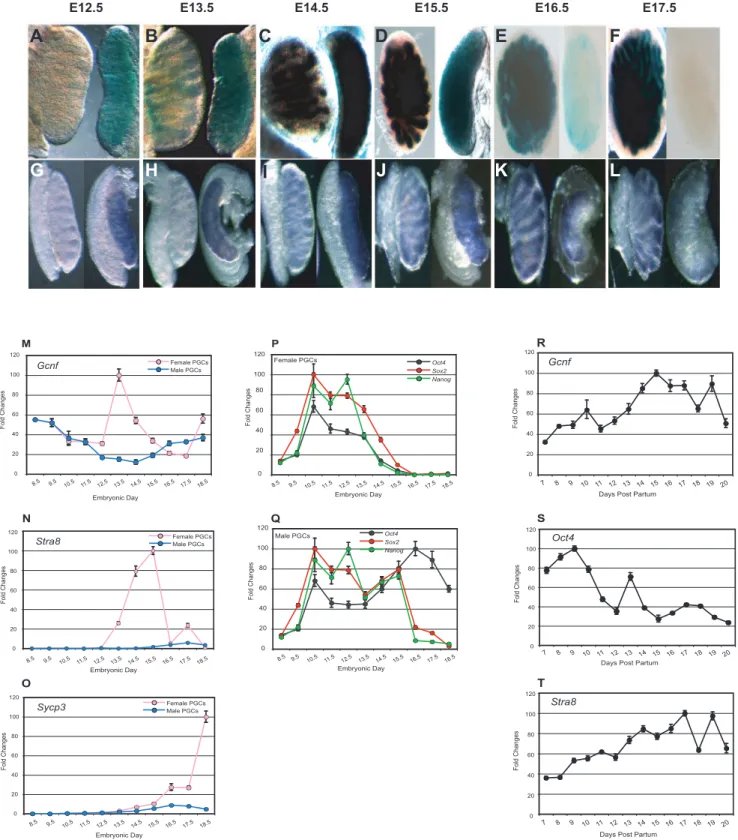
E17.5. In female gonads,Gcnfexpression was detected on E12.5, maintained through E15.5, decreased by E16.5, and completely turned off by E17.5. In contrast, in male gonads, LacZ reporter activity was not detected until E13.5. b-Galactosidase activity continued to increase through E15.5 and was maintained through E17.5 (Figures 2A–2F). To ensure that the sexually dimorphic expression of Gcnf detected in the Gcnf LacZ Knockin (GT) mouse model reflected the expression of the normal gene, we analyzed Gcnf expression in wt mice by whole-mount in situ hybridization (WMISH). A very similar pattern of expression was observed. At E12.5 and E13.5,Gcnfwas expressed in female, but not in male, gonads. By E14.5,Gcnfexpression was detected in both gonads, but by E17.5, Gcnf expression was turned off in female gonads but maintained in male gonads (Figures 2G–2L).
To confirm that theGcnf expression pattern observed in the male and female gonads is consistent withGcnfexpression in germ cells, we analyzed Gcnfexpression in purified germ cells. Germ cells from gonads of Oct4-GFP(DPE) mice at different stages of development were isolated by fluorescence-activated cell storing (FACS) for GFP-positive cells. Expression of Gcnf, as well as meiosis-related and pluripotency-associated genes in female and male PGCs during fetal development was assessed by real-time quantitative RT-PCR (q-RT-PCR). In female PGCs, expression of pluripotency-associated genes, such as Oct4, Sox2, and Nanog, was steadily downregulated, whereas Gcnf expression was upregulated starting on E12.5, one day before the onset of meiosis (Figures 2M and 2P). In contrast, in male PGCs, which undergo mitotic arrest on E13.5, the mRNA levels of Oct4 remained steadily high until birth (Figure 2Q), whereas expression ofGcnf and the meiosis-related genesStra8andSycp3remained low in all embryonic stages (Figures 2M, 2N, and 2O). To confirm the correlation of Gcnf and a regulatory role in meiosis, we determined the expression ofGcnf,Oct4, and the meiosis-related geneStra8in spermatogonial cells isolated from testes of 7 days post partum (dpp) to 20 dpp. We observed a steady upregulation of expression ofGcnfandStra8starting on 7 dpp, but a consistent downregulation ofOct4expression starting on 10 dpp, which is the onset of meiosis in the male mouse (Figures 2R–2T).
Taken together, these results show that Gcnf expression is upregulated, in contrast toOct4expression, which is downregu-lated, in both female and male germ cells upon entry into meiosis. The temporal expression ofGcnfin male and female PGCsin vivo suggests a potential role for Gcnf in the downregulation ofOct4 expression at the onset of meiosis and an involvement in either initiation of meiosis or activation of meiosis-related genes in both male and female germ cells.
Knockdown of Gcnfin vivoimpairs spermatogenesis
determine the functionality of lentiviral-infected cells, GSCs containing shGcnf or shLacZ were transplanted into the seminif-erous tubules of germ cell–depleted, busulfan-treated C57BL/6 mice. Only one testis per mouse was transplanted, with the non-transplanted testis serving as a control for endogenous spermato-genesis. Three months later, we observed colonization of the transplanted GSCs in both groups (Figures S2C–S2H).
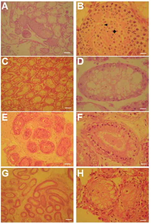
Furthermore, histological analysis of testicular sections of more than 10 transplanted testes from each group revealed that the transplanted shLacZ GSCs (control) had colonized the seminifer-ous tubules and restored spermatogenesis (Figures 3A and 3B). These tubules contained developing male germ cells of all stages, spermatids, and spermatozoa. In contrast, transplanted shGcnf GSCs had only colonized the tubules, but had failed to restore spermatogenesis (Figures 3E and 3F). Both groups of transplanted GSCs did not form teratomas in the host mice. In addition, histological sections of the non-transplanted testes from the same mice showed no restoration of spermatogenesis 3 months after transplantation, demonstrating that endogenous recovery of spermatogenesis had not occurred in germ cell–depleted mice (Figures 3C, 3D, 3G, and 3H). These results clearly demonstrate that knockdown ofGcnfblocks, or represses, further differentiation of GSCs into functional sperm within the seminiferous tubules of busulfan-treated mice.
Impairment of the differentiation of Gcnf-deficient ESCs into PGCsin vitro
To assess the role played by GCNF in the formation of germ cells, we assessed the ability ofGcnf-deficient ESCs to generate PGCsin vitro. Wt and mutantGcnfESC lines were derived from Gcnf+/+; Oct4-GFP(DPE) andGcnf2/2; Oct4-GFP(DPE) embryos from the same mouse model used in the studies in Figure 1. We analyzed the global gene expression patterns of PGCs derivedin vitroon days 12 and 15 of differentiation ofGcnf-deficient ESCs (mutant) compared with those ofin vitro–derived PGCs on days 12 and 15 of differentiation of Oct4-GFP(DPE) ESCs (wt). We had
previously assessed the dynamics of gene expression and the formation ofin vitro–derived PGCs from ESCs in a time-course analysisin vitro, and determined that cells on days 12 and 15 of differentiation exhibited the most PGC-like structural character-istics.
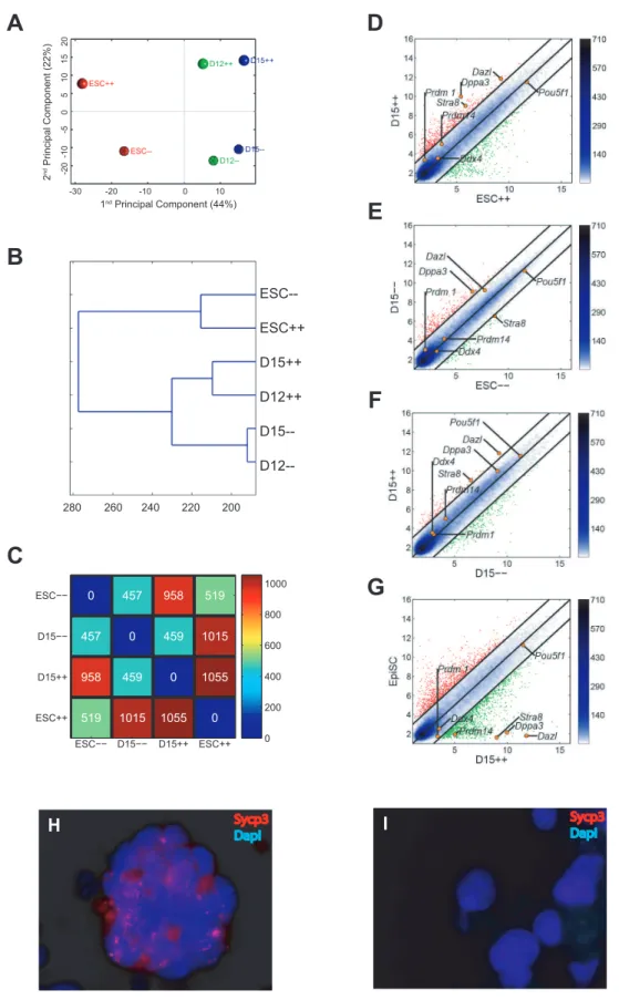
Principal component analysis (PCA) revealed that Oct4-GFP(DPE) ESC-derived PGCs exhibited a different global gene expression pattern compared with Gcnf-deficient ESC-derived PGCs on days 12 and 15 of differentiationin vitro. The second PCA component revealed a developmental difference between wt andGcnf-deficient PGCsin vitro(Figure 4A). Hierarchical cluster analysis of the global gene expression pattern revealed that wtin vitro–derived PGCs clustered separately from Gcnf-deficient in vitro–derived PGCs (Figure 4B). A scatter plot of the global gene expression of ESCs and day-15 in vitro–derived PGCs (corre-sponding of final stage ofin vitro–derived PGCs) showed a more dramatic increase in differentially expressed genes between ESCs and wt in vitro–derived PGCs than between ESCs and Gcnf -deficientin vitro–derived PGCs (Figures 4D and 4E). Specifically, ESCs differed from wt day-15 PGCs by 1,055 genes and from Gcnf-deficient day-15 PGCs by 457 genes (Figure 4C). In addition, a scatter plot of the global gene expression of wt and Gcnf-deficient PGCs derived on day 15 ofin vitrodifferentiation showed that many genes were differentially expressed, particularly PGC-specific and meiosis-related genes (Figure 4F). Finally, comparison of the global gene expression pattern of wt day-15 in vitro–derived PGCs with epiblast stem cells (EpiSCs) showed a dramatic number of differentially expressed genes, thus demon-strating proper germ cell differentiation in vitro (Figure 4G). These results suggest that in the absence of Gcnf, the derivation of in vitroPGCs is impaired.
To confirm these findings, we determined the expression of day-15in vitro–derived PGCs by real-time q-RT-PCR. Expression of all known germ cell and meiotic markers was found to be upregulated to moderate levels in Oct4-GFP(DPE) ESC-derived PGCs compared with Oct4-GFP(DPE) ESCs, whereas the expression of these genes was upregulated to a lesser extent in
Figure 1. Detection of PGCs expressing green fluorescence from Oct4-GFP(DPE) in wt andGcnf2/2embryos.(A–B) wt E8.5 embryos. (C–D)Gcnf2/2E8.5 embryos. (E–F) wt E9.5 embryos. (G–H)Gcnf2/2E9.5 embryos. Arrowheads indicate migratory PGCs. Note that the scale bars are 50 uM. Figures A, C, E and G are 256and figures B, D, F, and H are 406.
Figure 2. Analysis ofGcnf, pluripotency-associated genes, and meiosis-related gene expression profiles in PGCs in male and female embryonic gonads and in spermatogonial cells.(A–F) Analysis ofb-galactosidase activity in male (left-hand side of each panel) and female (right-hand side of each panel) gonads inGcnfLacZ KI embryos on E12.5 to E17.5. (G–L) WMISH analysis ofGcnfexpression in the gonads of wt male (left-hand side of each panel) and female mice (right-hand side of each panel) on E12.5 to E17.5. (M–Q) Time-course analysis of gene expression profiles of PGCs from male and female genital ridges and embryonic gonads. Real-time q-RT-PCR analysis of (M)Gcnf. (P–Q)Oct4, Nanog,and Sox2. (N)Stra8, and (O)Sycp3in PGCs isolated by FACS sorting Oct4-GFP–positive cells from the genital ridges (E8.5 to E11.5) and from male and female fetal gonads on E12.5 to E18.5. (R–T) Time-course analysis of gene expression profiles of spermatogonial cells in newborn testes. Real-time q-RT-PCR analysis of (R)Gcnf. (S)Oct4, and (T)Stra8in spermatogonial cells isolated by FACS sorting Oct4-GFP–positive cells from the testes at 7 dpp to 20 dpp, i.e., during meiosis in male mice.
doi:10.1371/journal.pone.0103985.g002
Gcnf-deficient ESC-derived PGCs compared withGcnf-deficient ESCs (Figures S3A and S3B). We identified 11 genes that were expressed in PGCs but not in pluripotent stem cells [26]. The majority of these genes were found to be expressed in wt day-15in vitro–derived PGCs but not in day-15Gcnf-deficientESC-derived PGCs (Figures S3C and S3D). We examined the localization of synaptonemal complex protein 3 (Sycp3) in both groups of in vitro–derived PGCs on day 15 of differentiation, and showed the positive immunostaining for this protein in Oct4-GFP(DPE)
ESC-derived PGCs but not in Gcnf-deficient ESC-derived PGCs (Figures 4H and 4I).
To exclude the possibility that day-15 PGCs are functionally pluripotent, we subcutaneously injected both wt and mutant day-15in vitro–derived PGCs into severe combined immunodeficiency (SCID) recipients. One month after the injection, we did not observe any teratomasin vivo, further confirming that these cells were not pluripotent ESCs. Taken together, our observations suggest that Gcnf is essential for the differentiation of PGCs and initiation of meiosisin vitro.
Gcnf is required for development of female fetal PGCs into oocytes
To examine whetherGcnf-deficient ESCs have the develop-mental potential to differentiate into oocytes in vivo, we used a blastocyst injection strategy with XX Oct4-GFP(DPE) ESCs and XX Gcnf2/2; Oct4-GFP(DPE) ESCs (Figure 5A) to generate chimeras. We then transferred the injected blastocysts into pseudopregnant recipients and subsequently observed germline contribution in the gonads of an E14.5 embryo by means of Oct4-GFP expression. The germline contribution of Gcnf2/2; Oct4-GFP(DPE) ESCs was estimated to be 15% compared with 45% for control ESCs (wt Oct4-GFP(DPE) ESCs), a difference of more than 60%. As more than 30% of chimeric embryos generated from Gcnf-deficient ESCs have brain and neural tube deficiency owing to the involvement of Gcnf in neurogenesis [9,19] (Figure 5B), we rescued female chimeric gonads by transplanting them under the kidney capsule of SCID mice and examined them after 4 weeks to determine whether wt and Gcnf2/2 chimeric gonads had the same potential to develop from early meiotic germ cells into mature oocytes. We observed that both groups of PGCs could develop ectopically into an ovarian-like structure (54% for control ESCs compared with 51% forGcnf2/2ESCs) under the kidney capsule (Figures 5C and 5D), but we were only able to isolate fully developed MII oocytes from the ovarian-like structures of chimeric gonads generated by blastocyst injection with Oct4-GFP(DPE) ESCs (35–40 MII oocytes from each ovarian-like structure) (Figure 5E). We did not observe fully developed oocytes from the ovarian-like structures ofGcnf2/2chimeric gonads, but only a few degenerated oocytes (5–10 degenerated oocytes from each ovarian-like structure), confirming thatGcnf-deficient ESCs have no developmental potential and differentiate into oocytesin vivo. Although, for unknown reasons, we could not monitor GFP signal in fully developed oocytes under the fluorescence microscope, we were able to detect GFP expression by nested PCR in cumulus cell–free oocytes derived from control ESCs (Figure 5F).
Gcnf is required for the repression ofOct4expression in embryonic gonads
During embryonic development,Oct4expression is regulated in a sexually dimorphic manner. In female gonads, the expression of Oct4is silenced upon entry of cells into meiosis. However, in male gonads, Oct4 expression is also repressed later on E17.5. The inverse correlation between Gcnf and Oct4 expression in developing gonads suggests that the retinoid-induced repression ofOct4during the differentiation of ESCs may reflect a role for Gcnf in the repression of Oct4 expression during gonadal development [6]. To test this hypothesisin vivo, we generated a regulated KO ofGcnfby combiningGcnffl/fl
mouse model with an ERT2Cre mouse model [27] in which this regulator had been knocked into the ROSA locus. We treated pregnant female mice with 4-OH tamoxifen on E11.5 (a time point late enough not to affect the early developmental requirement for GCNF) and then harvested embryos on E17.5 to analyze Oct4 expression. The experiment was designed such that each embryo would be homozygous for the floxed allele and be either Cre positive or negative. The Cre-negative embryo would serve as control for tamoxifen treatment, which can slow embryonic development. An E17.5 litter was weighed, the gonads were dissected, and DNA was
isolated from the remainder of the embryos for genotyping. The genotyping identified the embryos as Cre positive or negative and revealed the status of the floxedGcnfallele (Figures 6A and 6B). In the Cre-negative embryos, theGcnffloxed allele was intact, while in Cre-positive littermates, the floxed allele was completely recombined to yield what we term a Type I deletion, a deletion in the DNA binding domain–encoding exon [3] (Figure 6B).
WMISH analysis of Oct4 expression in the male gonads on E17.5 showed that Cre-negative embryos exhibited low Oct4 expression, while Cre-positive embryos showed loss of repression of Oct4expression (10 of 10 embryos). In female embryos, two events were observed. In some embryos, there was repression of Oct4expression on E17.5, while in others, there was clear loss of repression of Oct4 expression on E17.5 (Figures 6C). The difference between the two types of female responses is most likely due to the timing of Cre recombination of the floxedGcnf alleles. In female gonads with abrogatedOct4repression, theGcnf allele is inactivated at an early enough time point to prevent significantGcnfexpression. In female gonads with silencedOct4 expression, theGcnfgene is not completely inactivated, resulting in induction ofGcnfexpression and production of enough Gcnf protein to silence Oct4 expression. However, Gcnf is clearly required to repressOct4expression during the critical stages of gonadal development.
To confirm the correlation of Gcnf and Oct4 expression in gonadal development and particularly the requirement for Oct4 levels in the initiation of meiosis, we first performed conditional ectopic overexpression ofOct4levels in 14.5-dpc female gonads. To assess the short-term effect ofOct4ectopic expressionin vivo, we crossed a Tet-Oct4 mouse [28] with an Oct4-GFP mouse. We fed pregnant female mice drinking water that contained doxycy-cline on E12.5 (a time point when meiosis-related genes just start to be overexpressed in developing female gonads) and then harvested the embryos on E14.5 to analyzeOct4,Stra8, andSycp3 expression.
Interestingly, ectopic activation of Oct4in developing female gonads results in the inhibition of expression of meiosis markers such as Stra8 and Sycp3 (Figure 6F) and the interruption of meiotic initiation as typically seen in female gonads at this time of PGC development (Figures 2N and 2O). Taken together, these results showed that downregulation ofOct4expression is clearly required to activateStra8andSycp3expression during the onset of meiosis in developing female gonads, and confirmed that meiosis markers are not upregulated in developing male germ cells before birth, asOct4levels are not reduced in male gonads.
Gcnf is required for enhancingStra8expression in embryonic gonads
Stra8 expression is regulated in a sexually dimorphic manner during embryonic development. In female gonads, the expression of Stra8 is activated upon entry of cells into meiosis on E12.5, increases gradually till E15.5, and is dramatically downregulated on E16.5 at prophase I arrest in female gonads (Figure 2N). However, in male gonads,Stra8expression is nearly completely repressed during embryonic development. In male gonads,Stra8 expression is activated in the testis during neonatal development, increases steadily starting on 7 dpp, and continues till 19 dpp, coincident with the onset of meiosis in male spermatogonial cells scattering density, the darker the blue color. Positions of some known PGC markers are shown as orange dots. Gene expression levels are log2scaled. Genes upregulated in ordinate samples compared with abscissa samples are shown in red circles; genes downregulated are shown in green. (H–I) The localization of Sycp3 was performed in (H) Oct4-GFP(DPE) ESC-derived PGCs and (I) Gcnf-deficient ESC-derived PGCs of day-15 in vitro– differentiation cultures.
(Figure 2T). The direct correlation between Gcnf and Stra8 expression in developing gonads (Figure 2M, 2N and 2T) suggests that the retinoid-induced activation ofStra8expression during the differentiation of ESCs may reflect a role for Gcnf in the activation of Stra8expression during gonadal development [7,29]. To test this hypothesisin vivo, we used the same KO model forGcnfby crossing theGcnffl/fl
mouse with an ERT2Cre mouse. We treated
pregnant female mice with 4-OH tamoxifen on E11.5 (a time point late enough not to affect the early developmental require-ment for Gcnf) and then harvested embryos on E15.5 to analyze Stra8expression.
WMISH analysis ofStra8expression in the female gonads on E15.5 showed that Cre-negative embryos exhibited high Stra8 expression, while Cre-positive embryos showed loss of activation of
Figure 5.(A) Schematic overview of blastocyst injection, germline contribution, and kidney capsule transplantation of Oct4-GFP ESCs andGcnf2/2; Oct4-GFP(DPE) ESCs. (B) E14.5-chimericGcnf2/2embryos; note that the neural tubes and brain are affected. (C–D) E14.5-chimeric female gonads developed an ovarian-like structure under the kidney capsule of SCID recipients 4 weeks after transplantation. (E) MII oocytes isolated from only the wt ovarian-like structure and (F) expression of GFP performed by nested PCR from an isolated MII oocyte (E1 to E4 depicts the experimental replicate numbers).
doi:10.1371/journal.pone.0103985.g005
Stra8 expression (4 of 4 embryos) (Figure 6D–6E). This means that in female gonads with loss ofGcnfexpression, theStra8gene is not completely activated, resulting in inhibition of meiotic initiation. These results showed that Gcnf is clearly required to activate Stra8 expression during onset of meiosis in the development of both male and female gonads.
Discussion
Previous studies [1,10,11,30–32] have shown that theGcnfgene is active in adult testes and ovaries and is required for reproduction in female mice [17]. In adult testis, Gcnf acts as a transcriptional regulator of the postmeiotic genes Protamine 1 and 2 (Prm 1, Prm 2), functionally inhibiting CREMtau [4,13,14,33], whereas in the adult ovary, Gcnf regulates the expression of bone morphogenetic protein 15 (Bmp15) and growth differentiation factor 9 (Gdf9) [17]. The Cre/Lox mutation of floxedGcnfby ZP3Cre leads to subfertility in female mice [17]. Our current study demonstrates that Gcnf is involved in male and female gametogenesis during mouse development. We show that loss of Gcnf results in failure to produce spermatogonia and oocytes in developing testes and ovaries, respectively. Our study also shows that Gcnf is required to repressOct4expression during gonadal development.
Segregation of germ cells in the mouse occurs on E7.25 of embryonic development, when PGCs arise as a small cluster of alkaline phosphate–positive cells in the extraembryonic mesoderm [34–38]. At this developmental time point, Gcnf is functionally active and is required to repress pluripotency genes such asOct4— as the pluripotent late epiblast is patterned during gastrulation— and Gcnf2/2 embryos begin displaying phenotypic alterations compared with wt embryos. Thus, to determine whether Gcnf is required in the formation of the PGC lineage, we crossed the PGC reporter Oct4-GFP(DPE) withGcnf2/2mice. We clearly observe GFP-positive PGCs in the posterior ofGcnf2/2 embryos. Thus, Gcnf is not required for the segregation of the germ cell lineage. The normal anterior migration of PGCs from the allantois to the midgut, however, is disrupted in the Gcnf2/2embryos, and some PGCs even migrate into the ectopic tail bud [8]. Although we cannot rule out that Gcnf plays a direct role in the migration and homing of PGCs, the defect is more likely a secondary effect of the developmental defects of KO embryos.
To determine whether Gcnf plays a role in later stages of PGC development, we analyzed Gcnf expression at various stages of germ cell development.GcnfLacZ Knockin (KI) mice displayed sexually dimorphic expression ofb-galactosidase activity, suggest-ing that Gcnf plays a role in gonadal development. To confirm that this reporter accurately reflected Gcnf expression, we performed WMISH hybridization, which confirmed the sexually dimorphic expression observed. We then isolated germ cells from transgenic mice harboring the Oct4-GFP(DPE) reporter to confirm that the expression ofGcnf was indeed confined to the germ cell compartment. In addition, we analyzed the expression of meiosis-related genes as well as pluripotency-associated genes, as Oct4 is the main pluripotency-related gene required for the maintenance and survival of PGCs [21].
In female embryonic gonads, upregulation ofGcnfexpression begins at around E12.5, concordant with the upregulation of the premeiosis-related gene Stra8. In contrast, downregulation of pluripotency-associated genes, such asOct4,Sox2, andNanog, is observed at the onset of meiosis. Gene expression analysis in spermatogonial cells of newborn testis and embryonic female gonads confirmed a correlation between Gcnf, Oct4, andStra8 expression at the onset of meiosis in both sexes. In contrast, we did not observe coincident upregulation ofGcnfwithStra8andSycp3 expression, and downregulation of Oct4, Sox2, and Nanog expression in male embryonic gonads at mitotic arrest on E13.5. As Gcnf represses the expression ofOct4[2], these results suggest a possible association between Gcnf, Oct4, and Stra8 in regulating the initiation of meiosis in germ cells. To confirm this hypothesis and the requirement forOct4downregulation in the activation of Stra8 and Sycp3 expression during onset of meiosis in the development of female gonads, we demonstrated that upregulation of Oct4levels alone inhibited the expression of meiosis markers such as Stra8 and Sycp3 and consequently interrupted meiotic initiation. This suggests that the concomitant activation of Gcnf expression and Oct4 repression is required for the activation of meiosis-related genes.
We previously reported that adult unipotent germline stem cells derived from mouse testes could colonize the seminiferous tubules of busulfan-treated mice and restore spermatogenesis [24]. Here, we observed that knockdown ofGcnfby using small hairpin RNA (shRNA) in GSCs inhibits the differentiation of GSCs into functional sperm and blocks the restoration of spermatogenesis after transplantation of these cells into the seminiferous tubules of busulfan-treated mice. This observation is consistent with those of previous reports [1,10,11,30–32], confirming a functional role for Gcnf in spermatogenesis in the mouse.
To support a potential role for Gcnf during germ cell development and onset of meiosis, we assessed the ability of Gcnf-deficient ESCs to differentiate into PGCsin vitro. By using global gene expression analysis, real-time q-RT-PCR of known and newly identified PGCs markers, and localization of meiosis-related proteins, we showed that in contrast to wt ESCs,Gcnf2/2 ESCs fail to differentiate into PGCs and initiate meiosisin vitro. Previous studies have shown thatGcnfis expressed in developing oocytes during folliculogenesis in the mouse [1,10,11]. Gcnf may be involved in female fertility by regulating the expression of Bmp15andGdf9[17]. In this study, we demonstrated that Gcnf is required for the development of female fetal PGCs into oocytes. Although both wt andGcnf-deficient ESCs were able to contribute to the germline, following the transplantation of a chimeric fetal ovary under the kidney capsule of SCID mice, Gcnf2/2 PGCs could not generate mature oocytes inside the grafted ovary. Our finding suggests that Gcnf is essential for further development and maturation of PGCs into oocytes.
Here we report that Gcnf is required for spermatogenesis and oogenesis in the mouse and that loss of Gcnf disrupts the formation of functional gametes. Taken together, our findings using (1) gene expression in PGC development, (2) disruption of spermatogenesis after downregulation of Gcnf, (3) impairment of in vitro differentiation of PGCs by using Gcnf2/2 ESCs, and (4) the
Figure 6. Analysis of the role of Gcnf and Oct4 in gonadal development in Cre/Lox mutation of theGcnfgene.(A–B) The genotyping determined whether the embryos were Cre positive or negative, and the status of the floxedGcnfallele. (C) WMISH analysis ofOct4expression on E17.5 in male and female gonads of controlGcnfwt (Cre2) treated with 4-OH tamoxifen and 4-OH tamoxifen–induced ERT2Cre-inactivatedGcnf2/2 (Cre+) embryos. (D) The status of the floxedGcnfallele. (E) WMISH analysis ofStra8expression on E15.5 in female gonads of controlGcnfwt (Cre2) treated with 4-OH tamoxifen and 4-OH tamoxifen–induced ERT2Cre-inactivatedGcnf2/2(Cre+) embryos. (F) Real-time q-RT-PCR analysis showed that two days of treatment with doxycycline resulted in the upregulation of Oct4 expression and the downregulation ofStra8andSycp3expression in 14.5-dpc female gonads of Tet-Oct4 pregnant mice (14.5-dpc female PGCs were considered as 1 for normalization).
doi:10.1371/journal.pone.0103985.g006
inability of ectopic development of a chimeric fetal ovary under the kidney capsule of host mice all provide a robust model for a regulatory role for Gcnf in germ cell development. Support for the role of Gcnf in the maintenance of germ cells has been provided by ex vivoexperiments using anin vivo–regulated Cre/Lox KO of Gcnfduring gestation. TheGcnfgene was successfully deleted in the gonads and the embryo as a whole, and defects in Oct4 expression were observed. These defects are due to inactivation of the Gcnf gene in PGCs, as opposed to a secondary effect on development, as the whole embryos were normal overall. These results contrast those of a similar KO strategy by the Page group, who reported no functional requirement for Gcnf or its ligand-binding domain in germ cell development [39]. The two Gcnf floxed models differ in the regions deleted; in addition, another significant difference between the two studies is the use of tamoxifen to activate ERT2Cre, which first has to be metabolized into the active form 4-hydroxy tamoxifen before it can activate ERT2Cre, which would cause a functional delay to generate defects in Oct4 expression. We used 4-hydoxy tamoxifen to activate ERT2Cre, which would allow for earlier deletion of Gcnf expression and thus readily generate a phenotype.
Clearly, like in ESCs, Gcnf plays an essential role in the repression ofOct4expression in female gonads as they exit mitosis and enter meiotic prophase I. Postnatally, Gcnf likely plays a role in maintaining germ cells in both male and female newborns, as shown in the various experiments detailed here. This functional window must lie between the ERT2Cre KO of Gcnf during embryonic development, as the gonads still contain germ cellsand the late-stage ZP3Cre KO of Gcnfin adult female mice, which showed fertility defects but no loss of germ cells. Other mouse models are needed to delineate this important function of Gcnf in early postnatal life.
Several research groups have shown that the Stra8 gene (stimulated by retinoic acid gene 8) is a premeiotic marker that is expressed in premeiotic germ cells of the testes and ovaries and is regulated by retinoic acid [40,41]. Loss ofStra8gene function in germ cells leads to inhibition of onset of meiosis and results in the failure of germ cells to undergo meiosis in the ovaries or testes of Stra82/2mice [42–44].
We hypothesize that Gcnf plays either a direct or indirect role in germ cell development, and suggest a possible function for Gcnf in the activation or initiation of meiosis by enhancing Stra8 expression and inhibiting the expression of pluripotency-associated genes. This knowledge should enhance our understanding of the key mechanisms underlying germ cell development and onset of meiosis as well as prove beneficial for the derivation of viable and fertile sperm and oocytes from pluripotent stem cellsin vitro.
Methods
Animal ethics
This study was performed in accordance with the recommen-dations of the Federation of Laboratory Animal Science Associ-ations (FELASA). The corresponding ethics protocol for mice works was approved by the Landesamt fu¨r Natur, Umwelt und Verbraucherschutz (LANUV) of the state of North Rhine-Westphalia in Germany, and the corresponding ethics protocol for testicular transplantation was approved by Center for Stem Cell Research, Institute of Biomedical Sciences and Technology, Konkuk University, Seoul, Republic of Korea.
Analysis of the Oct4-GFP(DPE) in Gcnf KO embryos
Transgenic mice harboring the Oct4-GFP(DPE) reporter were crossed withGcnf+/2mice to generateGcnf+/2; Oct4-GFP(DPE)
mice, which were subsequently crossed with Gcnf+/2 mice to generate embryonic litters with +/+, +/2, and 2/2. Timed pregnant female mice were euthanized and embryos were harvested on E8.5 and E9.5. The fluorescent Oct4-GFP(DPE) reporter was visualized in PGCs using a Zeiss Stemi SV11.
Reporter LacZ staining
LacZ staining of embryonic gonads, which were isolated from matings between Gcnf LacZ KI heterozygous mice on E12.5– E17.5 with X-Gal staining, was performed according to the manufacturer’s protocol (Millipore, Billerica, MA). The yolk sacs of the embryos were lysed for genotyping.
WMISH Gcnffl/fl
mice were crossed with ROSA ERT2Cre mice to generateGcnffl/+
ERT2Cre mice, which were then crossed with Gcnffl/fl
mice; female mice were checked daily for plugs and treated with 1 mg 4-OH tamoxifen (Sigma, H7904) once on E11.5. The gonads were isolated from embryos on E17.5 and fixed in 4% paraformaldehyde. The yolk sacs of embryos were lysed for genotyping. WMISHs were carried out as described previously [2]. Genotyping Primers:
GcnfLacZ KI:
Primer1: 59TCGAGCGATGTTTCGCTT39; Primer2: 59ATATGGGATCGGCCATTGA39; Primer3: 59CAGTGCTGACTTATCCATG39; Primer4: 59TTCCTGTTCATGCCCATCT39. Gcnffl:
PCR-A: 59CGAACTCAGAAATCCACC39; PCR-C: 59AATCACAAACACCACAACTC39. GcnfType I:
Type I-F: 59CCAATTCCCCCAAAGTGTC39 Type I-R: 59CAGGTCGAGGGACCTATAAC39. ERT2Cre:
Cre-F: 59CGGTCGATGCAACGAGTGAT39; Cre-R: 59CCACCGTCAGTACGTGAGAT39.
Isolation of PGCs from mouse fetus and flow cytometric analysis
Germ cells of E8.5 to E18.5 embryos were isolated from OG2 (Oct4DPE-GFP)/CD1 mice by disaggregating the genital ridges of E8.5 to E11.5 embryos and the fetal gonads of E12.5 to E18.5 embryos. Disaggregation was performed by enzymatic digestion with collagenase type 1A (Sigma) and trypsin-EDTA (0.25%) at a ratio of 1:2 with shaking at 700 rpm for 8 min at 37uC. Single-cell suspension was achieved more efficiently by triturating the cell aggregates. Enzymatic digestion was stopped by adding a two-fold excess of DMEM/F12+10% FBS. Cell suspensions were washed once in DMEM/F12+10% FBS and FACS sorted for GFP. GFP-positive PGCs were directly sorted into lysis buffer RLT (QIAGEN, Hilden, Germany) using a FACSAria cell sorter (BD Biosciences). Single-cell suspensions of GFP-positive PGCs were analyzed using a FACSAria cell sorter (BD Biosciences). Data analysis was done using FlowJo software (Treestar).
RNA isolation
RT-PCR and real-time quantitative RT-PCR
Total RNA (250 ng) was reverse transcribed into cDNA by using the High-Capacity cDNA Reverse Transcription Kit (Applied Biosystems, Darmstadt, Germany). q-RT-PCR was performed on 7900ht and ht fast devices (Applied Biosystems) with milliQ H2O and TaqMan Universal PCR Master Mix
(Applied Biosystems). Raw data was obtained by SDS 2.3 (Applied Biosystems). Gene expression was normalized to the mouse housekeeping geneHprt. Relative quantitation of gene expression was calculated using theDDCt method. Three technical replicates were used for each real-time q-RT-PCR reaction; a reverse transcriptase blank and a no-template blank served as negative controls. Primer sequences for real-time q-RT-PCR are listed as supplementary material (Tables S1 and S2).
Vector construction and transduction with lentiviral vectors
pLVTHM-Td tomato was constructed by replacing the GFP in pLVTHM with the Td-tomato coding sequence. DNA constructs designed to produce shRNA hairpins targeting Gcnf (59- GGAT- GAATTGGCAGAGCTTGATTCAAGAGATCAAGCTCTGC-CAATTCATCC –39) or LacZ (59-GTGGATCAGTCGCTGAT-TAAATTCAAGAGATTTAATCAGCGACTGATCCAC –39) were cloned in front of the H1 promoter into the pLVTHM-Td tomato vector to produce shGcnf and pLVTHM-shLacZ, respectively. The 293T cell line was cultured in DMEM supplemented with 15% FBS. The recombinant lentiviruses were produced by transient transfection of 293T cells with 12mg of pLVTHM-shGcnf or pLVTHM-shLacZ, 8.5mg of psPax2, and 3mg of pMD2.G using Lipofectamine 2000 (Invitrogen) according to the manufacturer’s protocol. The supernatant was collected at 24 and 48 hr of transfection and concentrated by ultracentrifugation at 26,000 rpm for 2 hr at 4uC using an SW41 rotor (Beckman Coulter). After ultracentrifugation, the supernatant was decanted, and the viral pellet was resuspended in 200ml of DMEM. The suspension was stored at 280uC until use. pLVTHM and packaging plasmids were provided by D. Trono (Geneva, Switzer-land). GSCs were plated on 24-well plates (56104cells/well), and 20ml of the concentrated virus were added to the medium. Cells were washed after 16 hr of incubation.
Testicular transplantation
The transplantation experiments were performed as previously described [45]. Four-week-old germ-cell–depleted (40 mg/kg busulfan-treated) male mice were used as recipient mice. Approximately 36105 cells were injected with a micropipette (80-mm diameter tips) into the seminiferous tubules of the testes of recipient mice through the efferent duct. Three months after the transplantation, the mice were sacrificed, and the seminiferous tubules were dissociated by collagenase treatment and examined under a fluorescence microscope to detect transplanted GSCs by their RFP expression. To assess further colonization as well as restoration of spermatogenesis, the testes were fixed in Bouin’s solution and embedded in paraffin. Sections were stained with hematoxylin and eosin (H&E).
ESCs
Male and female Oct4-GFP(DPE) ESCs and female Gcnf2/2 ESCs [8] were grown on gamma-irradiated mouse embryonic fibroblasts (MEFs) in KNOCKOUT DMEM medium containing 4.5 g/l glucose and supplemented with 15% KNOCKOUT SR (Invitrogen), 2 mM L-glutamine (Invitrogen), 100mM nonessential amino acids (Invitrogen), 1mM 2-mercaptoethanol (Invitrogen),
and 50mg/ml each penicillin and streptomycin (Invitrogen) in the presence of 1,000 U/ml murine leukemia inhibitory factor (LIF) (ESGRO; Chemicon).
In vitrodifferentiation of PGCs from ESCs
Oct4-GFP(DPE) ESCs andGcnf2/2ESCs were grown in 0.1% gelatin-coated tissue culture plates in ESC medium supplemented with 15% FBS (Invitrogen) but without LIF and in the absence of MEFs at a density of approximately 8–96104 cells/cm2.
Non-adherent cells were removed after 3 days and the medium was replaced. GFP expression was monitored every 3 days in cells undergoing further differentiation until day 15. Approximately 25% of all cells exhibited GFP expression on day 15. The medium in the master culture plates was replaced every 2 days [46].
Isolation ofin vitro–derived PGCs from differentiation culture plates
Every 3 days, cells were dissociated with 0.25% trypsin-EDTA, neutralized with KNOCKOUT DMEM containing 15% FBS, washed twice with PBS, and then resuspended in KNOCKOUT DMEM containing 15% FBS. Approximately 26106cells/ml in DMEM/FBS were used for FACS sorting. Flow cytometry was performed on a FACSAria cell sorter. Putative PGCs derived from ESCs were sorted based on Oct4-GFP expression and then subjected to germ cell characterization, e.g., gene expression, ability for tumor formation, and localization of meiosis-related proteins.
Teratoma formation
Day-12 and day-15in vitro–derived PGCs were suspended in DMEM containing 15% FBS. A 200-ml aliquot (16106cells) of the cell suspension was injected subcutaneously into the dorsal flanks of SCID mice. Following injection of thein vitro–derived PGCs, wt and mutant ESCs were also subcutaneously injected into the SCID mice as controls. Four weeks after the injection, tumors were dissected from the mice. Samples were fixed in PBS containing 4% paraformaldehyde and embedded in paraffin. Tissue sections were stained with H&E.
Immunohistochemistry
Immunohistochemistry for Sycp3 was performed using the spreading technique, as previously described by others [47]. Slides were incubated with primary anti-Sycp3 (1:500; Abcam) for 2 hr at room temperature. After washing in blocking solution, slides were incubated with secondary fluorescent antibody (1:1,000; Alexa 568; Molecular Probes) for 1 hr at room temperature and then mounted in DAPI-containing mounting medium (Vecta-shield; Vector Laboratories Inc.).
Chimera formation
Six- to eight-week-old female mice (B6C3F1) were induced to superovulate (via intraperitoneal injection of 7.5 I.U. PMSG and 48 hr later of 7.5 I.U. hCG) and mated with CD1 male mice. Blastocysts were collected on day 3.5 after a vaginal plug check and flushed in HCZB medium containing 0.1% polyvinylpyrrol-idone (PVP). Blastocysts were washed extensively with HCZB medium and cultured in Alpha MEM medium containing 0.2% BSA at 37uC, 5% CO2in air until injection with ESCs.
HCZB medium containing 0.1% PVP and 0.2% BSA. Groups of 12–15 cells were injected into a single blastocyst. Injected embryos were then transferred into a drop of Alpha MEM-BSA and cultured at 37uC, 5% CO2in air. After 4 hr of culture, chimeric
blastocysts were transferred into the uterine horns of 2.5-dpc pseudopregnant CD1 recipient female mice.
Transplantation of chimeric ovaries under the kidney capsule
Fourteen days after transfer of the chimeric blastocysts into the uterine horns, the fetal ovaries of 14.5-dpc embryos were obtained from pregnant pseudopregnant CD1 recipient female mice and screened for germline contribution under fluorescent microscope. One to two chimeric fetal ovaries without attached mesonephroses were transplanted under the kidney capsule of ovariectomized female SCID recipient mice. Four weeks after transplantation, chimeric ovarian grafts were removed from the recipient mice and analyzed for both ovarian maturation and number and quality of existing oocytes.
Nested PCR to detect GFP signal in oocytes
The presence of the GFP signal was determined using nested PCR, as previously described by others [48]. Briefly, for primary reactions, we used 4ml of DNA template from a pool of isolated oocytes in a total volume of 15ml. Cycling conditions consisted of an initial 3-min denaturation at 95uC, followed by 25 cycles, each consisting of a 30-sec denaturation at 94uC, a 45-sec annealing at 55uC, and a 1-min extension at 72uC. These 25 cycles were followed by a 7-min extension at 72uC. For the nested amplifications, we used 1.0ml of the primary PCR product as the template in a total reaction volume of 12ml. Nested cycling conditions were as described for the primary amplification, except that 35 cycles were used. Reaction products were subsequently analyzed by 2% agarose gel electrophoresis. Negative controls of oocytes from non-transgenic mice as well as a reverse transcriptase blank and a no-template blank were included in each experiment.
Global gene expression analysis
For RNA isolation, PGCs were sorted by FACS, collected by centrifugation, lysed in RLT buffer (QIAGEN), and processed using the RNeasy micro and mini kits with on-column DNase digestion as per the manufacturer’s instructions. Integrity of RNA samples was quality checked using a 2100 Bioanalyzer (Agilent). When possible, 300 ng of total RNA per sample were used as starting material for linear amplification (Illumina TotalPrep RNA Amplification Kit, Ambion), which involved synthesis of T7-linked double-stranded cDNA and 14 hr of in vitro transcription incorporating biotin-labeled nucleotides. Purified and labeled cRNA was then quality checked on a 2100 Bioanalyser and hybridized as biological or technical duplicates for 18 hr onto MouseRef-8 v2 gene expression BeadChips (Illumina), following the manufacturer’s instructions. After being washed, the chips were stained with streptavidin-Cy3 (GE Healthcare) and scanned using an iScan reader and accompanying software.
Microarray data processing
Bead intensities were mapped to gene information using BeadStudio 3.2 (Illumina). Background correction was performed using the Affymetrix Robust Multi-array Analysis (RMA) back-ground correction model [49]. Variance stabilization was performed using log2scaling, and gene expression normalization
was calculated with the quantile method implemented in the lumi package of R-Bioconductor. Data post-processing and graphics
were performed with in-house developed functions in Matlab. Hierarchical clustering of genes and samples was performed with a correlation metric and the Unweighted Pair-Group Method using Average (UPGMA) linkage method.
All the raw and processed microarray data discussed in this publication have been deposited into NCBI’s Gene Expression Omnibus [50] and are accessible through GEO Series accession number GSE57506. (http://www.ncbi.nlm.nih.gov/geo/query/ acc.cgi$acc = GSE57506).
Supporting Information
Figure S1 Global gene expression shows the Gcnf
expression in different lines of mouse GSCs, compared with ESCs and germline-derived pluripotent stem cells (gPSCs).
(EPS)
Figure S2 Light microscopy of transplanted and non-transplanted GSCs into the testis.(A–B) Tomato lentivirus– infected GSCs before transplantation into the seminiferous tubules of germ cell–depleted busulfan-treated mice. (C–D) GSCs without GcnfsiRNA (controls, only with tomato lentivirus); note the red GSCs have colonized the tubules. (E–F) GSCs withGcnfsiRNA; note the red GSCs have colonized the tubules. (G–H) Non-transplanted testicular tubules, showing no recovery of germ cells in germ cell–depleted testes 3 months after treatment with busulfan. Note that the scale bars are 20 uM.
(TIF)
Figure S3 Expression of known germ cell markers and the newly identified genes in day-15 in vitro–derived PGCs. Real-time q-RT-PCR of pluripotency and known germ cell markers in (A) Oct4-GFP(DPE) ESC-derived PGCs and (B) Gcnf-deficient ESC-derived PGCs compared with ESCs. D denotes days. Real-time q-RT-PCR of the newly identified genes in (C) Oct4-GFP(DPE) ESC-derived PGCs and (D)Gcnf-deficient ESC-derived PGCs compared with ESCs.
(EPS)
Figure S4 Efficiency of shRNA transfection in GSCs.(A– B) FACS sorting plots, showing that more than 80% of the infected GSCs exhibited a positive tomato signal. (C) Depict the efficiency of shGcnf and showed the downregulation of gene in cells after 48 and 72 hours (75% downregulation of Gcnf after 72 hours).
(TIF)
Table S1 Assay codes for TaqMan real-time q-RT-PCR. (TIF)
Table S2 List of primers used for SYBR Green real-time q-RT-PCR.
(TIF)
Acknowledgments
We thank David Obridge, Martina Sinn, and Claudia Ortmeier for technical assistance. We also thank Jeanine Mu¨ller-Keuker for help with the figures and Martin Stehling for assistance with FACS sorting.
Author Contributions
References
1. Chen F, Cooney AJ, Wang Y, Law SW, O’Malley BW (1994) Cloning of a novel orphan receptor (GCNF) expressed during germ cell development. Mol Endocrinol 8: 1434–1444.
2. Fuhrmann G, Chung AC, Jackson KJ, Hummelke G, Baniahmad A, et al. (2001) Mouse germline restriction of Oct4 expression by germ cell nuclear factor. Dev Cell 1: 377–387.
3. Lan ZJ, Chung AC, Xu X, DeMayo FJ, Cooney AJ (2002) The embryonic function of germ cell nuclear factor is dependent on the DNA binding domain. J Biol Chem 277: 50660–50667.
4. Yan ZH, Medvedev A, Hirose T, Gotoh H, Jetten AM (1997) Characterization of the response element and DNA binding properties of the nuclear orphan receptor germ cell nuclear factor/retinoid receptor-related testis-associated receptor. J Biol Chem 272: 10565–10572.
5. Schmitz TP, Susens U, Borgmeyer U (1999) DNA binding, protein interaction and differential expression of the human germ cell nuclear factor. Biochim Biophys Acta 1446: 173–180.
6. Gu P, LeMenuet D, Chung AC, Mancini M, Wheeler DA, et al. (2005) Orphan nuclear receptor GCNF is required for the repression of pluripotency genes during retinoic acid-induced embryonic stem cell differentiation. Mol Cell Biol 25: 8507–8519.
7. Gu P, Xu X, Le Menuet D, Chung AC, Cooney AJ (2011) Differential recruitment of methyl CpG-binding domain factors and DNA methyltransfer-ases by the orphan receptor germ cell nuclear factor initiates the repression and silencing of Oct4. Stem Cells 29: 1041–1051.
8. Chung AC, Katz D, Pereira FA, Jackson KJ, DeMayo FJ, et al. (2001) Loss of orphan receptor germ cell nuclear factor function results in ectopic development of the tail bud and a novel posterior truncation. Mol Cell Biol 21: 663–677. 9. Susens U, Aguiluz JB, Evans RM, Borgmeyer U (1997) The germ cell nuclear
factor mGCNF is expressed in the developing nervous system. Dev Neurosci 19: 410–420.
10. Katz D, Niederberger C, Slaughter GR, Cooney AJ (1997) Characterization of germ cell-specific expression of the orphan nuclear receptor, germ cell nuclear factor. Endocrinology 138: 4364–4372.
11. Lan ZJ, Gu P, Xu X, Cooney AJ (2003) Expression of the orphan nuclear receptor, germ cell nuclear factor, in mouse gonads and preimplantation embryos. Biol Reprod 68: 282–289.
12. Agoulnik IY, Cho Y, Niederberger C, Kieback DG, Cooney AJ (1998) Cloning, expression analysis and chromosomal localization of the human nuclear receptor gene GCNF. FEBS Lett 424: 73–78.
13. Hummelke GC, Cooney AJ (2001) Germ cell nuclear factor is a transcriptional repressor essential for embryonic development. Front Biosci 6: D1186–1191. 14. Hummelke GC, Meistrich ML, Cooney AJ (1998) Mouse protamine genes are
candidate targets for the novel orphan nuclear receptor, germ cell nuclear factor. Mol Reprod Dev 50: 396–405.
15. Braat AK, Zandbergen MA, De Vries E, Van Der Burg B, Bogerd J, et al. (1999) Cloning and expression of the zebrafish germ cell nuclear factor. Mol Reprod Dev 53: 369–375.
16. Joos TO, David R, Dreyer C (1996) xGCNF, a nuclear orphan receptor is expressed during neurulation in Xenopus laevis. Mech Dev 60: 45–57. 17. Lan ZJ, Gu P, Xu X, Jackson KJ, DeMayo FJ, et al. (2003) GCNF-dependent
repression of BMP-15 and GDF-9 mediates gamete regulation of female fertility. EMBO J 22: 4070–4081.
18. Chung AC, Cooney AJ (2001) Germ cell nuclear factor. Int J Biochem Cell Biol 33: 1141–1146.
19. Chung AC, Xu X, Niederreither KA, Cooney AJ (2006) Loss of orphan nuclear receptor GCNF function disrupts forebrain development and the establishment of the isthmic organizer. Dev Biol 293: 13–24.
20. Akamatsu W, DeVeale B, Okano H, Cooney AJ, van der Kooy D (2009) Suppression of Oct4 by germ cell nuclear factor restricts pluripotency and promotes neural stem cell development in the early neural lineage. J Neurosci 29: 2113–2124.
21. Kehler J, Tolkunova E, Koschorz B, Pesce M, Gentile L, et al. (2004) Oct4 is required for primordial germ cell survival. EMBO Rep 5: 1078–1083. 22. Yeom YI, Fuhrmann G, Ovitt CE, Brehm A, Ohbo K, et al. (1996) Germline
regulatory element of Oct-4 specific for the totipotent cycle of embryonal cells. Development 122: 881–894.
23. Yoshimizu T, Sugiyama N, De Felice M, Yeom YI, Ohbo K, et al. (1999) Germline-specific expression of the Oct-4/green fluorescent protein (GFP) transgene in mice. Dev Growth Differ 41: 675–684.
24. Ko K, Tapia N, Wu G, Kim JB, Bravo MJ, et al. (2009) Induction of pluripotency in adult unipotent germline stem cells. Cell Stem Cell 5: 87–96. 25. Ogawa T, Dobrinski I, Avarbock MR, Brinster RL (2000) Transplantation of
male germ line stem cells restores fertility in infertile mice. Nat Med 6: 29–34.
26. Sabour D, Arauzo-Bravo MJ, Hubner K, Ko K, Greber B, et al. (2011) Identification of genes specific to mouse primordial germ cells through dynamic global gene expression. Hum Mol Genet 20: 115–125.
27. Ventura A, Kirsch DG, McLaughlin ME, Tuveson DA, Grimm J, et al. (2007) Restoration of p53 function leads to tumour regression in vivo. Nature 445: 661– 665.
28. Hochedlinger K, Yamada Y, Beard C, Jaenisch R (2005) Ectopic expression of Oct-4 blocks progenitor-cell differentiation and causes dysplasia in epithelial tissues. Cell 121: 465–477.
29. Barreto G, Borgmeyer U, Dreyer C (2003) The germ cell nuclear factor is required for retinoic acid signaling during Xenopus development. Mech Dev 120: 415–428.
30. Hirose T, O’Brien DA, Jetten AM (1995) RTR: a new member of the nuclear receptor superfamily that is highly expressed in murine testis. Gene 152: 247– 251.
31. Yang G, Zhang YL, Buchold GM, Jetten AM, O’Brien DA (2003) Analysis of germ cell nuclear factor transcripts and protein expression during spermato-genesis. Biol Reprod 68: 1620–1630.
32. Zhang YL, Akmal KM, Tsuruta JK, Shang Q, Hirose T, et al. (1998) Expression of germ cell nuclear factor (GCNF/RTR) during spermatogenesis. Mol Reprod Dev 50: 93–102.
33. Hummelke GC, Cooney AJ (2004) Reciprocal regulation of the mouse protamine genes by the orphan nuclear receptor germ cell nuclear factor and CREMtau. Mol Reprod Dev 68: 394–407.
34. Anderson R, Copeland TK, Scholer H, Heasman J, Wylie C (2000) The onset of germ cell migration in the mouse embryo. Mech Dev 91: 61–68.
35. Ginsburg M, Snow MH, McLaren A (1990) Primordial germ cells in the mouse embryo during gastrulation. Development 110: 521–528.
36. Lawson KA, Hage WJ (1994) Clonal analysis of the origin of primordial germ cells in the mouse. Ciba Found Symp 182: 68–84; discussion 84–91. 37. Molyneaux KA, Stallock J, Schaible K, Wylie C (2001) Time-lapse analysis of
living mouse germ cell migration. Dev Biol 240: 488–498.
38. Saitou M, Barton SC, Surani MA (2002) A molecular programme for the specification of germ cell fate in mice. Nature 418: 293–300.
39. Okumura LM, Lesch BJ, Page DC (2013) The ligand binding domain of GCNF is not required for repression of pluripotency genes in mouse fetal ovarian germ cells. PLoS One 8: e66062.
40. Koubova J, Menke DB, Zhou Q, Capel B, Griswold MD, et al. (2006) Retinoic acid regulates sex-specific timing of meiotic initiation in mice. Proc Natl Acad Sci U S A 103: 2474–2479.
41. Oulad-Abdelghani M, Bouillet P, Decimo D, Gansmuller A, Heyberger S, et al. (1996) Characterization of a premeiotic germ cell-specific cytoplasmic protein encoded by Stra8, a novel retinoic acid-responsive gene. J Cell Biol 135: 469– 477.
42. Anderson EL, Baltus AE, Roepers-Gajadien HL, Hassold TJ, de Rooij DG, et al. (2008) Stra8 and its inducer, retinoic acid, regulate meiotic initiation in both spermatogenesis and oogenesis in mice. Proc Natl Acad Sci U S A 105: 14976– 14980.
43. Baltus AE, Menke DB, Hu YC, Goodheart ML, Carpenter AE, et al. (2006) In germ cells of mouse embryonic ovaries, the decision to enter meiosis precedes premeiotic DNA replication. Nat Genet 38: 1430–1434.
44. Mark M, Jacobs H, Oulad-Abdelghani M, Dennefeld C, Feret B, et al. (2008) STRA8-deficient spermatocytes initiate, but fail to complete, meiosis and undergo premature chromosome condensation. J Cell Sci 121: 3233–3242. 45. Ogawa T, Arechaga JM, Avarbock MR, Brinster RL (1997) Transplantation of
testis germinal cells into mouse seminiferous tubules. Int J Dev Biol 41: 111– 122.
46. Hubner K, Fuhrmann G, Christenson LK, Kehler J, Reinbold R, et al. (2003) Derivation of oocytes from mouse embryonic stem cells. Science 300: 1251– 1256.
47. Peters AH, Plug AW, van Vugt MJ, de Boer P (1997) A drying-down technique for the spreading of mammalian meiocytes from the male and female germline. Chromosome Res 5: 66–68.
48. Wu G, Hao L, Han Z, Gao S, Latham KE, et al. (2005) Maternal transmission ratio distortion at the mouse Om locus results from meiotic drive at the second meiotic division. Genetics 170: 327–334.
49. Irizarry RA, Bolstad BM, Collin F, Cope LM, Hobbs B, et al. (2003) Summaries of Affymetrix GeneChip probe level data. Nucleic Acids Research 31. 50. Edgar R, Domrachev M, Lash AE (2002) Gene Expression Omnibus: NCBI
gene expression and hybridization array data repository. Nucleic Acids Research 30: 207–210.