Associated with Mild Neurological and Behavioral
Impairments
Kimberly B. Zumbrennen-Bullough1, Lore Becker2,3, Lillian Garrett2,4, Sabine M. Ho¨lter2,4, Julia Calzada-Wack2,5, Ilona Mossbrugger2,5, Leticia Quintanilla-Fend2,5, Ildiko Racz2,6, Birgit Rathkolb2,7,
Thomas Klopstock8,13,14, Wolfgang Wurst4,11,12,13,14, Andreas Zimmer6, Eckhard Wolf7, Helmut Fuchs2,3, Valerie Gailus-Durner2,3, Martin Hrabeˇ de Angelis2,3,9,10, Steven J. Romney15, Elizabeth A. Leibold15* 1Program in Anemia Signaling Research, Division of Nephrology, Program in Membrane Biology, Center for Systems Biology, Massachusetts General Hospital, Harvard Medical School, Boston, Massachusetts, United States of America,2German Mouse Clinic, Helmholtz-Zentrum Mu¨nchen, German Research Center for Environmental Health, Neuherberg, Germany,3Institute of Experimental Genetics, Helmholtz-Zentrum Mu¨nchen, German Research Center for Environmental Health, Neuherberg, Germany,4Institute of Development Genetics, Helmholtz-Zentrum Mu¨nchen, German Research Center for Environmental Health, Neuherberg, Germany,5Institute of Pathology, Helmholtz-Zentrum Mu¨nchen, German Research Center for Environmental Health, Neuherberg, Germany,6Institute of Molecular Psychiatry, Life & Brain Center, University of Bonn, Bonn, Germany,7Institute of Molecular Animal Breeding and Biotechnology, Gene Center, Ludwig-Maximilians-Universitat, Munich, Germany, 8Department of Neurology, Friedrich-Baur-Institute, Klinikum der Ludwig-Maximilians-Universitat, Munich, Germany,9Chair of Experimental Genetics, Center of Life and Food Sciences Weihenstephan, Technische Universitat Mu¨nchen, Freising, Germany,10German Center for Diabetes Research, Neuherberg, Germany,11Chair of Developmental Genetics, Technische Universitat Mu¨nchen, Freising-Weihenstephan, Germany,12Max Planck Institute of Psychiatry, Munich, Germany,13Deutsches Zentrum fu¨r Neurodegenerative Erkrankungen, Munich, Germany,14Munich Cluster for Systems Neurology, Munich, Germany,15University of Utah, Department of Medicine, Division of Hematology and Hematological Malignancies, Salt Lake City, Utah, United States of America
Abstract
Iron Regulatory Protein 2 (Irp2, Ireb2) is a central regulator of cellular iron homeostasis in vertebrates. Two global knockout mouse models have been generated to explore the role of Irp2 in regulating iron metabolism. While both mouse models show that loss of Irp2 results in microcytic anemia and altered body iron distribution, discrepant results have drawn into question the role of Irp2 in regulating brain iron metabolism. One model shows that aged Irp2 deficient mice develop adult-onset progressive neurodegeneration that is associated with axonal degeneration and loss of Purkinje cells in the central nervous system. These mice show iron deposition in white matter tracts and oligodendrocyte soma throughout the brain. A contrasting model of global Irp2 deficiency shows no overt or pathological signs of neurodegeneration or brain iron accumulation, and display only mild motor coordination and balance deficits when challenged by specific tests. Explanations for conflicting findings in the severity of the clinical phenotype, brain iron accumulation and neuronal degeneration remain unclear. Here, we describe an additional mouse model of global Irp2 deficiency. Our agedIrp22/2mice show marked iron deposition in white matter and in oligodendrocytes while iron content is significantly reduced in neurons. Ferritin and transferrin receptor 1 (TfR1, Tfrc), expression are increased and decreased, respectively, in the brain fromIrp22/2
mice. These mice show impairments in locomotion, exploration, motor coordination/balance and nociception when assessed by neurological and behavioral tests, but lack overt signs of neurodegenerative disease. Ultrastructural studies of specific brain regions show no evidence of neurodegeneration. Our data suggest that Irp2 deficiency dysregulates brain iron metabolism causing cellular dysfunction that ultimately leads to mild neurological, behavioral and nociceptive impairments.
Citation:Zumbrennen-Bullough KB, Becker L, Garrett L, Ho¨lter SM, Calzada-Wack J, et al. (2014) Abnormal Brain Iron Metabolism inIrp2Deficient Mice Is Associated with Mild Neurological and Behavioral Impairments. PLoS ONE 9(6): e98072. doi:10.1371/journal.pone.0098072
Editor:Kostas Pantopoulos, Lady Davis Institute for Medical Research/McGill University, Canada
ReceivedMarch 12, 2014;AcceptedApril 27, 2014;PublishedJune 4, 2014
Copyright:ß2014 Zumbrennen-Bullough et al. This is an open-access article distributed under the terms of the Creative Commons Attribution License, which permits unrestricted use, distribution, and reproduction in any medium, provided the original author and source are credited.
Data Availability:The authors confirm that all data underlying the findings are fully available without restriction. Due to size restrictions, lower resolution image data are in File S1. High resolution images of the same data can be obtained from the corresponding author.
Funding:This work was supported by the NIH grant R01GM45201 to EAL and the NIH Hematology training grant T32DK007115 to KZB. German Mouse Clinic researchers (SMH, WW, BR, EW, LG, LB, IM, JCW, LQF, IR and AZ) were supported by the German Federal Ministry of Education and Research (Infrafrontier grant 01KX1012); the German Center for Vertigo and Balance Disorders (grant 01EO 0901); by the Helmholtz Alliance HelMA-Helmholtz Alliance for Mental Health in an Ageing Society, through the Initiative and Network Fund of the Helmholtz Association; by the Helmholtz Portfolio Theme ‘Metabolic Dysfunction and disease’, by the DFG grant ‘DJ-1 Linked Neurodegeneration Pathways in New Mouse Models of Parkinson’s Disease’(WU 164/5-1). TK was supported by the Seventh Framework Programme of the European Commission (FP7/2007-2013, HEALTH-F2-2011), grant agreement no. 277984 to TIRCON (Treat Iron-Related Childhood-Onset Neurodegeneration). The funders had no role in study design, data collection and analysis, decision to publish, or preparation of the manuscript.
Competing Interests:The authors have declared that no competing interests exist.
Introduction
Iron is essential for growth and proliferation of mammalian cells due to its role as a protein cofactor for hemoglobin synthesis, DNA synthesis and mitochondrial respiration. Regulation of cellular iron content is crucial since excess cellular iron catalyzes the generation of reactive oxygen species that damage DNA and proteins, while cellular iron deficiency causes cell cycle arrest and cell death. Dysregulation of iron homeostasis caused by iron excess or iron deficiency leads to hematological, metabolic and neuro-degenerative diseases [1–3].
The central nervous system is particularly vulnerable to altered iron metabolism. Iron deficiency perinatally or postna-tally can cause permanent neurocognitive and motor impair-ments in humans [4–6] and in rodent models [7]. Abnormally high brain iron is associated with common neurodegenerative disorders, including Parkinson’s and Alzheimer’s diseases, as well as rare inherited diseases known as Neurodegeneration with Brain Iron Accumulation (NBIA) [3,8], which manifest as movement disorders. Whether brain iron accumulation is the primary pathologic event causing neurodegeneration or whether iron accumulation is a secondary event caused by neuronal death is unclear. However, two NBIA diseases, hereditary ferritinopathy and aceruloplasminemia, caused by mutations in the ferritin-L subunit gene (FTL) [9] and in the ceruloplasmin (CP) gene [10], respectively, suggest that abnormal iron metabolism is the pathologic event leading to neurodegeneration in these disorders. These studies highlight the importance of maintaining brain iron within a physiological range to avoid the adverse consequences of iron depletion or excess.
Vertebrate cellular iron metabolism is regulated post-tran-scriptionally by iron regulatory protein 1 (Irp1, also known as Aco1) and Irp2 [11,12]. Irps are cytosolic RNA-binding proteins that bind to iron-responsive elements (IREs) located in the 59or 39untranslated regions of mRNAs encoding proteins involved in iron sequestration (ferritin) and iron uptake (TfR1), respectively. When cells are iron-deficient, Irps bind IREs with high affinity inhibiting ferritin translation while stabilizing TfR1 mRNA. When cells are iron-sufficient, Irp1 is converted to an [4Fe-4S]-containing aconitase and Irp2 is degraded by iron-mediated proteasomal degradation [13–15], increasing ferritin translation and promoting TfR1 mRNA degradation. Irps thus regulate the amount of iron sequestered by ferritin and acquired by TfR1 ensuring that cells acquire adequate iron for their needs without it reaching toxic levels. Ferritin and TfR1 are the primary Irp-regulated target mRNAs; however, Irps regulate other IRE-containing mRNAs that encode proteins involved in the tricarboxylic acid cycle, heme biosynthesis, iron export, hypoxia and the cell cycle [12,16].
Mouse models of Irp1 and Irp2 deficiency have been generated [17–19]. Irp12/2 mice display polycythemia due to derepression of the Irp1-specific target mRNA hypoxia-inducible factor 2a (Hif-2a; also known as Epas1) [20–22]. Two mouse models of global Irp2 deficiency display dysregulation of ferritin and TfR1and abnormal iron content in several tissues, and develop microcytic anemia and erythropoietic protoporphyria [17,19,23,24]. Irp22/2 mice generated by LaVaute et al. [17] developed a progressive late-onset neurodegenerative disorder (.6 months old) characterized by tremors, abnormal gait, subtle kyphosis and hind-limb weakness. Neurological tests showed impaired neuromuscular performance and grooming activity in these mice and histochemical studies showed evidence of axonpathy in white matter that is associated with increased ferric iron and ferritin expression [17]. The severity of
neurodegeneration and microcytic anemia is worse in Irp22/2
mice lacking one copy of Irp1 (Irp22/2;Irp1+/2), indicating a
dosage effect [25]. In contrast, fourteen month old Irp2 2/2
mice generated by Galy et al. [26] showed no overt signs of neurodegeneration, abnormal brain iron accumulation or evidence of neuronal degeneration, and displayed only mild motor coordination/balance impairments and reduced grooming activity when assessed by neurological and behavioral tests [26]. The cause of the differences in the severity of clinical phenotypes and neuropathology in these two Irp22/2 mouse models remains unclear.
Here, we describe neurological, behavioral and brain iron phenotypes in an additional global Irp2 deficient mouse model. We find that ourIrp22/2mice recapitulate the salient features of otherIrp22/2models, including microcytic anemia, erythropoietic protoporphyria, altered body iron distribution and dysregulation of ferritin and TfR1 in several tissues. Our agedIrp22/2mice do not display overt signs of neurodegeneration, but show mild impairments in motor coordination/balance, locomotion and nociception.Irp22/2 mice have marked iron deposition in white matter and in oligodendrocytes, and iron deficiency in neurons without pathological evidence of neurodegeneration. We conclude that alterations in brain iron caused by Irp2 deficiency likely disrupts cellular function, and causes neurological, behavioral and nociception impairments.
Materials and Methods
Mice
The generation of Irp22/2 mice is described in Figure S1.
Irp22/2 mice were generated on a C57BL/6J and 129/Sv background and backcrossed with C57BL/6J for five generations.
Irp22/2and WT littermates were obtained from intercrosses from
Irp2+/2 parents. Only male mice were used for this study. Mice
used for neurological and behavioral experiments were 57– 63 weeks (WT) and 38–45 weeks (Irp22/2) and mice used for hematological/clinical analysis were 64–75 weeks (WT) and 49– 71 weeks (Irp22/2). Mice were kept in accordance with the recommendations in the Guide for the Care and Use of Laboratory Animals of the National Institutes of Health. The protocol was approved by the Institutional Animal Care and Use Committee (IACUC) of the University of Utah (Protocol Number: 13-02012). All mice were housed in a pathogen free environment with water and fed mouse breeder diet containing 270 mg Fe/kg (Teklad 8626) provided ad libitum. Mice were euthanized according to AVMA Guidelines for the Euthanasia of Animals. At the German Mouse Clinic, mice were maintained in IVC cages with free access to water and standard mouse chow containing 183 mg Fe/kg (Altromin no.1324) according to the GMC housing conditions and German laws. All tests performed at the GMC were approved by the responsible authority of the Regierung von Oberbayern.
Immunoblot analysis and RNA electrophoretic mobility shift assay (RNA-EMSA)
antibod-ies: chicken anti-Irp1 polyclonal antibody [27]; rabbit anti-Irp2 antibody [28]; rabbit anti-ferritin-L (Ftl1) antibody (Santa Cruz); TfR1 monoclonal antibody (Zymed); b-actin monoclonal anti-body (Calbiochem). Horseradish peroxidase-conjugated second-ary antibodies were bound and proteins were visualized using Western Lighting Chemiluminescence Reagent Plus (PerkinEl-mer Life Sciences). RNA-EMSA was performed by incubating tissue lysate (12 ug) isolated from Irp22/2 and WT mice with a
32
P-labeled ferritin-L IRE probe (pGL-66 linearized with Sma1) followed by analysis of the RNA-protein complexes on nondenaturing 5% polyacrylamide gels according to Leibold and Munro [29]. The gels were dried and exposed to a PhosphorImager screen for analysis.
Tissue iron content
Tissue iron content was determined by digesting tissue (20– 30 mg) in 40% metal-free nitric acid at 95uC. Samples were diluted in water and analyzed by PerkinElmer Optima 3100XL ICP-OES Spectrometer.
Immunohistochemistry
Male WT mice at 68–78 weeks old (n = 8) andIrp22/2mice at 52–73 weeks old (n = 10) were perfused with Wash Buffer (0.8% NaCl, 0.4% dextrose, 0.8% sucrose, 0.023% CaCl2, and 0.034%
sodium cacodylate) until liver was cleared, and then fixed with Fix Buffer (4% paraformaldehyde, 4% sucrose, and 1.4% sodium cacodylate, pH 7.4) by cardiac perfusion. Heads were amputated and stored overnight in Fix buffer. Brains were removed and stored in Cacodylate buffer (14.3% sodium cacodylate, pH 7.4) before all 18 brains were embedded together in a solid block matrix using Multi-Brain Technology (NeuroScience Associates (NSA), Knoxville, TN). Cryostat sections (35 um) were stained with 3,39-diaminobenzidine (DAB)-enhanced Perls’ iron stain to detect ferric iron. Whole brain images were viewed with a Zeiss Stemi SV6 microscope and captured with a MTI 3CCD camera. Perls’ stained brain sections were imaged with the EVOS-FL imaging system (Life Technologies). Quantification of Perls’ stain in CA1 pyramidal cells and Purkinje cells was carried out by measuring staining intensity in a defined subsection of brain sections fromIrp22/2(n = 10) and WT (n = 8) mice using Image J software.
Immunofluorescence
For immunofluorescence, cryostat sections were mounted on slides and incubated in a boiled citrate-based antigen retrieval solution (10 mM citrate pH 6.0, 0.05% Tween-20) until cooled (20 min). Sections were blocked using 5% goat serum in Tris-buffered saline plus 0.3% Triton X-100 for 1 h. Double immunofluorescence staining was performed by costaining sections with rabbit anti-rat ferritin antibody (UT106, generated in our laboratory against rat-H- and -L liver ferritin) overnight at 4uC and the following antibodies: mouse anti-NeuN for neuronal nuclei (1:500, MAB377, Millipore) and mouse calbindin for Purkinje cells (1:1000, Sigma). Sections were incubated with secondary antibodies Alexa Fluor 488 goat anti-rabbit IgG and Alexa Fluor 594 goat anti-mouse for 1 h at room temperature. Images were viewed with an Olympus IX81 microscope and captured with a DP71 camera.
Quantitative RT-PCR (qRT-PCR)
Total RNA was extracted from 9.5–10.5 day embryos using TRIzol reagent (Invitrogen). cDNA synthesis was performed with total RNA (200 ng) using Super SuperScript III First -Strand
synthesis SuperMix for qRT-PCR (Invitrogen). qRT-PCR was performed using TaqMan assay probes (Table S6) on an Applied Biosystems 7900HT Sequence Detection System. All experiments were performed using 8–10 mice/genotype with each sample assayed in triplicate. The fold change for each gene was calculated using the nnCt method normalized to Actin with data represented as average 6 SEM. Statistical significance was determined using a Student’st-test.
Hematology and clinical chemistry
Blood samples were collected under isoflurane anesthesia by retrobulbar puncture. Blood samples were divided in two portions collected Li-heparin-coated sample tubes (Kabe, Nu¨mbrecht-Elsenroth, Germany) for clinical chemistry of plasma samples and EDTA-coated sample tubes (Kabe, Nu¨mbrecht-Elsenroth, Germany) for hematological analyses, respectively.
EDTA-blood samples were analyzed for the complete blood count using an abc-animal blood counter (Scil animals care company, Viernheim, Germany) using predefined settings for C57BL/6 mice. Number and size of red blood cells, white blood cells, and platelets were measured by electrical impedance and hemoglobin by spectrophotometry. Mean corpuscular volume (MCV), mean platelet volume (MPV) and red blood cell distribution width (RDW) were calculated directly from the cell volume measurements. The hematocrit (HCT) was assessed by multiplying the MCV with the red blood cell count. Mean corpuscular hemoglobin (MCH) and mean corpuscular hemo-globin concentrations (MCHC) were calculated from hemoglo-bin/red blood cell count (MCH) and hemoglobin/hematocrit (MCHC), respectively. Li-Heparin plasma was separated from cells by centrifugation within two hours after collection. Samples were diluted 1:2 with deionized water and analyzed for 24 clinical-chemical parameters using an AU400 autoanalyzer (Olympus Germany, Hamburg, Germany) and reagents for human samples provided Olympus Germany (Hamburg, Germany) or Wako Chemicals GmbH (Neuss, Germany) in case of non-esterified fatty acid concentrations (NEFA). Param-eters measured included plasma iron concentration, ferritin levels, transferrin concentration and unsaturated iron binding capacity (UIBC). Transferrin saturation was calculated from iron and UIBC values as percentage of iron level on total iron binding capacity (TIBC = iron+UIBC). Data were statistically analyzed with the level of significance set at p,0.05 by pair-wise comparisons of the means by Welsh-Student’s t-test.
Protoporphyrin IX (PPIX) analysis
Tissue samples were collected in 150mL water, sonicated, and then an equal volume of 3 M HCl was added for a final concentration of 1.5 M HCl. Acidified samples were then incubated for 1 h at 37uC, spun at 13,000 RPM for 10 min, and the supernatant quantified by HPLC using the method for regular porphyrins [30].
Modified Hole Board (mHB)
200 lux in the middle of the test arena). After each trial, the arena was cleaned and disinfected. All trials were videotaped and tracked by Ethovision 2.3 (Noldus, Wageningen, NL) for calculation of horizontal locomotor activity parameters. The movement detection threshold was set at a shift of the center of gravity of the animal for at least 1 cm in a horizontal direction. A hand-held computer was used by a trained observer to assess line crossings, board entries, rearings on board, rearings in the box, hole exploration, familiar and unfamiliar object explora-tion, immobility, stretched attends (i.e. risk assessment behav-iour), defecation and grooming. Data were analysed by use of the Observer software 4.1 (Noldus, Wageningen, NL) with respect to frequency, latency of first occurrence and duration in % of total observation time. Any behaviour that did not occur within the 5 min observation time was given the maximal latency of 300 s. The object index was calculated as total investigation time (s) at the unfamiliar object divided by the sum of the total investigation time (s) at both objects.
Neurological testing
Neurological analysis was performed as previously described [33]. Grip strength was measured by measuring maximal force the mice applied to grid attached a force-meter (Bioseb, France) in three consecutive trials. Fore paws and combined fore and hind paw measurements were performed by allowing the mice to grasp the grid before being slowly pulled away. Motor coordination was evaluated by the latencies the animals stayed on a rotating rod, acceleration from 4 to 40 rpm in five minutes. Mice were tested four times with 15 minutes in between. For statistical analysis linear models were used also considering body mass variations.
Nociception methods
In nociception, 18Irp22/2mice (9 males, 9 females) and 20 WT animals (10 males and 10 females) were screened using a hot-plate assay. A mouse was placed on a 28 cm diameter metal surface maintained at 52+0.2uC surrounded by a 20 cm high Plexiglass wall (TSE, Bad Homburg, Germany). Mice remained for 30 seconds on the plate or until they performed one of three behaviors regarded as indicative of nociception: hind paw licking, hind paw shake/flutter or jumping [34]. The latency of the first sign of pain was compared for sex and genotype using a factorial ANOVA.
Transmission electron microscopy
Animals (a subset of 5 Irp2KO and 4 WT mice) were transcardially perfused with 50 ml ice cold PBS, prior to perfusion with 70 ml of fixing solution (2.5% PFA, 2.5% glutaraldehyde in PBS). Different brain regions were removed and cut into 1 mm3 cubes, which were postfixed in 2.5% glutaraldehyde containing 0.1 M sodium cacodylate buffer (pH 7.4) at 4uC overnight. Tissues were fixed in 2.5% electron microscopy grade glutaraldehyde in 0.1 M sodium cacodylate buffer pH 7.4 (Science Services, Munich, Germany), postfixed in 2% aqueous osmium tetraoxide, dehydrated in gradual ethanol (30–100%) and propylene oxide, embedded in Epon (Merck, Darmstadt, Germany) and baked for 24 hours at 60uC. Semi thin sections were cut and stained with toluidine blue. Ultrathin sections of 50 nm were collected onto 200 mesh copper grids, contrasted with uranyl acetate and lead citrate before examination by transmission electron microscopy (Zeiss EM 10 CR electron microscope, Carl Zeiss NTS GmbH, Oberkochen, Germany).
Results and Discussion
Phenotypic characterization ofIrp22/2mice
Irp22/2mice were generated by inserting a self-excision cassette containing neomycin (Neor) linked to Cre-recombinase (Cre) into exon 3 of the mouse Irp2 gene (Figure S1). Irp22/2 mice were fertile and indistinguishable from wildtype (WT) littermates in weight or appearance (data not shown).Irp22/2mice showed mild microcytic anemia characterized by reduced hemoglobin, hemat-ocrit and mean corpuscular volume (Table S1). Transferrin saturation, serum iron levels and total iron binding capacity were unchanged inIrp22/2 and WT mice while serum ferritin levels were markedly increased inIrp22/2mice (WT, 58.1612.5 ng/ml (n = 9);Irp22/2, 218.667.3 ng/mL (n = 10),p,0.001) (Table S2).
Irp22/2 mice also showed elevated levels of serum and liver protoporphyrin IX (PPIX) and PPIX-containing aggregates in the cystic, hepatic, and common bile ducts (Table S3 and Figure S2).
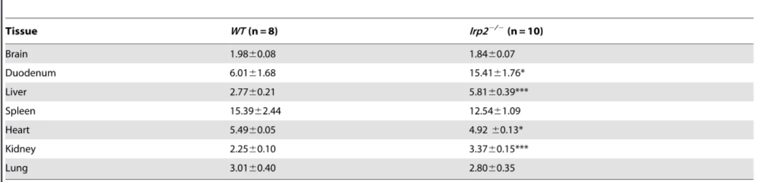
Irp22/2 mice displayed altered body iron distribution with total iron content increased in duodenum, kidney and liver and decreased in heart while total brain iron content is unchanged (Table 1).
Ferritin and TfR1 have been shown to be dysregulated in liver, duodenum and brain in two other distinctIrp22/2mouse strains [17,23,24]. Similarly, we find increased ferritin-L chain (Ftl1) levels in brain (forebrain and cerebellum), liver, kidney and duodenum and decreased TfR1 levels in brain, heart, liver and kidney in Irp22/2 mice compared to WT mice (Figure 1A). Ferritin-L and TfR1 levels are similar inIrp22/2and WT spleen. Irp2 RNA-binding activity and protein are not detected inIrp22/2
tissue lysates (Figure 1 A and B). A non-specific band is seen by Western blot analysis that migrates close to Irp2 in some tissue lysates (Figure 1A). Taken together, these phenotypes are in agreement with those described for otherIrp22/2mouse strains in
which Irp2 deficiency causes microcytic anemia, erythropoietic protoporphyria, and altered body iron distribution and expression of ferritin and TfR1 [18,19,23].
Motor coordination/balance, locomotion and nociception are impaired inIrp22/2mice
The Irp22/2 strain generated by LaVaute et al. [17,35] developed an adult-onset neurodegenerative movement disorder at .6 months of age characterized by tremors, hind-limb weakness, subtle kyphosis and abnormal gait. These mice displayed poor self-grooming activity, and impaired muscular strength and motor coordination when assayed by the hang test and rotarod [17,35]. In contrast,Irp22/2mice generated by Galy et al. [19] showed no overt signs of neurodegeneration or impaired muscle strength at 13–14 months of age. These mice displayed reduced self-grooming activity consistent with a tendency toward reduced rearings (vertical locomotor activity) and impaired motor coordination/balance when challenged by the modified-Hole Board and rotarod tests, respectively [26].
As our Irp22/2 mice at 45–63 weeks old did not display tremors, kyphosis, or abnormal gait, we performed a battery of tests to assess behavioral and neurological function. We used the modified-Hole Board test to assay locomotor and exploratory activities, arousal, memory and social affinity, the modified SHIRPA test to assess neurological function, the grip strength test to quantify muscular strength and the accelerating rotarod to measure motor coordination and balance (Tables S4–S5).
mice compared to WT mice, which was not observed in 20-week old Irp22/2 mice, suggesting a progressive phenotype (Figure 2C and data not shown). The grip strength test (2-paws and 4-paws) revealed that muscular strength was not impaired in Irp22/2 mice (Figure 2D).
Nociception was also assessed in Irp22/2 and WT mice.
Irp22/2 mice showed no significant difference in the first pain reaction (hind paw shaking) since they present with the same reaction latency to heat stimuli as WT mice (Figure 2E). However,Irp22/2 mice displayed a significantly longer reaction latency in the second pain reaction (hind paw licking) (Figure 2E). This finding indicated that Irp22/2 mice tolerated heat better than WT mice when it was used as noxious stimulus. Increased hind paw licking was also observed in 20-week oldIrp22/2mice, indicating that this reaction was independent of age (data not
shown). Retarded reaction to thermal stimuli on the hotplate could be a secondary effect of reduced motor abilities. If impaired motor abilities influenced the pain reaction, however, the same effect in both reactions, or at least in the first reaction and not in the second reaction, would be observed. Altered nociception heat tolerance was not reported for other Irp22/2
mice.
Impaired motor coordination and balance are consistent with theIrp22/2neurological phenotypes reported by Galy et al. [26] and LaVaute et al. [17], althoughIrp22/2mice generated by Galy et al. [26] did not display horizontal locomotor impairments that we observed in ourIrp22/2mice. Taken together, our data show that Irp2 deficiency is associated with mildly impaired horizontal locomotion, exploration, motor coordination/balance and noci-ceptive heat tolerance.
Table 1.Tissue iron content of aged maleIrp22/2and WT mice.
Tissue WT(n = 8) Irp22/2(n = 10)
Brain 1.9860.08 1.8460.07
Duodenum 6.0161.68 15.4161.76*
Liver 2.7760.21 5.8160.39***
Spleen 15.3962.44 12.5461.09
Heart 5.4960.05 4.9260.13*
Kidney 2.2560.10 3.3760.15***
Lung 3.0160.40 2.8060.35
Total iron content was determined by inductively-coupled plasma optical emission spectroscopy (ICP-OES). Statistical analysis performed by Student’s paired t-test (*p,
0.05; ***p,0.001, mean (mg Fe/mg wet tissue weight)61006SEM). Ages of mice: WT, 64–75 weeks;Irp22/2, 49–71 weeks. doi:10.1371/journal.pone.0098072.t001
Figure 1. Expression of iron homeostasis proteins inIrp22/2and WT mice.A)Western blot analysis of tissue extracts from maleIrp22/2and
WT mice (n = 3 mice/genotype) using antibodies to detect Irp2, Irp1, ferritin (Ftl1), TfR1 andb-actin (loading control). A non-specific band migrating near Irp2 is observed in someIrp22/2lysates.B)Irp1 andIrp2RNA-binding activity in lysates was assayed by RNA electrophoretic mobility shift assay
Iron accumulation in oligodendrocytes and axons in the brain ofIrp22/2mice
Marked ferric iron accumulation was observed in oligoden-drocyte soma and white matter axons in aged Irp22/2 mice generated by LaVaute et al. [17,25]. Iron accumulated in the cerebellum, caudate putamen, thalamus, substantia nigra and colliculi, while the frontal cortex, globus pallidus and corpus callosum were not affected [17,25,36]. Iron accumulation in axons in cerebellar white matter was associated with axono-pathy. Iron was also detected in neuronal cell bodies in gray matter, including the thalamus, colliculi and deep cerebellar neurons, and in Purkinje axons. In contrast, Galy et al. [26] did not detect abnormal brain iron accumulation in their aged
Irp22/2 mice. Both groups analyzed aged mice (12–14 months old) and used DAB-enhanced Perls’ iron staining method to detect ferric iron. Both groups also reported no significant differences in total brain iron [23] or total non-heme iron [26] between Irp22/2 and WT mice.
Similar to previously reported global Irp22/2 mice, we found no significant differences in total brain iron content between
Irp22/2 and WT mice using inductively-coupled optical emission spectroscopy (Table 1). We next assessed ferric iron histochemically in serial brain sections prepared from Irp22/2
(n = 10) and WT (n = 8) mice using DAB-enhanced Perls’ stain (Files S1 and S2). Marked iron deposition was observed in cortex, caudate putamen, thalamus, superior colliculus and cerebellum as well as in other brain regions in Irp22/2 mice
(Figure 3 and Files S1 and S2). In cortex, iron accumulated in small cells that have an eccentric nucleus characteristic of oligodendrocyte morphology [37] and in axons (Figure 4A). Iron accumulation was also observed in axon tracts and in oligodendrocyte soma associated with striosomes, as well as in oligodendrocytes scattered throughout the caudate putamen (Figure 4B). Significant iron deposition was observed in the superior colliculus (Figure 4C) and in the cerebellar white matter and oligodendrocytes associated with white matter in
Irp22/2 mice (Figure 4E). Iron deposition in caudate putamen, cerebellar white matter and colliculus, as well as other regions (Files S1 and S2), is in agreement withIrp22/2 mice generated by LaVaute et al. [17,25]. Unlike Irp22/2 mice generated by LaVaute et al. [17,25], we did not detect increased iron deposition in the substantia nigra of our Irp22/2 mice (Figure 4D). WT and Irp22/2 mice used in our study were older than mice used by Smith et al. [25] (,12 months old), and it is possible that the high iron content in the substantia nigra of our WT mice made it difficult to discern iron differences between WT and Irp22/2 mice. We also found significant iron deposition in the cerebral cortex and corpus collosum ofIrp22/2 mice, which was not observed by LaVaute et al. [17] (Figures 3C and 4A).
Iron content is reduced in CA1 pyramidal neurons and in Purkinje neurons ofIrp22/2mice
We next assessed Perls’ iron staining in hippocampal CA1 pyramidal neurons and in cerebellar Purkinje neurons inIrp22/2
and WT mice. Perls’ staining was significantly reduced in CA1 pyramidal neuronal soma and in their apical dendrites (Figure 5 and Figure S3). Purkinje neuronal soma inIrp22/2mice showed a slight, but significant reduction in iron content compared to WT mice (Figure 5 and Figure S3).
A reported phenotype ofIrp22/2 mice generated by LaVaute et al. [17] is the degeneration and partial loss of Purkinje neurons although this was not found inIrp22/2 mice generated by Galy Figure 2. Locomotion, motor coordination and nociception are
impaired inIrp22/2mice.Irp22/2mice display reduced horizontal
locomotor activity (total distance traveled, number of turns, number of total line crossings, mean velocity and angular velocity), andB) reduced vertical exploratory activity (number of rearing and rearing latency) assessed by the modified-Hole Board test [31]. C) Left panel,
performance of Irp22/2and WT mice on the accelerating rotarod in four trials on four consecutive trials with 15 min inter-trial-interval;right panel, decreased mean latency ofIrp22/2mice to fall off the rotarod (n = 4 trial;p= 0.055).D)4-paw grip force test shows no difference in muscular strength betweenIrp22/2 and WT mice.E) Hot plate test shows increased hind paw licking inIrp22/2mice. Data are given as the
mean6SEM; *p,0.05;**p
,0.01, ***p,0.001, relative to WT; WT (n = 9) andIrp22/2(n = 10).
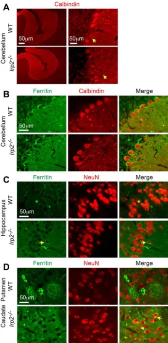
et al. [26]. We stainedIrp22/2and WT cerebellar sections with calbindin antibody (Purkinje cell specific) and found no abnor-malities in Purkinje cell morphology or cell number in Irp22/2
mice (WT, 3.16 cells/inch 60.267; Irp22/2, 3.57 cells/inch
60.232; p= 0.264) (Figure 6A). Ultrastructural analysis of substantia nigra, caudate putamen, cerebellum, cortex, hippo-campus and hypothalamus did not reveal pathological alterations in these regions except for the presence of age-related lipofuscin deposits in neurons that are detected in aged WT and Irp22/2
mice (Figure S4 and data not shown). Myelinization also appeared normal in Irp22/2 mice evaluated by Luxol Fast Blue staining (Figure S5).
The reduction in Perls’ staining in Irp22/2 CA1 pyramidal and Purkinje neurons suggests that these neurons may be iron
deficient. Because neurons acquire iron primarily by TfR1 [38,39], the reduction in TfR1 abundance inIrp22/2cerebellar and forebrain lysates compared to WT is consistent with reduced transferrin-dependent iron uptake (Figure 1A). In contrast to TfR1, ferritin expression is increased in Irp22/2
cerebellar and forebrain lysates (Figure 1A). We next assessed ferritin expression in neurons inIrp22/2 and WT sections from cerebellum, caudate putamen and hippocampus by double immunofluorescence using a ferritin antibody in combination with antibodies specific to neurons (NeuN) and to Purkinje neurons (calbindin). Ferritin immunoreactivity was similar in
Irp22/2 and WT Purkinje neurons (Figure 6B), but was increased in Irp22/2 CA1 pyramidal neurons and in neurons
in the caudate putamen (Figure 6C and D). We were not able to assess TfR1 immunostaining in these sections due to technical difficulties. Taken together, these data suggest that the reduction in TfR1 expression and increased ferritin expression in Irp22/2 mice could lead to reduced iron uptake and increased iron storage, thus causing cellular iron deficiency. While notable neuronal pathology was not observed inIrp22/2
brain, we suggest that neuronal iron deficiency may cause cellular dysfunction that contributes to the neurological and behavioral deficits in Irp22/2 mice.
Conclusions
In conclusion, our data show that mice with a global Irp2
deficiency recapitulate the main features of other Irp2 deficient mouse models such as microcytic anemia, erythropoietic proto-porphyria, altered body iron distribution and altered expression of ferritin and TfR1 in tissues. Our aged Irp22/2 mice did not exhibit, tremor, ataxia, bradykinesia and postural abnormalities as described by LaVaute et al. [17], but did display mild impairments in horizontal locomotion, rearing activity, balance/motor coordi-nation and nociceptive heat tolerance when assayed by specific tests. Our data are consistent with impaired motor and balance coordination reported for the other two strains ofIrp22/2 mice and with reduced rearing activity reported for Irp22/2 mice generated by Galy et al. [26]. The reduced rearing activity in our
Irp22/2mice is consistent with a slight reduction in self-grooming (which could imply a reduction in vertical locomotion), although this did not reach statistical significance, and is similar to other
Irp22/2 mice that exhibited a significant reduction in self-grooming activity [17,26].
Our Irp22/2 mice showed distinctive features not reported
for other Irp22/2 mice. The modified hole board test revealed that Irp22/2 mice displayed an overall reduction in locomotor activity characterized by decreased total distance moved, line crossings, turning frequency and movement velocity. This combination of mild hypoactivity and reduced rearing in our
Irp22/2 mice suggests reduced exploratory motivation. Galy et al. [26] also used the modified hole board test, but did not observe impaired horizontal locomotion in their 13–14 month old Irp22/2 mice. We also found that Irp22/2 mice showed a hypoalgesic phenotype as assessed by the hot plate test that was independent of age. Our Irp22/2 mice develop diabetes (EAL, SN, CPA and KBZ, unpublished observation), and it is possible that hypoalgesia is a consequence of impaired glucose tolerance. Another novel phenotype in ourIrp22/2mice is the presence of PPIX containing aggregates in the cystic, hepatic, and common bile ducts. Cooperman et al. [23] showed increased synthesis of the heme biosynthetic enzyme erythroid aminolevulinate synthase 2 (Alas2), which contains an IRE in the 59untranslated region, and reduced expression of TfR1 in Irp22/2 erythroid Figure 3. Iron accumulates in specific brain regions ofIrp22/2
precursor cells. Loss of translational repression of Alas2 by Irp2 deficiency and reduced Tf-dependent iron uptake likely results in elevated PPIX levels we observed in the serum, liver and bile ducts. Whether other Irp22/2 mice show PPIX aggregates in
the bile ducts remains to be determined.
An explanation for the varied neurodegenerative phenotypes in the differentIrp22/2 models is not clear, but it is known that neurological and behavioral phenotypes can be sensitive to genetic background differences and gene targeting strategies [40–42]. Our mice were generated using 129/Sv-CP ES cell line and were backcrossed to C57BL/6J mice for five generations, and those of Galy et al. [19,26] were generated using the 129P2/OlaHsd ES cell line and backcrossed to C57BL/6J mice for three generations. Irp22/2 mice generated by LaVaute et al. [17] are mixed genetic background consisting of C57BL/6 and B129S4/SVJ. In addition, different targeting approaches were used to generate Irp22/2 mice. LaVaute et al. [17] generated a global model of Irp2 deficiency by insertion of
a PGK-neomycin gene into exon 3/4 ofIrp2 gene. Galy et al. [19] generated conditional alleles ofIrp2by insertion of ab-Geo cassette flanked by Frt sites into intron 2 of the Irp2 gene and was co-inserted with LoxP sites flanking exon 3. Cre-mediated excision of exon 3 generated a null allele. Our Irp22/2 mice
were generated by the insertion of a self-excision cassette containing neomycin linked to Cre-recombinase into exon 3 of the mouse Irp2 gene. Targeting strategies can lead to different phenotypes due to silencing or activation of flanking genes caused by the retention of selection cassettes, generation of truncated gene products with biological activity or inactivation of non-coding RNAs with the targeted locus [43,44]. The expression of neighboring genes close to theIrp2 locus was not altered inIrp22/2 mice [19] and we did not identify annotated non-coding RNAs located within theIrp2locus. The differences in neurological and behavioral phenotypes in Irp22/2 mice could also be affected by environmental conditions (food, altitude or environmental stimulation) [44] and natural variation Figure 4. Iron accumulates in axons and oliogodendrocyte cell bodies ofIrp22/2 mice.Increased DAB-enhanced Perls’ iron stain is
observed in small cells that have an eccentric nucleus characteristic of oliogodendrocyte morphology [37] and in axons inA)cerebral cortex,B)
caudate putamen,C)superior colliculus,D)substantia nigra, andE) cerebellum.Arrows, oligodendrocytes;S, striosomes;wm, white matter;gl,granule layer. Scale bars: 50mm and 500mm.
in iron metabolism in different strains of inbred mice [45]. The reasons for the different neurological and behavioral phenotypes observed inIrp22/2 mice are not completely understood, but it
is likely that the genetic background and the targeting strategies used to generate Irp22/2 are important components.
Ultrastructural analyses of several regions of brain fromIrp22/2
mice reveal normal neuronal morphology and calbindin staining of cerebellar sections show no loss of Purkinje cells. However, marked iron accumulation is observed in white matter and in oligodendrocytes associated with white matter throughout the
brain consistent with studies from LaVaute et al. [17]. Oligoden-drocytes normally contain high levels of iron and express high levels of ferritin. The high iron content of oligodendrocytes is thought to be required for myelin production [46]. Myelinization Figure 5. Iron is reduced in Purkinje neurons and in CA1
pyramidal neurons ofIrp22/2 mice.A) Perls’ DAB-enhanced iron
staining in CA1 pyramidal neurons and Purkinje neurons inIrp22/2and
WT mice (Pkj, Purkinje; gl, granular layer; wm, white matter). B)
Quantification of Perls’ staining of images in(A)was carried out by measuring staining intensity in a defined subsection of brains from
Irp22/2(n = 10) and WT (n = 8) (Figure S3). Data are given as the mean
6 SEM; *p,0.05; ***p,0.001 relative to WT. Scale bars: 25mm and 50mm.
doi:10.1371/journal.pone.0098072.g005
Figure 6. Ferritin expression is increased inIrp22/2 neurons.
Double immunofluorescence labeling of brain sections from maleIrp22/ 2and WT mice using ferritin antibody with either calbindin (Purkinje
specific) or NeuN (neuronal nuclei) antibodies.A)Calbindin immuno-fluorescence shows that Purkinje cell number and morphology (yellow arrows, processes), are normal inIrp22/2 cerebellum. B)Ferritin and calbindin double immunofluorescence of cerebellum shows similar ferritin expression inIrp22/2and WT Purkinje neurons.C)Ferritin and NeuN double immunofluorescence of hippocampal CA1 pyramidal cell layer, andD)caudate putamen shows increased ferritin expression in neuronal cell bodies (yellow arrows) ofIrp22/2mice compared to WT
mice. Increased ferritin staining is observed in the neuropil in the hippocampus and caudate putamen ofIrp22/2mice. Scale bars: 50mm.
is not affected inIrp22/2brain, suggesting that high iron content is not detrimental to oligodendrocyte function.
A notable difference between ourIrp22/2 mice and those of LaVaute et al. [17] is the significant iron accumulation in oligodendroycytes in the cortex ofIrp22/2mice. We also found that CA1 pyramidal neurons and Purkinje neurons are iron deprived. Since neurons primarily acquire iron by TfR1, reduced TfR1 and increased ferritin expression in Irp22/2
brain would reduce transferrin-dependent iron uptake in parallel with an increase ferritin iron sequestration, eventually leading to cellular iron deficiency. Although we did not find pathological evidence of neurodegeneration, it is possible that cellular iron deficiency in neurons causes cellular dysfunction that leads to mild deficits in locomotion, motor coordination and nociception. This idea is consistent with a recent study showing thatIrp22/2
mice develop motor neuron disease characterized by increased ferritin and decreased TfR1 expression in motor neurons, reduced spinal cord iron and impaired mitochondrial function [47]. Taken together, these studies suggest that abnormal brain iron metabolism due to Irp2 deficiency disrupts cellular function and leads to mild neurological, behavioral and nociception impairments.
Supporting Information
Figure S1 Generation of Irp22/2 mice. (A1) Schematic representation of exons I-VII of the murineIrp2(Ireb2) gene. Exons (black boxes) and introns (black lines). (A2) Schematic diagram of the targeting vector. A genomic clone (p11.1) containing exons III-VII encoding amino acids 37–233 in Irp2 was isolated from a mouse 129 genomic library. A ScaI-PstI fragment from p11.1 (dotted lines) was subcloned into Bluescript SK to generate the targeting vector. A self-excision cassette containing neomycin (Neor) linked to Cre-recombinase (Cre) was inserted into exon III of the mouseIrp2gene [48]. This cassette (pACN) contains theCregene (driven by the testes-specific angiotensin-converting enzyme (tACE promoter) linked to the Neor gene (driven by the polymerase II promoter), and is flanked by loxP sites allowing for excision ofNeor
as it passes through the male germ line. This cassette also contains humanhIrp1-Flagsequence fused in-frame to exon III ofIrp2for experiments related to functional replacement of Irp2 with Irp1. For reasons discussed below, Irp1-Flag was not detected in any tissues examined. Thymidine kinase (Tk1 and Tk2) genes were inserted at the 59 and 39 ends of the targeting vector to select against random insertion. (A3) Predicted structure of the Irp2
replacement allele after homologous recombination and germ-line induced self-excision. The targeting vector was electroporated into R1 ES cell line (129/Sv-CP) and heterozygous cells in which a homologous recombination event occurred were identified by PCR and Southern blot analysis of ScaI-digested DNA probed with 39EXT and Neo probes (Figure S1B, left panels). A targeted ES cell clone (F4) was microinjected into C57BL/6J blastocysts to generate chimeric animals. A chimera was bred to C57BL/6J mice and resulting chimeric progeny was identified by coat color and for the presence of theIrp2+/2allele by PCR and Southern
blotting. Heterozygous offspring were backcrossed five times to the C57BL/6J background and intercrossed to generate homozygous
Irp22/2mice. (B) Germline transmission of theNeo-deleted allele was confirmed by Southern blot analysis ofSca1-digested genomic DNA fromIrp2+/+
,Irp2+/2andIrp22/2mice probed with the 39
EXT probe and shows a WT band (10 kb) inIrp2+/+, a WT and a
mutant band (7 kb) in Irp2+/2 and a mutant band in Irp22/2
(7 kb). The absence of the Neor/Cre cassette was confirmed by probing the blot with a Neo probe (not shown). PCR genotyping
was performed using tail DNA biopsies (Table S9). (C) Quanti-tative RT-PCR showedIrp2mRNA was not detected inIrp22/2
E9.5-E10.5 lysates and reduced 2-fold inIrp2+/2lysates compared
to WT lysates (TaqMan assay:Irp2exons 3–4).hIrp1-FlagmRNA was not expressed inIrp2+/+
lysates, but is expressed inIrp2+/2and
Irp22/2 lysates (TaqMan assay: hIrp1). A TaqMan assay corresponding to exons 1–2 (common to bothIrp2andhIrp1-Flag) showed thathIrp1-FlagmRNA expression is,80% of endogenous
Irp2 mRNA. (D) Although hIrp1-Flag mRNA was detected in
Irp22/2 lysates, no hIrp1-Flag protein or RNA-binding activity was detected by Western blot analysis or by RNA-EMSA, respectively, in E9.5-E10.5 lysates or any tissue examined (data not shown). The lack of hIrp1-Flag expression is likely due to faulty translation and/or increased turnover of the hIrp1-Flag protein. If hIrp1-Flag is expressed at low levels, the RNA-binding activity of this recombinant protein is not sufficient to compensate for loss of Irp2 protein since Irp22/2 mice display altered body iron distribution (Table 1), microcytic anemia (Table S1), and inappropriate regulation of Irp2 targets ferritin and TfR1 (Figure 1) in agreement with published phenotypes of Irp22/2
mice [17,23,24]. TaqMan assays used in these experiments can be found in Table S6.
(TIF)
Figure S2 Presence of PPIX-containing granules in the
common bile duct ofIrp22/2 mice. Black arrows indicate
PPIX-containing granules in Irp22/2 (C) mice. B and D are
magnified images of A and C. PPIX-containing granules are found in all male and female Irp22/2 mice examined at 2.5- to 18-months of age.
(TIF)
Figure S3 DAB-enhanced Perls’ iron staining of
hippo-campus and cerebellum of agedIrp22/2 and WT mice.
Coronal sections of hippocampus (A) (slide 41) and cerebellum (B) (slide 58) used for quantification of iron in CA1 pyramidal neurons and in Purkinje neurons in Figure 5. Dotted boxes indicate the WT (6B-4) and theIrp22/2(6A-3) mice used for DAB-enhanced Perls’ staining in File S1 and File S2. Ages of male mice: WT, 65– 71 weeks;Irp22/2, 46–53 weeks.
(TIF)
Figure S4 Ultrastructual analysis of brain regions of
aged Irp22/2 and WT mice shows no evidence of
neurodegeneration. Electron micrographs of the caudate
putamen, substantia nigra and cerebellum ofIrp22/2 and WT mice. Myelinized (white arrows) and non-myelinized (black arrow) axons are shown. No pathological alterations were evident in these brain regions. Similar amounts of lipofuscin deposits (L) were detected in bothIrp22/2and WT mice. This can be accounted as a normal finding since lipofuscin accumulates with age in many organs including brain. Representative images are shown from WT (n = 4) andIrp22/2(n = 5) mice. Ages of male mice: WT, 65– 71 weeks;Irp22/246–53 weeks. Scale bars are indicated. (TIF)
Figure S5 Luxol Fast Blue staining of brains of aged
Irp22/2and WT mice shows no difference in
myeliniza-tion. Representative images are shown from WT (n = 4) and
Irp22/2(n = 5) mice. Ages of male mice: WT, 65–71 weeks;Irp22/
2, 46–53 weeks.
(TIF)
Table S1 Hematological parameters of aged male WT
andIrp22/2mice.
Table S2 Serum iron parameters of aged male WT and
Irp22/2mice.
(DOCX)
Table S3 Protoporphyrin IX (PPIX) levels of 12-month
old male WT andIrp22/2mice.
(DOCX)
Table S4 Results of behavioral observation in the modified-Hole Board test.
(DOCX)
Table S5 Video-tracking results regarding locomotor behavior.
(DOCX)
Table S6 List of primers and TaqMan assays used for genotyping and qRT-PCR.
(DOCX)
File S1 DAB-enhanced Perls’ iron staining of coronal
sections of WT (6B-4) brains.Images (10–66) are from rostral
to caudal. High resolution files are available from the correspond-ing author.
(ZIP)
File S2 DAB-enhanced Perls’ iron staining of coronal
sections ofIrp22/2(6A-3) brains.Images (10–66) are from
rostral to caudal. High resolution files are available from the corresponding author.
(ZIP)
Acknowledgments
We thank Dr. Scott Rogers and Dr. Lorise Gahring for assistance with mouse perfusions and brain imaging, and neuroanatomy and Dr. John Phillip for assistance with PPIX assays. We also thank Ms. Diana Lim for assistance with manuscript and image preparation.
Author Contributions
Conceived and designed the experiments: EAL KZB MHA VGD HF SMH WW. Performed the experiments: KZB SJR BR LB. Analyzed the data: EAL KZB SJR BR EW LG SMH LB IM JCW LQF IR TK. Wrote the paper: EAL KZB. Reviewed the manuscript: TK EW AZ.
References
1. Ganz T, Nemeth E (2012) Hepcidin and iron homeostasis. Biochim Biophys Acta 1823: 1434–1443.
2. Simcox JA, McClain DA (2013) Iron and diabetes risk. Cell Metab 17: 329–341. 3. Rouault TA (2013) Iron metabolism in the CNS: implications for
neurodegen-erative diseases. Nat Rev Neurosci.
4. Rao R, Georgieff MK (2007) Iron in fetal and neonatal nutrition. Semin Fetal Neonatal Med 12: 54–63.
5. Lozoff B, Georgieff MK (2006) Iron deficiency and brain development. Semin Pediatr Neurol 13: 158–165.
6. Youdim MB (2008) Brain iron deficiency and excess; cognitive impairment and neurodegeneration with involvement of striatum and hippocampus. Neurotox Res 14: 45–56.
7. Fretham SJ, Carlson ES, Wobken J, Tran PV, Petryk A, et al. (2012) Temporal manipulation of transferrin-receptor-1-dependent iron uptake identifies a sensitive period in mouse hippocampal neurodevelopment. Hippocampus 22: 1691–1702.
8. Dusek P, Jankovic J, Le W (2012) Iron dysregulation in movement disorders. Neurobiol Dis 46: 1–18.
9. Burn J, Chinnery PF (2006) Neuroferritinopathy. Semin Pediatr Neurol 13: 176– 181.
10. Kono S, Miyajima H (2006) Molecular and pathological basis of aceruloplasmi-nemia. Biol Res 39: 15–23.
11. Hentze MW, Muckenthaler MU, Galy B, Camaschella C (2010) Two to tango: regulation of Mammalian iron metabolism. Cell 142: 24–38.
12. Anderson CP, Shen M, Eisenstein RS, Leibold EA (2012) Mammalian iron metabolism and its control by iron regulatory proteins. Biochim Biophys Acta 1823: 1468–1483.
13. Vashisht AA, Zumbrennen KB, Huang X, Powers DN, Durazo A, et al. (2009) Control of iron homeostasis by an iron-regulated ubiquitin ligase. Science 326: 718–721.
14. Salahudeen AA, Thompson JW, Ruiz JC, Ma HW, Kinch LN, et al. (2009) An E3 ligase possessing an iron-responsive hemerythrin domain is a regulator of iron homeostasis. Science 326: 722–726.
15. Moroishi T, Nishiyama M, Takeda Y, Iwai K, Nakayama KI (2011) The FBXL5-IRP2 axis is integral to control of iron metabolism in vivo. Cell Metab 14: 339–351.
16. Sanchez M, Galy B, Schwanhaeusser B, Blake J, Bahr-Ivacevic T, et al. (2011) Iron regulatory protein-1 and -2: transcriptome-wide definition of binding mRNAs and shaping of the cellular proteome by iron regulatory proteins. Blood 118: e168–179.
17. LaVaute T, Smith S, Cooperman S, Iwai K, Land W, et al. (2001) Targeted deletion of the gene encoding iron regulatory protein-2 causes misregulation of iron metabolism and neurodegenerative disease in mice. Nat Genet 27: 209– 214.
18. Meyron-Holtz EG, Ghosh MC, Iwai K, LaVaute T, Brazzolotto X, et al. (2004) Genetic ablations of iron regulatory proteins 1 and 2 reveal why iron regulatory protein 2 dominates iron homeostasis. EMBO J 23: 386–395.
19. Galy B, Ferring D, Hentze MW (2005) Generation of conditional alleles of the murine iron regulatory protein (Irp)-1 and -2 genes. Genesis 43: 181–188. 20. Anderson SA, Nizzi CP, Chang YI, Deck KM, Schmidt PJ, et al. (2013) The
IRP1-HIF-2alpha axis coordinates iron and oxygen sensing with erythropoiesis and iron absorption. Cell Metab 17: 282–290.
21. Ghosh MC, Zhang DL, Jeong SY, Kovtunovych G, Ollivierre-Wilson H, et al. (2013) Deletion of iron regulatory protein 1 causes polycythemia and pulmonary hypertension in mice through translational derepression of HIF2alpha. Cell Metab 17: 271–281.
22. Wilkinson N, Pantopoulos K (2013) IRP1 regulates erythropoiesis and systemic iron homeostasis by controlling HIF2alpha mRNA translation. Blood 122: 1658–1668.
23. Cooperman SS, Meyron-Holtz EG, Olivierre-Wilson H, Ghosh MC, McCon-nell JP, et al. (2005) Microcytic anemia, erythropoietic protoporphyria, and neurodegeneration in mice with targeted deletion of iron-regulatory protein 2. Blood 106: 1084–1091.
24. Galy B, Ferring D, Minana B, Bell O, Janser HG, et al. (2005) Altered body iron distribution and microcytosis in mice deficient in iron regulatory protein 2 (IRP2). Blood 106: 2580–2589.
25. Smith SR, Cooperman S, Lavaute T, Tresser N, Ghosh M, et al. (2004) Severity of neurodegeneration correlates with compromise of iron metabolism in mice with iron regulatory protein deficiencies. Ann N Y Acad Sci 1012: 65–83. 26. Galy B, Holter SM, Klopstock T, Ferring D, Becker L, et al. (2006) Iron
homeostasis in the brain: complete iron regulatory protein 2 deficiency without symptomatic neurodegeneration in the mouse. Nat Genet 38: 967–969; discussion 969–970.
27. Guo B, Yu Y, Leibold EA (1994) Iron regulates cytoplasmic levels of a novel iron-responsive element-binding protein without aconitase activity. J Biol Chem 269: 24252–24260.
28. Yu Y, Radisky E, Leibold EA (1992) The iron-responsive element binding protein. Purification, cloning, and regulation in rat liver. J Biol Chem 267: 19005–19010.
29. Leibold EA, Munro HN (1988) Cytoplasmic protein binds in vitro to a highly conserved sequence in the 59untranslated region of ferritin heavy- and light-subunit mRNAs. Proc Natl Acad Sci U S A 85: 2171–2175.
30. Phillips JD, Bergonia HA, Reilly CA, Franklin MR, Kushner JP (2007) A porphomethene inhibitor of uroporphyrinogen decarboxylase causes porphyria cutanea tarda. Proc Natl Acad Sci U S A 104: 5079–5084.
31. Ohl F, Holsboer F, Landgraf R (2001) The modified hole board as a differential screen for behavior in rodents. Behav Res Methods Instrum Comput 33: 392– 397.
32. Kallnik M, Elvert R, Ehrhardt N, Kissling D, Mahabir E, et al. (2007) Impact of IVC housing on emotionality and fear learning in male C3HeB/FeJ and C57BL/6J mice. Mamm Genome 18: 173–186.
33. Neff F, Flores-Dominguez D, Ryan DP, Horsch M, Schroder S, et al. (2013) Rapamycin extends murine lifespan but has limited effects on aging. J Clin Invest 123: 3272–3291.
34. Mogil JS (1999) The genetic mediation of individual differences in sensitivity to pain and its inhibition. Proc Natl Acad Sci U S A 96: 7744–7751.
35. Ghosh MC, Tong WH, Zhang D, Ollivierre-Wilson H, Singh A, et al. (2008) Tempol-mediated activation of latent iron regulatory protein activity prevents symptoms of neurodegenerative disease in IRP2 knockout mice. Proc Natl Acad Sci U S A 105: 12028–12033.
36. Grabill C, Silva AC, Smith SS, Koretsky AP, Rouault TA (2003) MRI detection of ferritin iron overload and associated neuronal pathology in iron regulatory protein-2 knockout mice. Brain Res 971: 95–106.
38. Dickinson TK, Connor JR (1998) Immunohistochemical analysis of the transferrin receptor: regional and cellular distribution in the hypotransferrinemic (hpx) mouse brain. Brain Research 801: 171–181.
39. Leitner DF, Connor JR (2012) Functional roles of transferrin in the brain. Biochim Biophys Acta 1820: 393–402.
40. Gerlai R (1996) Gene-targeting studies of mammalian behavior: is it the mutation or the background genotype? Trends Neurosci 19: 177–181. 41. Watase K, Zoghbi HY (2003) Modelling brain diseases in mice: the challenges of
design and analysis. Nat Rev Genet 4: 296–307.
42. Yoshiki A, Moriwaki K (2006) Mouse phenome research: implications of genetic background. Ilar J 47: 94–102.
43. Pham CT, MacIvor DM, Hug BA, Heusel JW, Ley TJ (1996) Long-range disruption of gene expression by a selectable marker cassette. Proc Natl Acad Sci U S A 93: 13090–13095.
44. Crusio WE (2004) Flanking gene and genetic background problems in genetically manipulated mice. Biol Psychiatry 56: 381–385.
45. Leboeuf RC, Tolson D, Heinecke JW (1995) Dissociation between tissue iron concentrations and transferrin saturation among inbred mouse strains. J Lab Clin Med 126: 128–136.
46. Connor JR, Menzies SL (1996) Relationship of iron to oligodendrocytes and myelination. Glia 17: 83–93.
47. Jeong SY, Crooks DR, Wilson-Ollivierre H, Ghosh MC, Sougrat R, et al. (2011) Iron insufficiency compromises motor neurons and their mitochondrial function in Irp2-null mice. PLoS One 6: e25404.