Comprehensive Analysis of Disease-Related
Genes in Chronic Lymphocytic Leukemia by
Multiplex PCR-Based Next Generation
Sequencing
Claudia Vollbrecht1,2,3, Fabian Dominik Mairinger4, Ulrike Koitzsch1,2,3, Martin Peifer3,5, Katharina Koenig1,2,3, Lukas Carl Heukamp1,2¤a¤b, Giuliano Crispatzu2,6,7, Laura Wilden2,6,
Karl-Anton Kreuzer2,6, Michael Hallek2,6, Margarete Odenthal1,2,3☯, Carmen Diana Herling2,6☯*, Reinhard Buettner1,2,3☯*
1Institute of Pathology, University Hospital Cologne, Cologne, Germany,2Center for Integrated Oncology (CIO) Cologne-Bonn, University Hospital Cologne, Cologne, Germany,3Center of Molecular Medicine Cologne, University of Cologne, Cologne, Germany,4Institute of Pathology, University Hospital Essen, University of Duisburg-Essen, Essen, Germany,5Department of Translational Genomics, Cologne Center of Genomics, University of Cologne, Cologne, Germany,6Department I of Internal Medicine, University Hospital Cologne, Cologne, Germany,7Excellence Cluster for Cellular Stress Response and Aging-Associated Diseases (CECAD), University of Cologne, Cologne, Germany
☯These authors contributed equally to this work.
¤a Current address: Institute for Hematopathology Hamburg, Hamburg, Germany ¤b Current address: New Oncology, Cologne, Germany
*reinhard.buettner@uk-koeln.de(RB);carmen.herling@uk-koeln.de(CDH)
Abstract
Background
High resolution molecular studies have demonstrated that the clonal acquisition of gene mutations is an important mechanism that may promote rapid disease progression and drug resistance in chronic lymphocytic leukemia (CLL). Therefore, the early and sensitive detection of such mutations is an important prerequisite for future predictive CLL diagnos-tics in the clinical setting.
Material & Methods
Here, we describe a novel, target-specific next generation sequencing (NGS) approach, which combines multiplex PCR-based target enrichment and library generation with ultra-deep high-throughput parallel sequencing using a MiSeq platform. We designed a CLL spe-cific target panel, covering hotspots or complete coding regions of 15 genes known to be re-currently mutated and/or related to B-cell receptor signaling.
Results
High-throughput sequencing was performed using as little as 40 ng of peripheral blood B-cell DNA from 136 CLL patients and a dilution series of twoATM- orTP53-mutated cell lines, the latter of which demonstrated a limit of mutation detection below 5%. Using a
OPEN ACCESS
Citation:Vollbrecht C, Mairinger FD, Koitzsch U, Peifer M, Koenig K, Heukamp LC, et al. (2015) Comprehensive Analysis of Disease-Related Genes in Chronic Lymphocytic Leukemia by Multiplex PCR-Based Next Generation Sequencing. PLoS ONE 10(6): e0129544. doi:10.1371/journal.pone.0129544
Academic Editor:Christos Hatzis, Yale University, UNITED STATES
Received:December 16, 2014
Accepted:May 11, 2015
Published:June 8, 2015
Copyright:© 2015 Vollbrecht et al. This is an open access article distributed under the terms of the Creative Commons Attribution License, which permits unrestricted use, distribution, and reproduction in any medium, provided the original author and source are credited.
Data Availability Statement:All relevant data are within the paper and its Supporting Information files.
Funding:This study was supported by the Deutsche Forschungsgemeinschaft (KFO 286, Speaker MH) to RB, KAK, CDH, and by the German Cancer AID (Onkologisches Spitzenzentrum Köln-Bonn).
stringent functional assessment algorithm, 102 mutations in 8 genes were identified in CLL patients, including hotspot regions ofTP53,SF3B1,NOTCH1,ATM,XPO1,MYD88,DDX3X and the B-cell receptor signaling regulatorPTPN6. The presence of mutations was signifi-cantly associated with an advanced disease status und molecular markers of an inferior prognosis, such as an unmutatedIGHVmutation status or positivity for ZAP70 by flow cytometry.
Conclusion
In summary, targeted sequencing using an amplicon based library technology allows a re-source-efficient and sensitive mutation analysis for diagnostic or exploratory purposes and facilitates molecular subtyping of patient sets with adverse prognosis.
Introduction
Chronic lymphocytic leukemia (CLL) is an incurable and common type of adult leukemia with significant variability in clinical prognosis that is hard to predict [1,2]. The current biological understanding is that variable courses of the disease are predominantly caused by molecular inter- and intrapatient heterogeneity of leukemic cells and the possibility of clonal disease evo-lution over time [2–4].
Recent whole-exome and genome sequencing studies have deciphered the mutational land-scape in CLL and discovered a variety of somatic mutations and small indels inNOTCH1,
SF3B1, and other candidate genes, which encode for putative and previously unknown drivers of CLL tumorigenesis [5–10]. Some of these mutations seem to be associated with prognosis, however, except for mutations and other genomic aberrations in theTP53gene, the clinical consequences to be taken in case a patient presents with one of these mutations, are not clari-fied [6,7,10–14].
Future risk assessment in CLL is now confronted with the need of prospective clinical trials, which systematically integrate mutation and traditional biomarker assessment to determine the parameters with a retained prognostic or predictive value, relevant to clinical practice. This has become of particular importance as new drugs, e.g. inhibitor to protein kinases PI3K and BTK, are entering clinical practice and conveying new mechanisms of treatment resistance compared to standard chemoimmunotherapy [15].
The aim of our study presented here was to develop a targeted genomic sequencing assay, being able to meet such diagnostic and clinical research needs in CLL. Targeted sequencing versus whole-genome or exome-wide massive parallel sequencing (i.e. next generation se-quencing, NGS) offers the opportunity to assess genomic changes in areas of specific interest at a coverage as high as deemed appropriate for diagnostic reporting.
In comparison to traditional Sanger sequencing currently used for routine assessment of the
TP53orIGHVgenes, NGS allows multiplexing of samples and gene targets in one experimen-tal setup. In addition, the possibility of automation for high-throughput sample processing further minimizes clinical laboratory efforts and final costs per gene and sample [16]. So far, only few studies have implemented targeted NGS technologies for mutation screening in CLL [17–20].
FBXW7,MYD88,NOTCH1,SF3B1,TP53,XPO[6,7,10,21–23], we chose target genes directly or indirectly involved in the B-cell receptor (BCR) signaling pathway (BTK,MAPK1,PIK3CA,
PIK3CD,PTEN,PTPN6). Using a modified chemistry setup for target enrichment and library preparation in a test cohort of 136 CLL patients and two mutated cell lines, we were able to ob-tain a high sequencing coverage and a low limit of mutation detection. Previously known and new mutations were detected in coding or hotspot regions of the genesATM,DDX3X,MYD88,
NOTCH1,SF3B1,TP53,XPO1andPTPN6(SHP-1), and associations between mutations and adverse prognostic markers were investigated.
Overall, our targeted NGS approach resembles a sensitive and resource efficient method for simultaneous mutation analysis of multiple gene regions on a high-throughput sequencing platform and is highly suitable to future diagnostic and clinical research purposes in CLL.
Materials and Methods
Clinical Samples
The study was approved by the ethical commission of the medical faculty of the University of Cologne (reference no. 13–091) and an informed written consent was obtained from all pa-tients. Between 2012 and 2013, 136 blood samples from CLL patients were collected at the Uni-versity of Cologne, Germany. All cases demonstrated typical features of CLL as defined by the International Workshop on CLL [24]. Clinical and routine laboratory parameters were re-trieved from medical records. CLL-related chromosomal abnormalities were assessed by inter-phase fluorescence-in-situ hybridization (FISH) using commercially available probes, detecting trisomy 12 and deletions on chromosomes 6q21 (SEC63), 11q22.3 (ATM), 13q34 (D13S319) and 17p13.1 (TP53) (Abbott, Abbott Park, IL, USA). In addition, CLL immunophenotypes in-cluding CD38 and ZAP70 surface expression and the somatic mutation status ofIGHVgenes was determined as described previously [25].
B-cells were enriched by negative selection using RosetteSep-based cell removal (Stemcell Technologies, Vancouver, BC, Canada) followed by Pancoll human density centrifugation (Pan Biotech, Aidenbach, Germany).
Genomic DNA was extracted from B-cell fractions by standard column based purifica-tion (DNeasy, Qiagen, Hilden, Germany). DNA quality and quantity was assessed by gel electrophoresis.
Library Construction and Deep Sequencing
In order to selectively amplify either hotspot or complete coding regions of the following genes
ATM,BTK,CD79B,DDX3X,FBXW7,MAPK1,MYD88,NOTCH1,PIK3CA,PIK3CD,PTEN,
including 1% PhiX control library were prepared for sequencing according to the MiSeq Sys-tem User Guide (Illumina, San Diego, CA, US). Subsequently, sequencing was carried out on a MiSeq instrument (Illumina) using the v2 chemistry as recommended by the manufacturer.
Estimation of Lowest Detection Rate Using Cell Line DNA Dilutions
The mantle cell lymphoma cell line, Mino (kindly provided by M. Herling, Cologne, Germany), carrying a known homozygousTP53mutation (c.440T>G; p.V147G; NM_000546) [26], and the AT45RM B-cell line (kindly provided by L. Chessa, Rome, Italy) containing an heterozygousATMmutation (c.7792C>T; p.R2598; NM_000051) [27] were used to evaluate the limit of de-tection (LoD) of our NGS approach. Cells were cultured according to standard protocols. DNA was extracted and sequenced as described above. 200 to 9,000 genomic copies of each cell line DNA were diluted in wild type DNA from human embryonic kidney cells (HEK-293, obtained from the American Type Culture Collection ATCC) harboring no known gene mutations.
Sequencing Data Analysis
Fastq files generated by the MiSeq Reporter Software (Illumina) were analyzed with an in-house developed bioinformatics pipeline, based on the general cancer genome analysis algo-rithm, which was further optimized for the diagnostic workflow [28]. Briefly, adaptor se-quences were first removed from raw sequencing reads. The resulting data was then aligned against NCBI build 37 (hg19) using the Burrows-Wheeler Aligner (BWA, version 0.6.1-r104) [29] with its default settings. In order to capture longer insertion and deletions we realigned unmapped reads with the BLAST-like alignment tool (BLAT) [30,31]. For variant calling we first determined the background error rate of the sequencer using known single nucleotide polymorphisms (SNPs): Bases diverting other than the possible two variants were counted and set into the relation to the total coverage at the location of the SNP. Finally, variants were called by testing if a mutation was not compatible with the afore mentioned error rate. For this pur-pose, we set the significance threshold to 0.01, which leads to a slight overcalling of the se-quencing data. Spurious calls were subsequently filtered out by the following strategy: Detected
Table 1. Overview of the genes covered by the CLL panels.
Gene Biological Process Exons Transcript ID n Amplicons
ATM DNA damage/ cell cycle control Complete (62) NM_000051 117
BTK B-cell receptor signaling pathway 14–16 NM_000061 5
CD79B B-cell receptor signaling pathway 4–5 NM_021602 2
DDX3X RNA splicing and processing 7–9, 11, 14 NM_001356 6
FBXW7 Protein ubiquitination 6–9 NM_033632 7
MAPK1 MAP kinase signaling pathway 7 NM_002745 1
MYD88 Toll-like receptor signaling pathway Complete (5) NM_002468 9
NOTCH1 Notch signaling pathway Complete (34) NM_017617 71
PIK3CA B-cell receptor signaling pathway 9–11, 20–21 NM_006218 10
PIK3CD B-cell receptor signaling pathway 21–24 NM_005026 7
PTEN AKT-mTOR signaling pathway 5–6, 9 NM_000314 7
PTPN6(SHP-1) B-cell receptor signaling pathway 11–12 NM_080548 2
SF3B1 RNA splicing and processing Complete (25) NM_012433 52
TP53 DNA damage/ cell cycle control Complete (9) NM_000546 16
XPO1 RNA splicing and processing 12–13, 15 NM_003400 7
Total number of amplicons 338
variants were annotated by using the databases dbSNP (http://www.ncbi.nlm.nih.gov/SNP/) and the exome variant server (http://evs.gs.washington.edu/EVS/). Furthermore, obtained vari-ants were analyzed for their functional impact on the protein by the MutationAssessor (http:// mutationassessor.org; release 2) [32] and by implementation of the ANNOVAR algorithm [33], which combines the bioinformatic tools SIFT [34], PolyPhen2 [35] and the Mutation Taster [36]. Variants with an allelic frequency below 5%, synonymous and variants without functional impact were removed (Fig 1). Additional, visual analysis of called variants was per-formed by means of the Integrative Genomic Viewer (IGV, Broad Institute, Cambridge, MA, USA). Potential false positive variants, particularly in repetitive or highly homologous regions of the genome, variants in high background noise, as well as single strand variants, were either eliminated when they were clearly recognizable as artifacts, or were further re-assessed by Sanger sequencing.
Variant Confirmation
A subset of variants, including variants with less than 100 reads, was confirmed by conventional Sanger sequencing using the BigDye Terminator v3.1 Cycle Sequencing Kit (Life Technologies) (S4andS5Tables). Variants that could not be confirmed were excluded from further analysis.
Statistical Analysis
Statistical analysis for associations with clinical and/or prognostic covariates was performed for genes with mutations in multiple samples (more than 10) with predicted impact on protein func-tion. Consequentially, the five genesTP53,SF3B1,ATM,NOTCH1andXPO1were tested for as-sociations with clinical and prognostic parameters (genomic aberration, age, gender, Binet-stage, white blood count (WBC), platelets, ZAP70 and CD38 positivity andIGHVmutation status) as available in our dataset. Associations between mutated patient subsets and covariates were as-sessed applying standard statistical tests (Fisher’s exact, Pearson’s chi-square, Wilcoxon Mann-Whitney rank sum test). Correlations between linear vectors were tested via Spearman’s rho co-efficients. Statistical calculations were computed in R version 3.1.0 (R Foundation for Statistical Computing, Vienna, Austria). All reported P-values were considered significant at P0.05.
Results
Patients
’
Characteristics
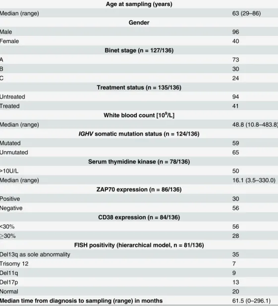
We performed target-specific sequencing on purified B-cell DNA obtained from 96 men and 40 women with confirmed CLL disease, treated and followed at the University of Cologne, Ger-many. The majority of patients presented with previously untreated (94/70%) and/or early stage disease (Binet stage A, 73 patients/58%), at the time the sample was obtained. A subset of 41 (31%) of all patients had received a median of 2 (1–11) CLL specific treatments prior to in-clusion into our study. The median time from diagnosis to sample was 41 months (0–209 months). Among patients from whom FISH analysis was available (81/60%), there was a sub-stantial subset with deletions in chromosome 17p (13 cases/16.0%), most probably due to refer-ral to our institution as a tertiary care center. Other prognostic markers, such as theIGHV
mutation status, ZAP70-, CD38-surface expression, and serum thymidine kinase were distrib-uted according to expected rates (Table 2andS6 Table).
Fig 1. Algorithm of variant analysis.A) Variants with an allelic frequency below 5% were discarded, resulting in 4,396 variants. B) Only the 3,322 non-synonymous variants were used for further analysis. The variant count per gene is represented in the bar chart. C) Variants located in areas of high background noise and/or in homopolymeric regions, and single strand variants were visually identified in the Integrative Genomic Viewer (IGV, Broad Institute) and removed. In doubtful cases, Sanger sequencing was performed to prove or disprove an alteration. Furthermore variants without functional impact on the protein determined by at least two of four applied program algorithms as described in material and method were removed. This resulted in 102 final mutations in 60 CLL specimens.
amplicons demonstrated a mean coverage per exon in a range of 0 to 7,156 reads. Only for five exons (3%;ATMexon 20,NOTCH1exon 27,SF3B1exon 5 and 11,TP53exon 11) the mean read count was less than 100, but 83% of targeted exons were covered by more than 500 reads (S1 Fig).
Two cell lines (Mino, AT45RM) with known mutations inTP53(exon 5) orATM(exon 53) were selected as positive controls to estimate the lowest detection rate of our targeted NGS method. Analyzing fractional dilutions of mutated cell line DNA (5% to 100%), the allelic fre-quency of theTP53andATMmutations detected by NGS followed a linear relationship with increasing amounts of tumor DNA (S2 Fig, P0.003, rho 1.000). We unambiguously identified the homozygousTP53mutation p.V147G in a background of 95% wild type DNA and the het-erozygousATMmutation p.R2598in up to 90% wild type DNA background (obtained allelic
Table 2. Patient characteristics.
Absolute
Age at sampling (years)
Median (range) 63 (29–86)
Gender
Male 96
Female 40
Binet stage (n = 127/136)
A 73
B 30
C 24
Treatment status (n = 135/136)
Untreated 94
Treated 41
White blood count [109/L]
Median (range) 48.8 (10.8–483.8)
IGHVsomatic mutation status (n = 124/136)
Mutated 59
Unmutated 65
Serum thymidine kinase (n = 78/136)
>10U/L 50
Median (range) 16.1 (3.5–330.0)
ZAP70 expression (n = 86/136)
Positive 30
Negative 56
CD38 expression (n = 84/136)
<30% 56
30% 28
FISH positivity (hierarchical model, n = 81/136)
Del13q as sole abnormality 35
Trisomy 12 7
Del11q 9
Del17p 13
Normal 20
Median time from diagnosis to sampling (range) in months 61.5 (0–296.1)
frequency: 2% and 8%, respectively). Therefore, our NGS method obtained an adequate low LoD to uncover small subsets of mutated CLL cells due to clonal heterogeneity.
Sequencing data analysis resulted in a total of 4,396 variants after raw data alignment and first background removal (Fig 1A and 1B). Exclusion of sequencing errors, synonymous vari-ants, variants without functional impact, and SNPs, led to 102 mutations predicted to affect protein function by at least two of four applied program algorithms as described in material and methods [32,33]. These 102 mutations including 83 missense mutations, 12 deletions, 6 nonsense mutations, and 1 insertion were detected in eight genes and 60 out of 136 CLL sam-ples (Fig 1CandS8 Table). In the remaining 76 patients (56%) no variants could be identified.
Fastq files are available at European Nucleotide Archive (ENA;http://www.ebi.ac.uk/ena/ data/view/PRJEB9036).
Multiplex PCR-based NGS Detects Variants in CLL-Related Genes
The highest frequency of mutations was obtained forTP53andSF3B1followed byNOTCH1,ATMandXPO1, whereasMYD88,PTPN6andDDX3Xshowed only two or one variant, re-spectively (Fig 1C). No mutations were found inBTK,CD79B,FBXW7,MAPK1,PIK3CA,
PIK3CDandPTEN.
A total of 16ATMmutations appeared in 15 of the 136 CLL patients (11%) and were evenly distributed over the entire gene (Fig 2). Interestingly, most of the samples harboring anATM
mutation showed at least one additional mutation in another gene (Fig 3). ThreeATMmutated patients had also a deletion ofATMin the second allele, as assessed by FISH.
Nearly 98% (2,719) of detectedNOTCH1variants turned out to be SNPs or sequencing er-rors. The remaining 17 mutations occurred in 17 patients (13%). Seven patients (5%) exhibited the previously reported p.P2514fs mutation, located in the PEST domain encoded by exon 34 [6]. Six variants were located in the NOTCH extracellular part (NEC, 6/17 variants, 35%). In-terestingly, one patient exhibited a missense mutation located in exon 26 (p.F1606L) affecting the heterodimerization domain (HD) of the NOTCH1 protein.
TP53was the second most frequently mutated gene with 20 of 136 patients (15%) harboring a total of 29 mutations. Most of them (21/29; 72%) occurred in exon 6 to 8 and were identified to disrupt the TP53 DNA binding function. TenTP53mutated patients had also a deletion of
TP53in the second allele, verified by FISH.
In agreement to previous data from Wanget al.SF3B1showed a mutation frequency of 15% (21/136 patients) clustering in exon 14 to 16 [10]. The most frequent mutation was determined as p.K700E in exon 15 (4/23, 17%). Typical and functionally relevant exon 15 mutations in
XPO1occurred in 13 patients (10%, p.E571I/K/Q) [6,18].
Furthermore, we found twoMYD88mutations (p.V217F in exon 3 and p.L265P in exon 5) in two of the 136 patients (2%).
Only one patient (1%) exhibited a mutation in exon 9 ofDDX3X(p.T275P) and one patient in exon 11 ofPTPN6(p.V451M). The latter one was located in the highly conserved catalytic protein-tyrosine phosphatase domain of the growth factor regulator SHP-1(PTPN6)and oc-curred with an allelic frequency of 51%.
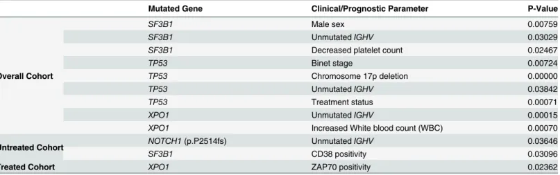
Variants Detected by NGS Associate with Clinical and Prognostic
Parameters
Genes with mutations in at least ten samples obtained by our analysis, i.e.TP53,SF3B1,
47/53, 89%; P = 0.03) or previously untreated CLL (42/53, 79%; P = 0.001) at the time of sam-pling. Presence of mutations inNOTCH1,SF3B1,TP53andXPO1was associated with at least one unfavorable prognostic marker such as an unmutatedIGHVgene status or positivity for ZAP70 or CD38 (Table 3).SF3B1mutated patients were significantly more frequent of male gender (20/96 males vs. 1/40 females, P = 0.008) andIGHVunmutated (15/21, 72% vs. 5/21, 24%, P = 0.03) thanSF3B1wild type cases (S3 Fig). In the untreated patient cohort, the pres-ence ofSF3B1mutations significantly correlated with positivity for CD38 assessed by flow cy-tometry (P = 0.03) (S4 Fig).
Similarly, an unmutatedIGHVstatus occurred more frequently inTP53mutants (15/20, 75% vs. 4/20, 20%, P = 0.04) and in untreated patients with aNOTCH1PEST domain mutation (4/4, 100% vs. 0/4, 0%, P = 0.04), compared to their wild type counterparts.
Fig 2. Alteration type, number of occurrence and location of detected mutations inTP53,SF3B1,NOTCH1and ATM are shown.TP53: AD activation domain (amino acid 1–50); PD proline-rich domain (amino acid 63–97); TD tetramerization domain (amino acid 323–356); ND negative regulation domain (amino acid 363–393);SF3B1: The majority ofSF3B1alterations were clustered in the region encoding the highly conserved HEAT (huntingtin, elongation factor 3, protein phosphatase 2A, target of rapamycin 1) repeats 5–8. Only one alteration occurred in the N-terminal (amino acids 1–450), domain, which is an important docking or binding domain for numerous splicing factor partners like U2AF1/2, and cyclin E.NOTCH1: (EGF)-like epidermal growth factor repeats (amino acid 20–1426), LNR Lin-12 NOTCH repeats (amino acid 1449–1571), HD-N/C heterodimerization domain (N-terminus; C-terminus), RAM RAM domain, ANK ankyrin repeat domain (amino acid 1927–2089); PEST Pro-Glu-Ser-Thr motif for degradation (amino acid 2507–2526);ATM: FAT
FRAP-ATM-TRRAP (amino acid 1960–2566), KD protein kinase domain (amino acid 2712–2962), PRD PIKK-regulatory domain (amino acid 2961–3025), FATC FAT-c-term domain (amino acid 3024–3056); aa amino acid
Patients with mutations inXPO1exhibited significantly increased WBC, possibly reflecting the proliferative capacity of CLL cells, compared to patients withoutXPO1mutations (mean: 134 vs. 65 x109/L,P<0.001). In treated patients, positivity for ZAP70 was significantly overrep-resented in patients with a mutatedXPO1gene (P = 0.02).
OnlyTP53mutations were found to be enriched in treated versus untreated patients (12/41, 29% vs. 8/95, 8%,P<0.001), indicating a possible selection of this genetic alteration due to prior treatments. Further, patients withTP53mutations exhibited significantly more frequent deletions in the second allele on chromosome 17p, resulting in a complete disruption of the TP53 protein function (P<0.001) (Table 3).
Discussion
CLL is a socioeconomically relevant disease of older adults with a currently rapidly changing field of new drugs entering clinical practice and an evolving discovery of genomic mutations with major clinical relevance [3,15]. Future diagnostics and research in CLL and cancer in gen-eral will require the implementation of mutational screening assays, which are resource effi-cient, sensitive, and rapidly adaptable to clinical and scientific needs.
Here, we present a targeted sequencing assay, which combines library and sequencing chemistry beyond the boundaries of manufacturers. For this approach we optimized target am-plification, sequencing output and data analysis for routine application. We performed a multi-plex PCR-based library amplification combining an Ion AmpliSeq primer design (Life
Technologies) with a modified library preparation chemistry that allows sequencing on an Illu-mina instrument. Our assay targeted the complete coding regions of five most frequently mu-tated“CLL-genes”(ATM,MYD88,NOTCH1,SF3B1andTP53) [6,7,10] and additionally ten genes with a more exploratory driven interest, e.g. the kinase domains of the drug targetsBTK
andPIK3CD[15]. Our method is performable within three days from sample DNA extraction to data analysis and offers suitable flexibility by the replacement or addition of target regions during primer design.
Fig 3. Genetic profile of 60 CLL samples carrying gene mutations determined by NGS.Each row represents the variants of one patient, each column summarizes the mutations occurring in one specific gene. Per each gene the number of mutations is given per patient. Dark blue samples indicate patients with aberration on chromosome 11 (del11q) forATMmutated cases or on chromosome 17 (del17p) forTP53 mutated cases, determined by FISH.
doi:10.1371/journal.pone.0129544.g003
Table 3. Statistical correlations between gene mutation status and clinical and biological parameters.
Mutated Gene Clinical/Prognostic Parameter P-Value
Overall Cohort
SF3B1 Male sex 0.00759
SF3B1 UnmutatedIGHV 0.03029
SF3B1 Decreased platelet count 0.02467
TP53 Binet stage 0.00724
TP53 Chromosome 17p deletion 0.00000
TP53 UnmutatedIGHV 0.03842
TP53 Treatment status 0.00071
XPO1 UnmutatedIGHV 0.00015
XPO1 Increased White blood count (WBC) 0.00070
Untreated Cohort NOTCH1(p.P2514fs) UnmutatedIGHV 0.03646
SF3B1 CD38 positivity 0.03096
Treated Cohort XPO1 ZAP70 positivity 0.02362
While targeted NGS offers the advantage to assess multiplexed samples and genes in one ex-perimental setup—thus being relatively cost-efficient compared to Sanger sequencing-, one disadvantage is that the ability to detect mutations in a distinct gene depends on the achieved coverage/depth of reads in this specific region. Coverage and sequencing depth can vary sub-stantially, depending on the gene region itself (e.g. GC rich, homopolymers, etc.), enzyme chemistry and sequencing platform. With our technology we were able to cover 83% of tar-geted exons with a minimum coverage of 500 reads. Only for five exons the mean number of reads was below 100, a threshold under which we would consider calling of mutations not pos-sible for diagnostic purposes and apply either repeated NGS or Sanger sequencing.
Most studies on targeted CLL sequencing published so far have implemented NGS methods without giving details on the performance of the technology. Suttonet al. are the first to report details on assay quality and analytical requirements of targeted NGS results using the HaloPlex probe technology from Agilent in CLL [20]. This technology offers the advantage of target spe-cific probe hybridization without PCR amplification. They investigated a set of 188 patients with poor prognostic features for gene alterations inATM,BIRC3,KLHL6,MYD88,NOTCH1,
POT1,SF3B1 TP53, andXPO1. For final analysis, they only included patients, for which they obtained at least 100 reads for 80% of the targeted bases (96% of their samples). Thus, their assay achieved reasonable quality results in terms of coverage and uniformity of read depth, comparable to ours. As discussed by the authors, cutoffs currently chosen for quality parame-ters to evaluate targeted NGS data are more or less arbitrary. Further studies are needed to standardize and harmonize such parameters for comparability of different datasets and clinical implementation.
BCR signaling. Therefore inactivating mutations could constitutively activate BCR signaling in CLL cells and therefore influence disease development and outcome [39,40].
In general, the mutation rates obtained by our NGS assay for genes known to be mutated in CLL (TP53,SF3B1,ATM,NOTCH1,XPO1,MYD88,DDX3X) are comparable to other studies [6–8,10,13,17–19]. Most of the mutations detected are located at typical hotspot locations, such as the p.K700E and p.G742D mutations inSF3B1(predominantly found in male patients), the p.L256P mutation inMYD88,thep.T275P mutation in exon 9 ofDDX3X, or the p.E571K mutation inXPO1[6,7,10,18,41]. One CLL case exhibited aMYD88p.V217F mutation, an alteration previously described in diffuse large B-cell lymphoma (DLBCL) by Ngoet al. [41].
Targeted NGS studies in CLL published to date frequently omitted sequencing ofATM, due to the lack of hotspots regions in this relatively large gene and size limitations of their assay [13,17–19]. In our hands,ATMsequencing within a larger gene panel was feasible and muta-tions detected at a rate of 11%, comparable to the 12% rate reported by Austen and colleagues [22]. Interestingly, we foundATMvariants occurring more frequently in combination with other variants, in particular withNOTCH1orTP53(Fig 3), an aspect also confirmed by the study of Suttonet al., described above. ForNOTCH1our data analysis obtained a high inci-dence of non-functional variants, which might be attributable to technical issues during target enrichment and/or sequencing, e.g. by polymerase reading errors in GC- or homopolymeric re-gions. Beside variants in the EGF-like and PEST domains, we detected a gain-of-function mu-tation (p.F1606L) in the HD domain of the NOTCH1 extracellular subunit. Only the p.P2514 frameshift deletion in the PEST domain revealed a significant correlation with an unmutated
IGHVstatus (Table 3), indicating that these mutations are preferentially enriched in CLL pa-tients with adverse prognosis.
One advantage of NGS technologies for mutation analysis is that the achievement of a high sequencing coverage allows the more sensitive determination of small subclones carrying mu-tations. It has been demonstrated that such subclones can evolve over time and drive CLL pro-gression and transformation [4,42]. In our assay, the allelic frequency of mutations ranged from 5% to 100%. The smallest clonal fraction was determined for twoNOTCH1mutations (p. W327R; 4,765 reads and p.F1606L; 166 reads) and oneTP53mutation (p.Y234C; 2,635 reads). Furthermore, sequencing of mutated cell lines allowed us to estimate the low LoD of our meth-od, which depicted 5% ofTP53mutated cells diluted in wild type background at a 2% allelic frequency. Thus, our method would be clearly able to pick up small mutated subclones in CLL, presumably beyond the detection limit of Sanger sequencing. This compares to other NGS studies reporting a sensitivity or LoD of targeted NGS at 2–3% [17,18].
In conclusion, we have developed a targeted NGS panel and high-throughput assay for mu-tation analysis in CLL, which is resource-efficient and highly sensitive for the detection of low frequency alleles and fast enough to be applicable to clinical decision processes. Applying a sys-tematic functional data assessment, we found various alterations including known hotspot mu-tations and one interestingPTPN6mutation in the BCR, without the need of non-tumor DNA sequencing. Our NGS methodology can be easily translated to molecular diagnostics of other types of cancer and may pave the way for a fast-throughput combination of morphological and molecular diagnostics in hematologic and non-hematologic malignancies.
Supporting Information
S1 Fig. Median and mean read count per exon for 167 exons of the 15 genes are shown. Re-sults reflect data from five NGS runs.
S2 Fig. Linear relationship of mutation rate and allele frequency detected by NGS. Sequenc-ing of two dilution series of cell line DNA with a known A) homozygousTP53c.440T>G; p. V147G mutation (Mino cell line) and B) heterozygousATMc.7792C>T; p.R2598mutation (AT45RM cell line) demonstrated a linear relationship of the fractional dilution rate and the mutation allele frequency obtained by NGS. Further, the data point to the detection limit achieved by our NGS approach by detecting at least 214ATMmutated AT45RM cells in a background of 2,036 wild type HEK-293 cells and 214TP53mutated Mino cells in a back-ground of 4,071 wild type HEK-293 cells.
(TIF)
S3 Fig. Associations ofSF3B1andTP53mutations with clinical and prognostic parameters
are shown.A)SF3B1mutated patients were mainlyIGHVunmutated, in contrast toSF3B1
wild type patients that showed a normal mutatedIGHVstatus (P = 0.03). B)SF3B1mutated patients were significantly more of male gender (P = 0.008). C)TP53mutations were found particularly more frequent in intermediate and advanced stage with a need for treatment (Binet stage B/C) compared with patients in an early stage (Binet stage A) (P = 0.008). D and E)TP53mutations were also frequently more detected in treated patients (P<0.001) and in pa-tients with genomic aberrations on chromosome 17 (del17p) (P<0.001). ND not determined. (TIFF)
S4 Fig. The presence of functional relevant mutations inSF3B1andXPO1was associated
with the unfavorable prognostic marker like positivity for ZAP70 or CD38.A)SF3B1 mu-tated untreated patients showed an increased CD38 expression (p<0.04). B and C) Patients harboringXPO1mutations showed an increased WBC (p<0.001) and treated patients pre-sented a higher ZAP70 expression compared to their wild type counterparts (p<0.03). (TIF)
S1 Table. Target regions of the CLL panel 1 and 2.
(DOCX)
S2 Table. Parameters for theHFEqPCR.DNA quantification was done using native DNA from HEK-293 (human embryonic kidney) cells without known gene mutations; all samples were measured in duplicates.
(DOCX)
S3 Table. PCR parameters for quantification of the constructed libraries by qPCR.A) PCR components; B) PCR conditions. Amplicon library quantification was performed with 5-fold dilutions of PhiX Control V3 (Illumina, San Diego, CA, USA) in a range from 0.064 up to 40 pM as reference standard. The library samples were diluted 1:4000 and measured in duplicates. (DOCX)
S4 Table. Primer used for Sanger sequencing validation.
(DOCX)
S5 Table. Parameters for PCR and Sanger sequencing.A1 and 2) Components and condi-tions for amplification of target regions by PCR; B1 and 2) Components and condicondi-tions for Sanger sequencing reaction.
(DOCX)
S6 Table. Clinical information of the patients analyzed in this study.
S7 Table. Run parameters from the five MiSeq sequencing runs.
(DOCX)
S8 Table. Complete list of detected mutations.Bold mutations were confirmed by Sanger se-quencing. fs frame shift;
stop gained; Freq frequency; Cov coverage; dbSNP single nucleotide variants database.
(DOCX)
Acknowledgments
We greatly appreciate the help of Thomas Landwehr (CLL Biobank, Department I of Internal Medicine, University of Cologne, Cologne, Germany) for sample collection and the support of Jasmin Bahlo from the German CLL Study Group (GCLLSG) in statistical data interpretation. We thank Christian Reinhardt (Department I of Internal Medicine, Center for Integrated On-cology (CIO) Cologne-Bonn, and Excellence Cluster for Cellular Stress Response and Aging-Associated Diseases (CECAD), University of Cologne, Cologne, Germany) for many helpful discussions.
Author Contributions
Conceived and designed the experiments: CV CDH MO KK. Performed the experiments: CV LW UK. Analyzed the data: CV FDM GC CDH MO. Contributed reagents/materials/analysis tools: MO CDH KAK MH MP LCH. Wrote the paper: CV MO CDH RB.
References
1. Dighiero G, Hamblin TJ. Chronic lymphocytic leukaemia. Lancet. 2008; 371(9617):1017–29. Epub 2008/03/25. doi:10.1016/S0140-6736(08)60456-0PMID:18358929.
2. Cramer P, Hallek M. Prognostic factors in chronic lymphocytic leukemia-what do we need to know? Nat Rev Clin Oncol. 2011; 8(1):38–47. Epub 2010/10/20. doi:10.1038/nrclinonc.2010.167PMID:
20956983.
3. Gruber M, Wu CJ. Evolving understanding of the CLL genome. Semin Hematol. 2014; 51(3):177–87. Epub 2014/07/23. doi:10.1053/j.seminhematol.2014.05.004PMID:25048782; PubMed Central PMCID: PMC4107366.
4. Landau DA, Carter SL, Stojanov P, McKenna A, Stevenson K, Lawrence MS, et al. Evolution and im-pact of subclonal mutations in chronic lymphocytic leukemia. Cell. 2013; 152(4):714–26. Epub 2013/ 02/19. doi:10.1016/j.cell.2013.01.019PMID:23415222; PubMed Central PMCID: PMC3575604. 5. Fabbri G, Rasi S, Rossi D, Trifonov V, Khiabanian H, Ma J, et al. Analysis of the chronic lymphocytic
leukemia coding genome: role of NOTCH1 mutational activation. The Journal of experimental medi-cine. 2011; 208(7):1389–401. Epub 2011/06/15. doi:10.1084/jem.20110921PMID:21670202; PubMed Central PMCID: PMC3135373.
6. Puente XS, Pinyol M, Quesada V, Conde L, Ordonez GR, Villamor N, et al. Whole-genome sequencing identifies recurrent mutations in chronic lymphocytic leukaemia. Nature. 2011; 475(7354):101–5. Epub 2011/06/07. doi:10.1038/nature10113PMID:21642962; PubMed Central PMCID: PMC3322590. 7. Quesada V, Conde L, Villamor N, Ordonez GR, Jares P, Bassaganyas L, et al. Exome sequencing
identifies recurrent mutations of the splicing factor SF3B1 gene in chronic lymphocytic leukemia. Nature genetics. 2012; 44(1):47–52. Epub 2011/12/14. doi:10.1038/ng.1032PMID:22158541.
8. Wan Y, Wu CJ. SF3B1 mutations in chronic lymphocytic leukemia. Blood. 2013; 121(23):4627–34. Epub 2013/04/10. doi:10.1182/blood-2013-02-427641PMID:23568491; PubMed Central PMCID: PMC3674664.
9. Villamor N, Lopez-Guillermo A, Lopez-Otin C, Campo E. Next-generation sequencing in chronic lym-phocytic leukemia. Semin Hematol. 2013; 50(4):286–95. Epub 2013/11/20. doi:10.1053/j.
seminhematol.2013.09.005PMID:24246696.
11. Rossi D, Rasi S, Fabbri G, Spina V, Fangazio M, Forconi F, et al. Mutations of NOTCH1 are an inde-pendent predictor of survival in chronic lymphocytic leukemia. Blood. 2012; 119(2):521–9. Epub 2011/ 11/15. doi:10.1182/blood-2011-09-379966PMID:22077063; PubMed Central PMCID: PMC3257017. 12. Villamor N, Conde L, Martinez-Trillos A, Cazorla M, Navarro A, Bea S, et al. NOTCH1 mutations identify
a genetic subgroup of chronic lymphocytic leukemia patients with high risk of transformation and poor outcome. Leukemia. 2013; 27(5):1100–6. Epub 2013/01/09. doi:10.1038/leu.2012.357PMID:
23295735.
13. Cortese D, Sutton LA, Cahill N, Smedby KE, Geisler C, Gunnarsson R, et al. On the way towards a 'CLL prognostic index': focus on TP53, BIRC3, SF3B1, NOTCH1 and MYD88 in a population-based co-hort. Leukemia. 2014; 28(3):710–3. Epub 2013/11/13. doi:10.1038/leu.2013.333PMID:24217197. 14. Rossi D, Fangazio M, Rasi S, Vaisitti T, Monti S, Cresta S, et al. Disruption of BIRC3 associates with
flu-darabine chemorefractoriness in TP53 wild-type chronic lymphocytic leukemia. Blood. 2012; 119 (12):2854–62. Epub 2012/02/07. doi:10.1182/blood-2011-12-395673PMID:22308293.
15. Woyach JA, Furman RR, Liu TM, Ozer HG, Zapatka M, Ruppert AS, et al. Resistance mechanisms for the Bruton's tyrosine kinase inhibitor ibrutinib. The New England journal of medicine. 2014; 370 (24):2286–94. Epub 2014/05/30. doi:10.1056/NEJMoa1400029PMID:24869598.
16. Chang F, Li MM. Clinical application of amplicon-based next-generation sequencing in cancer. Cancer Genet. 2013; 206(12):413–9. Epub 2013/12/18. doi:10.1016/j.cancergen.2013.10.003PMID:
24332266.
17. Jethwa A, Hullein J, Stolz T, Blume C, Sellner L, Jauch A, et al. Targeted resequencing for analysis of clonal composition of recurrent gene mutations in chronic lymphocytic leukaemia. British journal of hae-matology. 2013; 163(4):496–500. Epub 2013/09/17. doi:10.1111/bjh.12539PMID:24032483. 18. Jeromin S, Weissmann S, Haferlach C, Dicker F, Bayer K, Grossmann V, et al. SF3B1 mutations
corre-lated to cytogenetics and mutations in NOTCH1, FBXW7, MYD88, XPO1 and TP53 in 1160 untreated CLL patients. Leukemia. 2014; 28(1):108–17. Epub 2013/10/12. doi:10.1038/leu.2013.263PMID:
24113472.
19. Baliakas P, Hadzidimitriou A, Sutton LA, Rossi D, Minga E, Villamor N, et al. Recurrent mutations refine prognosis in chronic lymphocytic leukemia. Leukemia. 2014. Epub 2014/06/20. doi:10.1038/leu.2014. 196PMID:24943832.
20. Sutton LA, Ljungstrom V, Mansouri L, Young E, Cortese D, Navrkalova V, et al. Targeted next-genera-tion sequencing in chronic lymphocytic leukemia: a high-throughput yet tailored approach will facilitate implementation in a clinical setting. Haematologica. 2015; 100(3):370–6. Epub 2014/12/07. doi:10. 3324/haematol.2014.109777PMID:25480502; PubMed Central PMCID: PMC4349276.
21. Thompson AA, Talley JA, Do HN, Kagan HL, Kunkel L, Berenson J, et al. Aberrations of the B-cell re-ceptor B29 (CD79b) gene in chronic lymphocytic leukemia. Blood. 1997; 90(4):1387–94. Epub 1997/ 08/15. PMID:9269755.
22. Austen B, Powell JE, Alvi A, Edwards I, Hooper L, Starczynski J, et al. Mutations in the ATM gene lead to impaired overall and treatment-free survival that is independent of IGVH mutation status in patients with B-CLL. Blood. 2005; 106(9):3175–82. Epub 2005/07/15. doi:10.1182/blood-2004-11-4516PMID:
16014569.
23. Zenz T, Eichhorst B, Busch R, Denzel T, Habe S, Winkler D, et al. TP53 mutation and survival in chronic lymphocytic leukemia. J Clin Oncol. 2010; 28(29):4473–9. Epub 2010/08/11. doi:10.1200/JCO.2009. 27.8762PMID:20697090.
24. Hallek M, Cheson BD, Catovsky D, Caligaris-Cappio F, Dighiero G, Dohner H, et al. Guidelines for the diagnosis and treatment of chronic lymphocytic leukemia: a report from the International Workshop on Chronic Lymphocytic Leukemia up dating the National Cancer Institute-Working Group 1996 guide-lines. Blood. 1996; 111:5446–56.
25. Schweighofer CD, Huh YO, Luthra R, Sargent RL, Ketterling RP, Knudson RA, et al. The B cell antigen receptor in atypical chronic lymphocytic leukemia with t(14;19)(q32;q13) demonstrates remarkable ste-reotypy. International journal of cancer Journal international du cancer. 2011; 128(11):2759–64. Epub 2010/08/18. doi:10.1002/ijc.25605PMID:20715110.
26. Lai R, McDonnell TJ, O'Connor SL, Medeiros LJ, Oudat R, Keating M, et al. Establishment and charac-terization of a new mantle cell lymphoma cell line, Mino. Leukemia research. 2002; 26(9):849–55. Epub 2002/07/20. PMID:12127561.
27. Magliozzi M, Piane M, Torrente I, Sinibaldi L, Rizzo G, Savio C, et al. DHPLC screening of ATM gene in Italian patients affected by ataxia-telangiectasia: fourteen novel ATM mutations. Disease markers. 2006; 22(4):257–64. Epub 2006/11/25. PMID:17124347; PubMed Central PMCID: PMC3862285. 28. Peifer M, Fernandez-Cuesta L, Sos ML, George J, Seidel D, Kasper LH, et al. Integrative genome
29. Li H, Durbin R. Fast and accurate short read alignment with Burrows-Wheeler transform. Bioinformat-ics. 2009; 25(14):1754–60. Epub 2009/05/20. doi:10.1093/bioinformatics/btp324PMID:19451168; PubMed Central PMCID: PMC2705234.
30. Kent WJ. BLAT—the BLAST-like alignment tool. Genome Res. 2002; 12(4):656–64. Epub 2002/04/05. doi:10.1101/gr.229202Article published online before March 2002. PMID:11932250; PubMed Central PMCID: PMC187518.
31. Bhagwat M, Young L, Robison RR. Using BLAT to find sequence similarity in closely related genomes. Current protocols in bioinformatics / editoral board, Andreas D Baxevanis [et al]. 2012;Chapter 10: Unit10 8. Epub 2012/03/06. doi:10.1002/0471250953.bi1008s37PMID:22389010; PubMed Central PMCID: PMC4101998.
32. Reva B, Antipin Y, Sander C. Predicting the functional impact of protein mutations: application to cancer genomics. Nucleic acids research. 2011; 39(17):e118. Epub 2011/07/06. doi:10.1093/nar/gkr407
PMID:21727090; PubMed Central PMCID: PMC3177186.
33. Landau DA, Wu CJ. Chronic lymphocytic leukemia: molecular heterogeneity revealed by high-through-put genomics. Genome Med. 2013; 5(5):47. Epub 2013/06/05. doi:10.1186/gm451PMID:23731665; PubMed Central PMCID: PMC3706960.
34. Kumar P, Henikoff S, Ng PC. Predicting the effects of coding non-synonymous variants on protein func-tion using the SIFT algorithm. Nat Protoc. 2009; 4(7):1073–81. Epub 2009/06/30. doi:10.1038/nprot. 2009.86PMID:19561590.
35. Adzhubei IA, Schmidt S, Peshkin L, Ramensky VE, Gerasimova A, Bork P, et al. A method and server for predicting damaging missense mutations. Nat Methods. 2010; 7(4):248–9. Epub 2010/04/01. doi:
10.1038/nmeth0410-248PMID:20354512; PubMed Central PMCID: PMC2855889.
36. Schwarz JM, Rodelsperger C, Schuelke M, Seelow D. MutationTaster evaluates disease-causing po-tential of sequence alterations. Nat Methods. 2010; 7(8):575–6. Epub 2010/08/03. doi:10.1038/ nmeth0810-575PMID:20676075.
37. Malecki MJ, Sanchez-Irizarry C, Mitchell JL, Histen G, Xu ML, Aster JC, et al. Leukemia-associated mu-tations within the NOTCH1 heterodimerization domain fall into at least two distinct mechanistic classes. Mol Cell Biol. 2006; 26(12):4642–51. Epub 2006/06/02. doi:10.1128/MCB.01655-05PMID:16738328; PubMed Central PMCID: PMC1489116.
38. Gunnarsson R, Mansouri L, Rosenquist R. Exploring the genetic landscape in chronic lymphocytic leu-kemia using high-resolution technologies. Leuleu-kemia & lymphoma. 2013; 54(8):1583–90. Epub 2012/ 11/22. doi:10.3109/10428194.2012.751530PMID:23167608.
39. Tibaldi E, Brunati AM, Zonta F, Frezzato F, Gattazzo C, Zambello R, et al. Lyn-mediated SHP-1 recruit-ment to CD5 contributes to resistance to apoptosis of B-cell chronic lymphocytic leukemia cells. Leuke-mia. 2011; 25(11):1768–81. Epub 2011/06/28. doi:10.1038/leu.2011.152PMID:21701493.
40. Wu C, Sun M, Liu L, Zhou GW. The function of the protein tyrosine phosphatase SHP-1 in cancer. Gene. 2003; 306:1–12. Epub 2003/03/27. PMID:12657462.
41. Ngo VN, Young RM, Schmitz R, Jhavar S, Xiao W, Lim KH, et al. Oncogenically active MYD88 muta-tions in human lymphoma. Nature. 2011; 470(7332):115–9. Epub 2010/12/24. doi:10.1038/ nature09671PMID:21179087.
42. Landau DA, Carter SL, Getz G, Wu CJ. Clonal evolution in hematological malignancies and therapeutic implications. Leukemia. 2014; 28(1):34–43. Epub 2013/08/28. doi:10.1038/leu.2013.248PMID: