Apoptosis-Related Gene Expression in an
Adult Cohort with Crimean-Congo
Hemorrhagic Fever
Nil Guler1*, Cafer Eroglu2, Hava Yilmaz2, Adil Karadag3, Hasan Alacam4, Mustafa Sunbul2, Tom E. Fletcher2,5, Hakan Leblebicioglu2
1Department of Hematology, School of Medicine, Ondokuz Mayis University, Samsun, Turkey,
2Department of Clinical Microbiology and Infectious Diseases, School of Medicine, Ondokuz Mayis University, Samsun, Turkey,3Department of Medical Microbiology, School of Medicine, Ondokuz Mayis University, Samsun, Turkey,4Department of Medical Biochemistry, School of Medicine, Ondokuz Mayis University, Samsun, Turkey,5Liverpool School of Tropical Medicine, Liverpool L3 5QA, United Kingdom
*nilvecay@yahoo.com
Abstract
Crimean-Congo Hemorrhagic Fever (CCHF) is a life threatening acute viral infection char-acterized by fever, bleeding, leukopenia and thrombocytopenia. It is a major emerging infec-tious diseases threat, but its pathogenesis remains poorly understood and few data exist for the role of apoptosis in acute infection. We aimed to assess apoptotic gene expression in leukocytes in a cross-sectional cohort study of adults with CCHF. Twenty participants with CCHF and 10 healthy controls were recruited at a tertiary CCHF unit in Turkey; at admission baseline blood tests were collected and total RNA was isolated. The RealTime ready Human Apoptosis Panel was used for real-time PCR, detecting differences in gene expres-sion. Participants had CCHF severity grading scores (SGS) with low risk score (10 out of 20) and intermediate or high risk scores (10 out of 20) for mortality. Five of 20 participants had a fatal outcome. Gene expression analysis showed modulation of pro-apoptotic and anti-apoptotic genes that facilitate apoptosis in the CCHF patient group. Dominant extrinsic pathway activation, mostly related with TNF family members was observed. Severe and fatal cases suggest additional intrinsic pathway activation. The clinical significance of rela-tive gene expression is not clear, and larger longitudinal studies with simultaneous mea-surement of host and viral factors are recommended.
Introduction
Crimean Congo Hemorrhagic Fever (CCHF) is a life threatening acute viral infection caused by a RNA virus belonging to the familyBunyaviridae. The first documented infection was observed in 1944 in the Crimea region of Ukraine and was designated as Crimean haemorrha-gic fever. It was later observed in Congo in 1956 and the name was changed to CCHF [1]. CCHF virus (CCHFV) is an enveloped single-stranded RNA virus, the genome of which includes small (S), medium (M) and large (L) segments, and phylogenetic analysis of CCHFV
a11111
OPEN ACCESS
Citation:Guler N, Eroglu C, Yilmaz H, Karadag A, Alacam H, Sunbul M, et al. (2016) Apoptosis-Related Gene Expression in an Adult Cohort with Crimean-Congo Hemorrhagic Fever. PLoS ONE 11(6): e0157247. doi:10.1371/journal.pone.0157247
Editor:Tetsuro Ikegami, The University of Texas Medical Branch, UNITED STATES
Received:March 7, 2016
Accepted:May 26, 2016
Published:June 15, 2016
Copyright:© 2016 Guler et al. This is an open access article distributed under the terms of the Creative Commons Attribution License, which permits unrestricted use, distribution, and reproduction in any medium, provided the original author and source are credited.
Data Availability Statement:All relevant data are within the paper and its Supporting Information files.
Funding:This study was supported by Ondokuz Mayis University (Turkey) with project number of PYO.TIP.1901.11.020. TF is funded by Wellcome Trust (104480/Z/14/Z) and the UK Ministry of Defence. The funders had no role in study design, data collection and analysis, decision to publish, or preparation of the manuscript.
strains has revealed high degree of genetic diversity amongst strains, particularly between viruses from different geographic regions [2].
CCHF is a major emerging infectious disease threat and is widely distributed across Africa, Eastern Europe, Asia and the Middle East. Turkey reports up to 1000 cases per year and CCHFV is predominantly transmitted to humans viaHyalommaspp. ticks [3]. Clinical fea-tures of disease represent a spectrum of disease severity, characterized by fever, bleeding and lethargy, and has a case fatality rate of 5–50%. The most common hematologic findings in CCHF are leukopenia (60%) and thrombocytopenia (100%)[4], with evidence of haemophago-cytosis in the bone marrow [5]. At present there is no approved vaccine or therapeutic agent, although ribavirin and hyper immune serum are utilized by some clinicians.
The pathogenesis of CCHF remains poorly understood but apoptotic programmed cell death has been proposed to play a role [2]. Regulation of apoptosis through suppression or induction during acute viral infection is important for virus survival and dissemination [6]. Apoptosis can be started by an extrinsic (death receptor mediated) or intrinsic pathway (mito-chondria mediated). The external pathway is activated when ligands—TNF-α, TNF-related
apoptosis-inducing ligand (TRAIL), and Fas ligand (FasL) known as CD95L, bind to death receptors on the cell surface. Some TRAIL receptors do not contain death domains and are referred to as decoy receptors 1 (DcR1) (TNFRSF10C) and 2 (DcR2) (TNFRSF10D) and pre-vent apoptosis signals from initiating. The intrinsic pathway to cell death is controlled by inter-actions on the mitochondrial outer membrane between sub-groups of the BCL-2 family, including the effector proteins BAX and BAK [7]. Both pathways ultimately result in up-regu-lation of effector caspases (caspase-3 & caspase-7) resulting in fragmentation of the DNA, cyto-skeleton, nuclear proteins and expression of ligands for phagocytic cells [8].
Apoptosis has been demonstrated in a range of cells in other acute viral infections including Dengue [9]. Massive intravascular apoptosis, including leukocyte apoptosis, has also been dem-onstrated in Ebola virus infection [10], where it has been shown to be associated with a fatal outcome. CCHFV has been shown to induce caspase-3 and modulate both intrinsic and extrin-sic pathways in cell cultures [11,12]. Elevated apoptotic markers have also recently been dem-onstrated in a paediatric cohort of CCHF [13], and associations were found with the disease’s severity criteria. We have previously observed apoptotic transformation in leukocytes in peripheral blood smears of patients with CCHF, and in the present work, this was investigated further by studying up and down-regulation of genes which are involved in leukocyte apoptosis in a cohort of adult patients with CCHF.
Methods
Study Participants
importance. It has subsequently been validated and may be used to aid triage/risk stratification of patients and to improve the functionality of healthcare staff.
Patient sampling and RealTime ready Apoptosis Panel
Four milliliter blood samples were drawn into EDTA blood tubes on the first day of hospitali-zation and standard baseline blood tests took place (Table 1). Red Blood Cell Lysis Buffer (Roche Diagnostics, Germany) was added to the blood samples and total RNA was isolated uti-lizing a High Pure RNA Isolation Kit (Roche Diagnostics, Germany). RNA samples were then stored at kept at -70°C until sample processing when cDNAs was obtained using a Transcriptor First Strand cDNA Synthesis Kit (Roche Diagnostics, Germany). Expression levels of apopto-tic-related genes were determined using The RealTime ready Human Apoptosis Panel (Roche Diagnostics, Germany) and a LightCycler 480 Probes Master. The RealTime ready Apoptosis Panel consists of ready-to-use qPCR assays designed for expression profiling of genes involved in the apoptosis pathway. Each multi-well plate contains assays for 84 different apoptosis-related genes and seven housekeeping genes, as well as five controls. This study was approved by the Ondokuz Mayis University Ethical Committee (2010/178) and all patients provided written informed consent.
Statistical analyses
The REST 2009 program (Qiagen, Germany) was used to quantify changes in the expression levels of genes and to determine statistical differences between the CCHF and control groups, and between CCHF fatal and survivor groups. Differences in gene expression were also ana-lyzed between intermediate or high SGS risk score participants (Group 1) and low SGS risk score (Group 2). P values less than or equal to 0.05 were accepted as statistically significant. Descriptive analysis of patient characteristics was performed using the Statistical Package for the Social Sciences (SPSS) version 15 for Windows (SPSS Inc., Chicago, IL, USA).
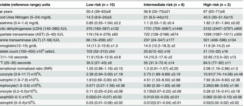
Table 1. Participant laboratory characteristics according to severity score: Mean (Minimum-Maximum)±Standard Deviation.
Variable (reference range) units Low risk (n = 10) Intermediate risk (n = 8) High risk (n = 2)
Age years 50.4 (39–63)±8 56.8 (20–73)±21 67 (63–71)±6
Blood Urea Nitrogen (5–24) mg/dL 14.3 (8.6–24)±5 21 (6.5–44)±12 40.5 (30–51.4)±15
Creatinine (0.4–1.4) mg/dL 0.85 (0.55–1.04)±0.2 1.1 (0.53–1.9)±0.4 1.82 (1.81–1.84)±0.02
Lactic dehydrogenase (LDH) (140–280) IU/L 370 (183–567)±132 1731 (705–5067)±1446 3122 (2447–3797)±955
Aspartate transaminase (AST) (5–40) IU/L 119 (16.4–279)±83 722 (128–2198)±674 1299 (1087–1511)±300
Alanine transaminase (ALT) (7–56) IU/L 66 (16–209)±57 237 (24–547)±177 501 (406–596)±134
Hemoglobin(12–15) gr/dL 14 (11.3–15.6)±1.3 14.5 (12.2–18.3)±2 11.6 (9–14.2)±4
Platelet count (150–450) x109cells/L 103 (52–212)±54 25.6(12–52)±16 21 (10–32)±16
PT(11–14) seconds 11.9 (10.8–12.9)±0.8 14 (10.2–17.4)±2 22.65 (13.3–32)±13
PTT (25–35) seconds 35.5 (27–43)±6 55 (31.3–72.4)±14 84.5 (77–92)±11
International normalized ratio (INR) 1.05 (0.96–1.16)±0.10 1.3 (0.91–1.57)±0.20 2.08 (1.19–2.96)±1.0
Leucocyte (3.9–11.7) x109/L 2.39 (0.94–5.00)±1.18 5.73 (1.89–9.89)±3.19 10.91(7.74–14.08)±4.48
Neutrophil (1.9–7.9) x109/L 1.61(0.59–3.00)±0.76 4.51 (1.53–8.50)±2.68 7.92 (6.24–9.60)±2.38
Lymphocyte(1.3–3.6) x109/L 0.571 (0.27–1.59)±0.38 0.80 (0.30–1.83)±0.58 2.26(0.88–3.63)±1.95
Monocyte (0.2–0.5)x109/L 0.11 (0.20–0.24)±0.08 0.135(0.07–0.22)±0.06 0.28 (0.15–0.41)±0.18
Basophil(0–0.1) x109/L 0.02(0.01–0.07)±0.02 0.01(0.02–0.03)±0.01 0.062 (0.02–0.10)±0.06
Eosinophil (0–0.4)x109/L 0.03 (0.01–0.06)±0.02 0.012(0.01–0.04)±0.01 0.02(0.02–0.02)±0.02
Results
Twenty patients (female: 6; male: 14) and 10 healthy controls (female: 5; male: 5) were included in this study. The patients mean age was 54.6 (20–73) years-old. At admission, participants were a mean 3.4 (1–6) days since onset of symptoms of disease. According to the SGS system 10 participants were low risk (score 0–4), 8 participants intermediate risk (5–8), and 2 partici-pants in the high risk group (9–14). Five of the 20 participants (25%) died of CCHF during the hospital admission. Three of the 5 fatal cases had an SGS score of 8, 1/5 a score of 9 and 1/5 a score of 13. Petechiae and/or ecchymosis were detected in 7/20 (35%) participants, and signifi-cant haemorrhage developed in a further 6/20 (30%) participants.
Detailed laboratory characteristics are displayed inTable 1stratified by severity group. Mean cohort data (n = 20) at baseline showed: leukocytes 4.58 x 10⁹cells/L (0.94–14.1); neutro-phils 3.4 x 10⁹cells/L (0.59–9.6); lymphocytes 0.83 x 10⁹cells/L (0.03–3.63); monocytes 0.14 x10⁹cells/L (0.02–0.41); basophils 0.11 x10⁹cells/L (0.01–1.01); alanine aminotransferase (ALT) 178 IU/L (16–596); aspartate transaminase (AST) IU/L 410 (16.4–2198); lactate dehy-drogenase (LDH) 1233 IU/L (183–5067); platelets 69.7 x 10⁹/L (10–212); prothrombin time (PT) 13.8 seconds (10.2–32); partial thromboplastin time (PTT) 48.2 seconds (26.6–92); inter-national normalized ratio (INR) 1.24 (0.91–2.96); blood urea nitrogen (BUN) 19.6 mg/dL (6.5–
51.4); and creatinine 1.05 mg/dL (0.53–1.9). The healthy control group exhibited a normal pro-file in laboratory parameters.
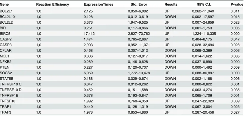
Expression analysis showed that the BCL2L1, BCL2L2, BIRC5, CASP2, CASP3, SOCS2, TNFSF10, and TRAF3 genes were up-regulated and the BCL2L10, BID, CFLAR, MCL1, NFKB2, PTEN, STAT5B, TNFRSF10C, TNFRSF10D, TNFRSF1B, and TRAF1 genes were down-regulated in the patient group compared with the healthy control group (Table 2).
There were no significant changes in the expression of AKT1, APAF1, AVEN, BAD, BAG1, BAK1, BAX, BBC3, BCL2, BCL2L11, BCL2L13, BIK, BIRC2, BIRC3, BOK, CAD, CASP1,
Table 2. Changes in gene expression level (up- or down-regulation) in the patient group compared with the healthy group.
Gene Reaction Efficiency ExpressionTimes Std. Error Results 95% C.I. P-value
BCL2L1 1,0 2,125 0,850–6,082 UP 0,262–11,940 0,011
BCL2L10 1,0 0,128 0,012–3,619 DOWN 0,002–17,597 0,015
BCL2L2 1,0 3,373 1,947–9,525 UP 0,007–24,859 0,028
BID 1,0 0,251 0,117–0,866 DOWN 0,001–1,753 0,005
BIRC5 1,0 17,412 2,827–70,762 UP 1,224–110,335 0,000
CASP2 1,0 1,474 0,765–2,667 UP 0,404–6,175 0,047
CASP3 1,0 2,903 0,952–11,071 UP 0,028–32,494 0,028
CFLAR 1,0 0,468 0,207–1,012 DOWN 0,068–2,369 0,003
MCL1 1,0 0,336 0,127–0,817 DOWN 0,014–1,622 0,005
NFKB2 1,0 0,289 0,146–0,628 DOWN 0,037–0,990 0,000
PTEN 1,0 0,227 0,120–0,707 DOWN 0,000–1,492 0,009
SOCS2 1,0 6,069 1,772–19,478 UP 0,688–86,897 0,000
STAT5B 1,0 0,188 0,029–0,674 DOWN 0,002–1,168 0,006
TNFRSF10 C 1,0 0,047 0,012–0,262 DOWN 0,000–0,822 0,001
TNFRSF10 D 1,0 0,452 0,151–1,588 DOWN 0,063–4,274 0,035
TNFRSF1B 1,0 0,378 0,193–0,847 DOWN 0,065–1,706 0,001
TNFSF10 1,0 1,992 0,768–4,350 UP 0,247–22,329 0,039
TRAF1 1,0 0,440 0,128–1,319 DOWN 0,067–3,004 0,023
TRAF3 1,0 1,978 0,853–4,860 UP 0,287–20,458 0,027
CASP10, CASP4, CASP5, CASP6, CASP7, CASP8, CASP8AP2, CASP9, CRADD, DFFA, DIA-BLO, endoG, FADD, FAM96A, FAM96B, FAS, FASLG, HMGB1, HRK, HSP90B1, HTRA2, LRDD, NFKB1, NGFR, PMAIP1, REL, RELA, RELB, SOCS3, STAT1, STAT5A, TNF,
TNFRSF10 A, TNFRSF10 B, TNRFSF1A, TNFRSF25, TNFSF8, TP53, TP53I3, TRAF2, TRAF5, TRAF6, and TRAF7 genes in the patient group compared with the healthy control group.
Changes in the expression of apoptosis-related genes between fatal and survivor group are shown inTable 3. Three genes showed significant down-regulation in expression in fatal cases compared to survivors (BBC3, BCL2L2 and CASP2), and one gene significant up-regulation (TNFRSF1A).
Changes in the expression of apoptosis-related genes between the intermediate/high SGS risk group and low SGS risk group participants are shown inTable 4. Expression analysis showed that the BAX, BCL2L13, CASP4, CASP9, CRADD, ReIA, TNFRSF10C and TNFRSF1A genes were up-regulated and the BCL2L10 and BCL2L11 genes were down-regulated.
Discussion
The pathogenesis of CCHF remains poorly understood despite significant recent scientific progress and research efforts [2]. Viral factors and an impaired host immune response includ-ing an exaggerated pro-inflammatory cytokine effect, all play a role in the severity and progno-sis of viral haemorrhagic fevers including CCHF [15,16]. Direct cytopathic effects of the virus contribute to disease severity, but the targeting of the innate immune response, combined with a subsequent failure in adaptive immunity, exacerbated by lymphocyte apoptosis, play key roles in pathogenesis and outcome [17]. In this study, we aimed to evaluate the role of apopto-sis in CCHF, through expression and regulation of apoptotic genes in an adult cohort in Tur-key. The cohort had an ideal balance of severe/fatal and moderate/mild CCHF cases to investigate apoptotic change and a large panel of apoptotic genes from different pathways were evaluated.
Table 3. Changes in gene expression level (up- or down-regulation) in fatal cases compared to survivors.
Gene Reaction Efficiency Expression times Std. Error Results 95% C.I. P-value
BBC3 1,0 0,423 0,137–1,093 DOWN 0,057–1,896 0,015
BCL2L2 1,0 0,121 0,002–0,862 DOWN 0,001–1,557 0,002
CASP2 1,0 0,546 0,325–0,916 DOWN 0,177–1,704 0,006
TNFRSF1A 1,0 3,681 0,909–17,450 UP 0,165–35,533 0,036
doi:10.1371/journal.pone.0157247.t003
Table 4. Changes in gene expression level (up- or down-regulation) in intermediate and high risk participants compared with low risk participants.
Gene Reaction Efficiency Expression times Std. Error Results 95% C.I. P-value
BAX 1,0 2,110 0,824–5,344 UP 0,340–14,112 0,027
BCL2L10 1,0 0,101 0,004–2,043 DOWN 0,001–26,704 0,030
BCL2L11 1,0 0,049 0,002–1,603 DOWN 0,000–28,436 0,013
BCL2L13 1,0 2,876 0,685–12,136 UP 0,215–44,091 0,033
CASP4 1,0 8,341 0,845–104,284 UP 0,036–611,645 0,024
CASP9 1,0 1,707 0,907–3,446 UP 0,465–6,325 0,030
CRADD 1,0 7,342 0,676–252,737 UP 0,273–1005,402 0,026
RelA 1,0 5,626 0,775–59,557 UP 0,085–130,134 0,018
TNFRSF10C 1,0 5,898 0,764–36,234 UP 0,150–2406,870 0,020
TNFRSF1A 1,0 3,984 1,077–17,458 UP 0,227–32,879 0,008
The gene of caspase-3, that is an important executioner caspase in apoptosis, was up-regu-lated in our study compared to healthy volunteers. With respect to the extrinsic pathway, one of its most important ligands TRAIL (TNF-related apoptosis inducing ligand), that induces apoptosis, was up-regulated. Decoy receptors of TRAIL, DcR1 and DcR2 were down-regulated resulting in cells being more sensitive to apoptosis [18]. Another important finding related to cell sensitivity to apoptosis was the down-regulation of c-FLIP expression. The c-FLIP protein structurally resembles caspase-8 and effects the extrinsic pathway in opposite ways dependent on the extent of its expression. Down-regulated and low levels of c-FLIP are thought to pro-mote caspase-8 activation, whereas in high concentrations c-FLIP may compete for binding, reduce caspase-8 activity and the cell’s susceptibility to apoptosis [7,19].
Analysis of the intrinsic apoptotic pathway related genes, showed up-regulation of anti-apo-ptotic BIRC5 [20] (an inhibitor of apoptosis protein) and BID, a pro-apoptotic member of the Bcl-2 family was down-regulated compared to controls. Protein kinase B (AKT) plays a key role in cell survival including inhibition of apoptosis and cell proliferation. Phosphatase and tensin homolog (PTEN) which is a critical regulator of AKT [21] was also found to be down-regulated. No significant differences in gene expression were noted in BAX or DIABLO genes, which are important components of the intrinsic pathway. These findings combined with BIRC5, PTEN and BID results do not support major intrinsic apoptotic pathway activation in CCHF patients compared to controls and the up-regulation demonstrated in caspase-2 may be independent [22].
NF-κB is a major rapidly acting transcription factor that regulates genes responsible for
both the innate and adaptive immune response, including apoptotic genes affecting both the intrinsic and extrinsic pathways [23]. NFκB exerts its anti-apoptotic effect via two pathways,
the canonical and non-canonical that both result in mature dimeric NFκB proteins that
trans-locate to the cell nucleus and activate anti-apoptotic genes. The NFκB system is composed of
five subunits, which are RelA, RelB, c-Rel, p50 monomer (encoded by the NFκB1 gene) and
p52 monomer (encoded by the NFκB2 gene) [24]. While the p50: RelA, NFκB dimer is
domi-nant in canonical pathway, the p52: RelB, NFκB dimer is dominant in non-canonical pathway.
There was significant down-regulation in NFκB2 gene expression whilst there were no
changes observed for RelA, RelB, c-Rel, and p50. We interpreted this result as inhibition of the non-canonical NFκB pathway because of down regulation of its dominant member NFκB2.
These finding are also consistent with our results as the canonical pathway is also thought to be predominantly involved in the regulatory process of the intrinsic apoptotic pathway [23]. Interactions also exist between NFκB and c-FLIP and the transcription of c-FLIP is positively
regulated by NFκB. NFκB levels are also increased by the interaction of c-FLIP with the
TRAF-1 (TNF receptor-associated factor-TRAF-1) and TRAF-2 [25,26]. TRAF-1 expression was found to be down-regulated in our study and TRAF 3, that has a critical negative regulatory role in non-canonical NFκB signaling [24,27], was up-regulated.
Our results support this hypothesis suggesting additional effects through cytokine mediated apoptosis.
Previous research has implicated important clinical and laboratory factors that are related to mortality in CCHF [31]. As a result, as well comparing gene expression in CCHF and healthy volunteers acting as controls, we wanted to evaluate the differences further through sub-group analysis of fatal and non-fatal CCHF cases and by stratifying for CCHF disease severity. There were fewer significant differences but TNFRSF1A (TNF receptor superfamily member 1A) was up-regulated in the fatal CCHF group. This receptor has a key role in TNF-related apoptosis [21,22]. PUMA (p53 up-regulated modulator of apoptosis), also known as BBC3 (BCL-2 binding component), was down-regulated and the down-regulation of caspase-2 gene expression in the fatal group may be related to p53 [32]. BCL-2 is a regulatory protein that may induce or inhibit apoptosis and Baize et al. elicited interesting results when evaluating BCL-2 levels in Ebola virus infection. Increased synthesis of BCL-2 mRNA was detected at the time of T-cell activation in survivors in their study, with disappearance of BCL-2 mRNA expression observed in fatal cases [10].
In our study, there was no difference between BCL-2 gene expression between fatal and non-fatal cases. However, BCL2L2 which is another anti-apoptotic protein was down-regulated in the fatal CCHF group compared to survivor group. In contrast it was up-regulated in the larger cohort when compared to the healthy volunteers. BLC2L2 (also known as BCL-W) is one of the pro-survival proteins of the BCL-2 family and these contrasting results, in combina-tion with the down-regulacombina-tion of PUMA, suggest modulacombina-tion of the intrinsic pathway in fatal cases, and are interesting targets for further research. Intrinsic pathway activation linked to CCHF disease severity was also suggested by gene expression analysis of the intermediate/high SGS risk group compared to the low SGS risk group. There was up-regulation of pro-apoptotic BAX, BCL2L13, CASP 9 and down-regulation of anti-apoptotic BCL2L10 (that suppresses apo-ptosis) in the intermediate and high risk groups. CRADD which acts with caspase-2 in intrinsic pathway activation [33] was found up-regulated. In the intermediate/high risk CCHF group there was also evidence of extrinsic pathway activation, with upregulated caspase-4, that has a role in apoptosis induced by TRAIL [34], and up-regulation of the pro-apoptotic TNFRSF1A gene, and decoy receptor TNFRSF10C gene.
difficulties and laboratory safety requirements of undertaking research in patients with viral haemorrhagic fever.
It must also be recognized that although the values we have reported may be statistically sig-nificant, the clinical significance of the relative expression of particular genes is not clear. A 2-fold increase in expression may be a very important change for some genes and relatively insignificant for others, regardless of statistical significance. Although we have investigated our results linked to patient outcomes and CCHF severity grading score (SGS), these are broad measures highlighted by the fatal cases that occurred in the intermediate group. It must also be appreciated that as per the clinical disease in CCHF there is probably a spectrum of apoptosis in CCHF dependent on disease stage and disease severity. Improved understanding of apopto-tic processes in CCHF could be achieved through simultaneous measurement of host and viral factors including cytokine response, quantitative viral load and in depth genomic sequencing in cohorts with a standardized platform of supportive care.
Further research is also required to determine the broader role of apoptosis in the pathogen-esis of acute viral infection and CCHF. In particular research to improve the understanding of the specific viral and host immune response factors that initiate apoptotic signaling are key, and must be correlated with clinical data. Until this relationship is more fully understood it is not clear if novel translational approaches targeting the pathways of apoptosis would improve immune mediated viral clearance or aid in viral replication and dissemination. In conclusion, our study of leucocytes in patients with CCHF provides further evidence of apoptosis in the disease and suggests dominant extrinsic pathway activation, mostly related with TNF family members, but additional intrinsic pathway activation in severe/fatal cases.
Supporting Information
S1 Table. Participant SGS groups.
(XLS)
S2 Table. Participant epidemiological and baseline clinical data.
(XLS)
Acknowledgments
This study was supported by Ondokuz Mayis University (Turkey) with project number of PYO.TIP.1901.11.020. TF is funded by Wellcome Trust (104480/Z/14/Z) and the UK Ministry of Defence. The funders had no role in study design, data collection and analysis, decision to publish, or preparation of the manuscript.
Author Contributions
Conceived and designed the experiments: NG CE HY AK HA MS HL. Performed the experi-ments: NG CE HY AK HA. Analyzed the data: NG CE HY AK HA TF. Contributed reagents/ materials/analysis tools: NG CE HY AK HA MS TF. Wrote the paper: NG CE HY AK HA MSTF HL.
References
1. Whitehouse C a. Crimean–Congo hemorrhagic fever. Antiviral Res. 2004; 64: 145–160. doi:10.1016/j. antiviral.2004.08.001PMID:15550268
2. AkıncıE, Bodur H, Leblebicioglu H. Pathogenesis of crimean-congo hemorrhagic Fever. Vector Borne
3. Leblebicioglu H, Ozaras R, Irmak H, Sencan I. Crimean-Congo hemorrhagic fever in Turkey: Current status and future challenges. Antiviral Res. 2016; 126: 21–34. doi:10.1016/j.antiviral.2015.12.003 PMID:26695860
4. Papa A, Drosten C, Bino S, Papadimitriou E, Panning M, Velo E, et al. Viral Load and Crimean-Congo Hemorrhagic Fever. Emerging Infectious Diseases. 2007. pp. 805–806. doi:10.3201/eid1305.061588 PMID:18044055
5. Tasdelen Fisgin N, Tanyel E, Doganci L, Tulek N. Risk factors for fatality in patients with Crimean-Congo haemorrhagic fever. Trop Doct. 2009; 39: 158–160. doi:10.1258/td.2008.080092PMID: 19535753
6. Teodoro JG, Branton PE. Regulation of apoptosis by viral gene products. J Virol. 1997; 71: 1739–46. PMID:9032302
7. Czabotar PE, Lessene G, Strasser A, Adams JM. Control of apoptosis by the BCL-2 protein family: implications for physiology and therapy. Nat Rev Mol Cell Biol. 2014; 15: 49–63. doi:10.1038/nrm3722 PMID:24355989
8. Elmore S. Apoptosis: a review of programmed cell death. Toxicol Pathol. 2007; 35: 495–516. doi:10. 1080/01926230701320337PMID:17562483
9. Martins S de T, Silveira GF, Alves LR, Duarte dos Santos CN, Bordignon J. Dendritic cell apoptosis and the pathogenesis of dengue. Viruses. 2012; 4: 2736–53. doi:10.3390/v4112736PMID:23202502 10. Baize S, Leroy EM, Georges-Courbot MC, Capron M, Lansoud-Soukate J, Debré P, et al. Defective
humoral responses and extensive intravascular apoptosis are associated with fatal outcome in Ebola virus-infected patients. Nat Med. 1999; 5: 423–6. doi:10.1038/7422PMID:10202932
11. Barnwal B, Karlberg H, Mirazimi A, Tan Y-J. The Non-structural Protein of Crimean-Congo Hemor-rhagic Fever Virus Disrupts the Mitochondrial Membrane Potential and Induces Apoptosis. J Biol Chem. 2016; 291: 582–92. doi:10.1074/jbc.M115.667436PMID:26574543
12. Rodrigues R, Paranhos-BaccalàG, Vernet G, Peyrefitte CN. Crimean-Congo hemorrhagic fever
virus-infected hepatocytes induce ER-stress and apoptosis crosstalk. PLoS One. 2012; 7: e29712. doi:10. 1371/journal.pone.0029712PMID:22238639
13. Güven AS, Sancakdar E, Uysal EB, Kaya A, Oflaz MB, Karapınar H, et al. Evaluation of serum
per-forin, caspase-3, sFasL and M-30 levels as apoptotic markers in children with Crimean-Congo hem-orrhagic fever. Pediatr Infect Dis J. 2015; 34: 208–13. doi:10.1097/INF.0000000000000530PMID: 25170551
14. Bakır M, Gözel MG, Köksal I, Aşık Z, Günal Ö, Yılmaz H, et al. Validation of a severity grading score
(SGS) system for predicting the course of disease and mortality in patients with Crimean-Congo hemor-rhagic fever (CCHF). Eur J Clin Microbiol Infect Dis. 2015; 34: 325–30. doi: 10.1007/s10096-014-2238-0PMID:25213721
15. Feldmann H, Geisbert TW. Ebola haemorrhagic fever. Lancet. Elsevier Ltd; 2011; 377: 849–862. doi: 10.1016/S0140-6736(10)60667-8PMID:21084112
16. Papa A, Tsergouli K, Çağlayık DY, Bino S, Como N, Uyar Y, et al. Cytokines as biomarkers of
Crimean-Congo hemorrhagic fever. J Med Virol. 2016; 88: 21–7. doi:10.1002/jmv.24312PMID:26118413 17. Bray M. Pathogenesis of viral hemorrhagic fever. Curr Opin Immunol. 2005; 17: 399–403. doi:10.1016/
j.coi.2005.05.001PMID:15955687
18. Mahalingam D, Szegezdi E, Keane M, de Jong S, Samali A. TRAIL receptor signalling and modulation: Are we on the right TRAIL? Cancer Treat Rev. 2009; 35: 280–8. doi:10.1016/j.ctrv.2008.11.006PMID: 19117685
19. Subramaniam K, Hirpara JL, Tucker-Kellogg L, Tucker-Kellogg G, Pervaiz S. FLIP: a flop for execution signals. Cancer Lett. 2013; 332: 151–5. doi:10.1016/j.canlet.2012.07.005PMID:22781394
20. Lamers F, van der Ploeg I, Schild L, Ebus ME, Koster J, Hansen BR, et al. Knockdown of survivin (BIRC5) causes apoptosis in neuroblastoma via mitotic catastrophe. Endocr Relat Cancer. 2011; 18: 657–68. doi:10.1530/ERC-11-0207PMID:21859926
21. Fiandalo M V, Kyprianou N. Caspase control: protagonists of cancer cell apoptosis. Exp Oncol. 2012; 34: 165–75. PMID:23070001
22. Tinel A, Tschopp J. The PIDDosome, a protein complex implicated in activation of caspase-2 in response to genotoxic stress. Science. 2004; 304: 843–6. doi:10.1126/science.1095432PMID: 15073321
23. Ghobrial IM, Witzig TE, Adjei AA. Targeting apoptosis pathways in cancer therapy. CA Cancer J Clin. 55: 178–94. PMID:15890640
24. Shih VF-S, Tsui R, Caldwell A, Hoffmann A. A single NFκB system for both canonical and
25. Kataoka T, Tschopp J. N-terminal fragment of c-FLIP(L) processed by caspase 8 specifically interacts with TRAF2 and induces activation of the NF-kappaB signaling pathway. Mol Cell Biol. 2004; 24: 2627– 36. PMID:15024054
26. Kataoka T, Budd RC, Holler N, Thome M, Martinon F, Irmler M, et al. The caspase-8 inhibitor FLIP pro-motes activation of NF-kappaB and Erk signaling pathways. Curr Biol. 2000; 10: 640–8. PMID: 10837247
27. Hauer J, Püschner S, Ramakrishnan P, Simon U, Bongers M, Federle C, et al. TNF receptor (TNFR)-associated factor (TRAF) 3 serves as an inhibitor of TRAF2/5-mediated activation of the noncanonical NF-kappaB pathway by TRAF-binding TNFRs. Proc Natl Acad Sci U S A. 2005; 102: 2874–9. doi:10. 1073/pnas.0500187102PMID:15708970
28. Krebs DL, Hilton DJ. SOCS proteins: negative regulators of cytokine signaling. Stem Cells. 2001; 19: 378–87. doi:10.1634/stemcells.19-5-378PMID:11553846
29. Hui D, Satkunam N, Al Kaptan M, Reiman T, Lai R. Pathway-specific apoptotic gene expression profil-ing in chronic lymphocytic leukemia and follicular lymphoma. Mod Pathol. 2006; 19: 1192–202. doi:10. 1038/modpathol.3800632PMID:16763612
30. Rojas-Cartagena C, Appleyard CB, Santiago OI, Flores I. Experimental intestinal endometriosis is char-acterized by increased levels of soluble TNFRSF1B and downregulation of Tnfrsf1a and Tnfrsf1b gene expression. Biol Reprod. 2005; 73: 1211–8. doi:10.1095/biolreprod.105.044131PMID:16093357 31. Ozturk B, Tutuncu E, Kuscu F, Gurbuz Y, Sencan I, Tuzun H. Evaluation of factors predictive of the
prognosis in Crimean-Congo hemorrhagic fever: new suggestions. Int J Infect Dis. 2012; 16: e89–93. doi:10.1016/j.ijid.2011.06.005PMID:22154082
32. Baptiste-Okoh N, Barsotti AM, Prives C. Caspase 2 is both required for p53-mediated apoptosis and downregulated by p53 in a p21-dependent manner. Cell Cycle. 2008; 7: 1133–8. PMID:18418048 33. Guo Y, Srinivasula SM, Druilhe A, Fernandes-Alnemri T, Alnemri ES. Caspase-2 induces apoptosis by
releasing proapoptotic proteins from mitochondria. J Biol Chem. 2002; 277: 13430–7. doi:10.1074/jbc. M108029200PMID:11832478
34. Mao ZG, Jiang CC, Yang F, Thorne RF, Hersey P, Zhang XD. TRAIL-induced apoptosis of human mel-anoma cells involves activation of caspase-4. Apoptosis. 2010; 15: 1211–22. doi: 10.1007/s10495-010-0513-9PMID:20514521