Diurnal Variation Has Effect on Differential
Gene Expression Analysis in the
Hippocampus of the Pilocarpine-Induced
Model of Mesial Temporal Lobe Epilepsy
Evelin Antonieli da Silva Santos1, Thalita Ewellyn Batista Sales Marques1, Heloísa de Carvalho Matos1, João Pereira Leite2, Norberto Garcia-Cairasco3, Maria Luisa
Paçó-Larson4, Daniel Leite Góes Gitaí1 *
1Department of Cellular and Molecular Biology, Institute of Biological Sciences and Health, Federal University of Alagoas, Maceio, Alagoas, Brazil,2Department of Neurology, Ribeirão Preto School of Medicine, University of São Paulo, Ribeirão Preto, São Paulo, Brazil,3Department of Physiology, Ribeirão Preto School of Medicine, University of São Paulo, Ribeirão Preto, São Paulo, Brazil,4Department of Cellular and Molecular Biology, Ribeirão Preto School of Medicine, University of São Paulo, Ribeirão Preto, São Paulo, Brazil
Abstract
The molecular mechanisms underlying epileptogenesis have been widely investigated by differential gene expression approach, especially RT-qPCR methodology. However, con-troversial findings highlight the occurrence of unpredictable sources of variance in the experimental designs. Here, we investigated if diurnal rhythms of transcript’s levels may impact on differential gene expression analysis in hippocampus of rats with experimental epilepsy. For this, we have selected six core clock genes (Per1,Per3,Bmal1,Clock,
Cry1andCry2), whose rhythmic expression pattern in hippocampus had been previously reported. Initially, we identifiedTubb2a/Rplp1andTubb2a/Ppiaas suitable normalizers for circadian studies in hippocampus of rats maintained to 12:12 hour light:dark (LD) cycle. Next, we confirmed the temporal profiling ofPer1,Per3,Bmal1,Cry1andCry2mRNA levels in the hippocampus of naive rats by both Acrophase and CircWave statistical tests for circadian analysis. Finally, we showed that temporal differences of sampling can change experimental results forPer1,Per3,Bmal1,Cry1andCry2, but not forClock, which was consistently decreased in rats with epilepsy in all comparison to the naive group. In conclu-sion, our study demonstrates it is mandatory to consider diurnal oscillations, in order to avoid erroneous conclusions in gene expression analysis in hippocampus of rats with epi-lepsy. Investigators, therefore, should be aware that genes with circadian expression could be out of phase in different animals of experimental and control groups. Moreover, our results indicate that a sub-expression ofClockmay be involved in epileptogenicity, although the functional significance of this remains to be investigated.
OPEN ACCESS
Citation:Santos EAdS, Marques TEBS, Matos HdC, Leite JP, Garcia-Cairasco N, Paçó-Larson ML, et al. (2015) Diurnal Variation Has Effect on Differential Gene Expression Analysis in the Hippocampus of the Pilocarpine-Induced Model of Mesial Temporal Lobe Epilepsy. PLoS ONE 10(10): e0141121. doi:10.1371/ journal.pone.0141121
Editor:Giuseppe Biagini, University of Modena and Reggio Emilia, ITALY
Received:March 19, 2015
Accepted:October 5, 2015
Published:October 16, 2015
Copyright:© 2015 Santos et al. This is an open access article distributed under the terms of the
Creative Commons Attribution License, which permits unrestricted use, distribution, and reproduction in any medium, provided the original author and source are credited.
Data Availability Statement:All relevant data are within the paper.
Introduction
Mesial temporal lobe epilepsy (MTLE) is a chronic disease characterized by spontaneous and recurrent seizures (SRS). The pathogenesis of MTLE involves structural and cellular reorgani-zation of the hippocampal formation, including neuron loss, neurogenesis, gliosis, axonal dam-age or sprouting, dendritic plasticity, inflammation and reorganization of the extracellular matrix [1,2]. The molecular mechanisms underlying these processes have been widely investi-gated by differential gene expression approaches [3,4]. Combining different terms, such as
‘‘gene expression”,‘‘pilocarpine”,‘‘epilepsy”and‘‘PCR”, we performed a PubMed search for articles published from January 1, 2005 to January 1, 2015 and got 57 available articles that evaluated gene expression changes by RT-PCR in the PILO-model. Surprisingly, when we con-fronted the data from independent studies carried out under similar experimental conditions, we found out that many genes showed controversial findings for the expression pattern during epileptogenic process, indicating unpredictable sources of variance in the experimental designs.
Real-time quantitative RT-PCR (RT-qPCR) is the dominant quantitative technique for analyzing RNA abundance because of its accuracy, sensitivity, specificity and speed [5–8]. However, in this type of analysis, the impact of experimental variations caused by technical (e.g., pipetting errors, reverse transcription efficiency, RNA quality and suitable normalizer) or biological (e.g., age, sex, tissue) factors can lead to inaccurate results and erroneous con-clusions [9,10]. The recognition of these factors, therefore, is essential for a proper experi-mental design.
Diurnal rhythm has been described as a factor in many aspects of mammalian function, including the regulation of gene expression [11,12]. A master pacemaker located in the suprachiasmatic nucleus (SCN) of the hypothalamus drives these rhythms. The SCN inte-grates environmental information, based in both photic and non-photic stimuli, to synchro-nize circadian oscillations found throughout the body [13,14]. Hippocampus is a peripheral brain oscillator known to show robust circadian rhythms in morphological and physiological properties [15]. The molecular basis for circadian oscillation has been described as inter-locked transcription-translation feedback loop, involving a set of clock genes, such asClock,
Bmal1,Period(Per)1,Per2,Per3,Cryptochrome(Cry) 1,Cry2and others [11,13,16–18].
These genes might control the circadian oscillation in genome-wide mRNA expression, which in turn regulate various biological processes. In fact, many studies using high through-put gene expression analysis have revealed that 9% to 30% of the transcriptome in cyanobac-teria,Arabidopsis,Drosophilaand mammals is under circadian control [19–23]. Despite this, most studies have underestimated the role of diurnal rhythms on transcript levels as an important source of variance in differential gene expression studies, especially those using RT-qPCR methodology.
Here, we evaluated the impact of diurnal variation on differential gene expression in hippo-campus of rats with epilepsy. For this, we selected six clock genes (Per1,Per3,Bmal1,Clock,
Cry1andCry2), whose circadian or diurnal expression pattern in hippocampus have been reported [24–32]. As a first step, we investigated the most reliable reference genes for normali-zation of circadian studies in hippocampus of rats maintained to 12:12 hour light:dark (LD) cycle. Next, we analyzed, systematically, the temporal profiling of clock mRNA levels in the hippocampus of naive rats. Finally, we examined if temporal differences have an effect on results of differential expression analysis in the hippocampus of the Pilocarpine-induced epi-leptic rats. This model has been widely used for the study of the pathogenesis of temporal lobe epilepsy and to evaluate potential antiepileptogenic drugs [33].
Diurnal Rhythm Affects Differential Gene Expression in Epileptic Rats
(FAPESP) and Fundação de Apoio ao Ensino Pesquisa e Assistência do Hospital das Clínicas da Faculdade de Medicina de Ribeirão Preto da Universidade de São Paulo (FAEPA-HCRP). The funders had no role in study design, data collection and analysis, decision to publish, or preparation of the manuscript.
Materials and Methods
Animals
Experiments were conducted on Wistar male rats (n = 49) from the main breeding stock of the Federal University of Alagoas, being 30 naive and 19 submitted to epilepsy induction protocol. All rats were 192–206 days-old and kept at 22± 2°C in groups of five per cage with free access to food and water. The animals were under a 12h light and 12h dark regimen, which was divided into 24-hourZeitgeber timeunits (ZT), where ZT0 is when light is turned on (6 a.m.) and ZT12 when light is turned off (6 p.m.).The rats were divided into naive and epileptic groups. Naive Rats were euthanized every 4 hours during a 24h period (five animals per time point) at the ZT 0, 4, 8, 12, 16 and 20. Epileptic rats were euthanized at ZT8 and ZT12, five ani-mal per time point. Since the choice of any one of the sacrificial points for epileptic rats would allow answering the purposes of this investigation, there was not a specific criterion for choos-ing ZT08 and ZT12, except for greater convenience of time of the day. All the animals were euthanized by decapitation using a guillotine within 20min of the eachZeitgeber times.
All animal experiments were performed in accordance with a protocol approved by the Research Ethics Committee of the Federal University of Alagoas (Permit number: 02/2012) and were consistent with the International guidelines of the ethical use of animals, such as those from the Society for Neuroscience. The research staff monitored rats at least twice every week for signs of illness or impairment by observing the general body condition, respiration rate, dehydration, posture, immobility, social interaction and response to manipulation. For the animals submitted tostatus epilepticus(SE), monitoring the health was carried out for 8–10 hours/day until the complete post-ictal recovery (lasted up to 2 days after SE). During this period, all efforts were made to minimize the suffering of the animals by electrolyte and nutri-ent replacemnutri-ent (i.p saline 0.9% and glucose 5%; and by feeding animals with pasty food). No animals presented clinical/behavior signal of pain or unexpected distress used as humane end-point criteria for euthanasia. All efforts were made to reduce the number of animals used and to avoid any unnecessary suffering.
Pilocarpine-induced model of mesial temporal lobe epilepsy
Animals were injected intra-peritoneally (i.p) with scopolamine butyl-bromide (1mg/kg) in order to reduce peripheral cholinergic effects, followed 30 min by PILO in a dose of 300mg/kg. All animals that had SE were rescued with diazepam (5mg/kg; i.p), 90 min after SE establish-ment. Out of 19 PILO-injected rats, 9 died during the SE, and 10 survived to SE. Indeed, the SE is associated with high mortality rates for male Wistar rats treated with 300–400 mg/kg of PILO, that is able to lead to the chronic phase of epilepsy [33–36]
From the third day after SE, animals were individually placed in acrylic cages and their behavior was recorded on videotapes for up to 6 hours per day, during 65 days. All the videos were analyzed by two independent observers and the seizure severity was classified according to Racine scale [37]. Only animals with two or more limbic seizures equal or greater than 3 val-ues in the Racine´s scale were included in the molecular analysis (5 and 4 rats for ZT8 and ZT12 groups, respectively). After 12 weeks from PILO-induced SE, the epileptic animals were euthanized to tissue collection.
RNA extraction and reverse transcription
CA, USA), following the manufacturers protocol. Total RNA was treated with DNase I (Ambion, TX, USA) for 30 min in order to avoid amplification of genomic DNA.
Total RNA (1μg) from the left hippocampus of each rat was reverse transcribed to single
stranded cDNA using the High-Capacity cDNA Reverse Transcription Kit (Applied Biosys-tems, Foster City, CA) according to manufacturer’s instructions. Once reverse-transcription was complete, samples were diluted (10X) in TE (Tris 10mM, pH 7,4; EDTA 0,1mM, pH 8,0) and stored at–80°C until further analysis.
Real time PCR
Real-time analysis was carried out on StepOnePlus™Real Time PCR systems (Applied Biosys-tem, CA, and USA). Amplifications were performed in 12.5μL volume reactions containing
cDNA (2.5μL), 0.2–0.6μM each of specific forward (F) and reverse (R) primers, and 6μl Power
Syber1Green PCR Master Mix (Applied Biosystem, CA, USA). For clock genes, all primer sequences and characteristics are listed inTable 1, whereas for candidate reference genes, see Marques et al. [38]. The amplification protocol used was as follows: initial 10min denaturation and 40 cycles of 95°C for 15s and 60°C for 1min. To ensure specificity of the PCR amplicon, a temperature controlled melting curve analysis was performed as a last step of the PCR reaction. As expected, each melting curve revealed a single peak, corresponding to the desired specific amplification product, with exception forPer1, which had the specificity of amplicon con-firmed on 8% polyacrylamide gels. For all genes, the absence of contamination was concon-firmed by PCR amplification using a no template control (NTC), with water in place of cDNA, on every plate. Each assay was performed in triplicate and the mean values were used for further analysis. To estimate the efficiencies of amplification, a standard curve was generated for each primer pair based on 5 points of serial dilution of pooled cDNA (1:20; 1:40; 1:80; 1:160 and 1:320). All calibration curves exhibited a real-time PCR efficiency ranging 90 to 110% (Table 1).
All the target gene expression was normalized to the most stable combination (Tubb2a/
Rplp1), as determined by geNorm and NormFinder analyses. Relative amounts of transcripts
were calculated using the 2-ΔΔCtmethod [39]. Values were expressed in quantities relative to the calibrator, which was run on each PCR plate through the entire experiment.
Table 1. Primer sequences and amplification summary.
Gene* Reference 5'-3' sequence Amplicon lenght (pb) PCR efficiency (%)
Clock AB019258.1 F-CTTCAGTTCAGCAGCCAGC 125 109,00
R-GCTCTGTTGTAGTGGAAAGGCA
Cry1 NM_198750.2 F-CAGTTGGGAAGAAGGGATGAAG 60 99,78
R-ATGCTCCAGTCGGCGTCAA
Per1 NM_001034125.1 F-GCAGAAACAACAGCCACGGT 115 103,93
R-GTCCACACAAGCCGTTACATCG
Per3 NM_023978.2 F-CCACAGCATCAGTACAGCAAG 142 90,22
R-GCTCTGTCTCTCTGTCTATCCT
Bma1l NM_024362.2 F-CCGTGGACCAAGGAAGTAGA 97 97,66
R-CTGTGAGCTGTGGGAAGGTT
Cry2 NM_133405.2 F-ATTGAGCGGATGAAGCAGAT 103 98,63
R-CCACAGGGTGACTGAGGTCT
*Clock, Circadian locomotor output cycle kaput; Cry1-2, Cryptochrome1-2; Per1-3, Period 1–3; Bmal1, Brain and muscle Arnt-like protein-1.
doi:10.1371/journal.pone.0141121.t001
Selection of reference gene
Eight commonly used reference genes beta-actin (Actb), beta-2-microglobulin (B2m), glyceral-dehyde-3-phospate dehydrogenase (Gapdh), beta-glucuronidase (Gusb), beta-tubulin
(Tubb2a), peptidylprolyl isomerase A (Ppia), ribosomal protein, large, P1 (Rplp1) and
poly-merase (RNA) I polypeptide A (Polr1a) were selected and their expression measured in the hippocampus of Wistar rats at different ZT of a 12:12 light-dark cycle.
Initially, we assessed the stability of candidate reference genes using two commonly and publicly available programs named geNorm and NormFinder. For this, Ct values were con-verted into relative quantities via the delta-Ct method using the sample with the lowest Ct as calibrator, in accordance with the method [39]. GeNorm calculates the stability of selected ref-erence genes according to the similarity of their expression profile by pair-wise comparison and then calculates M value, a gene expression stability factor, where a lower M value indicates higher stability of the reference gene. The program also estimates the pairwise variation between two sequential calculations of normalization factors (NF) including an increasing number of genes. This defines the minimal number of genes required to calculate a robust nor-malization factor. GeNorm defines a pairwise variation of 0.15 as the cutoff value, below which the inclusion of an additional reference gene is unnecessary. NormFinder uses an ANOVA-based model to estimate intra- and inter-group variation, and combines these estimates to pro-vide a direct measure of variation in expression levels for each gene.
In order to validate the best reference genes, we evaluatedPer1expression profile in hippo-campus of rats at differentZeitgebertimes after normalization with the most stable combina-tion reference genes.
Statistical analysis
Statistics were performed using GraphPad Prism 5.00 (GraphPad Software, Inc. San Diego, CA, USA). Unpaired Student’s t-test was used for comparison of gene expression results between epileptic (ZT8) and each ZT of naive group, separately. Mean differences were consid-ered statistically significant when P<0.05. For analysis of diurnal patterns of clock genes expression, both Acrophase (http://www.circadian.org/softwar.html) and CircWave (http:// www.euclock.org/results/item/circ-wave.html) softwares were used. The CircWave employs a forward linear harmonic regression to calculate the profile of the wave with a 24h period. A 24-hour rhythm was confirmed if the null amplitude hypothesis was rejected from an F test producing a significant value (p<0.05). The Acrophase uses the cosinor method that fits one (or several) cosine curve(s) by least squares to the data, yielding estimates for the amplitude (half the difference between the minimum and maximum of the fitted cosine function), mesor (middle value of the fitted cosine curve representing the rhythm adjusted mean) and acrophase (F, time of peak value of the fitted cosine function). Based on the residual sum of squares, a P value was derived for the zero-amplitude (no-rhythm) test and for the computation of confi-dence intervals of 95% for the parameters. A p<0.05 was taken as indicative of the presence of a rhythm with the 24h (anticipated) period.
Results
Selection of reference genes for circadian studies and temporal profiling
of clock transcript levels in hippocampus of naive rats
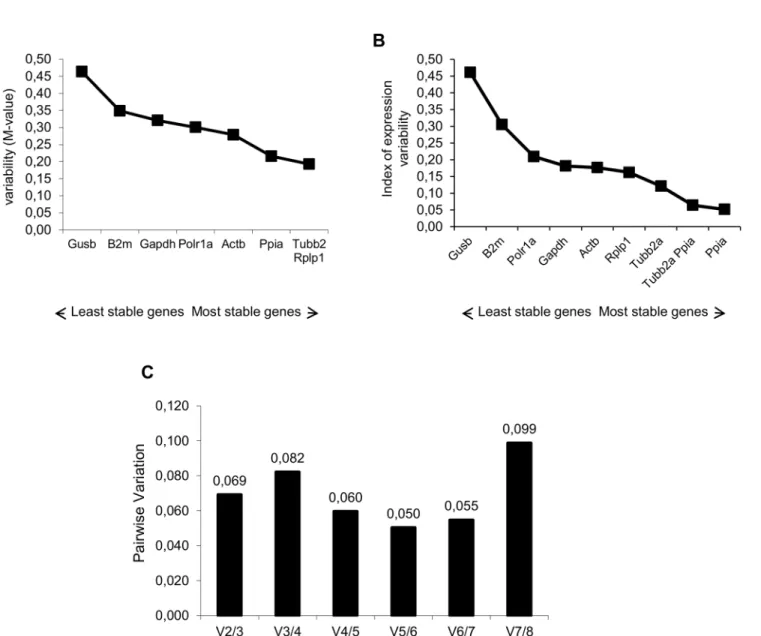
and Normfinder softwares. The average expression stability values (M values) of the reference genes in all tested samples are displayed inFig 1A. All the genes presented high expression sta-bility, with the M values varying between 0.21 (Ppia) and 0.46 (Gusb). The pairwise variation V2/3 was 0.069 (Fig 1C); thus, theTubb2a/Rplp1genes were indicated as the optimal pair to provide normalization of gene expression at the different photoperiods tested. Results of NormFinder analysis are shown inFig 1B. Also,PpiaandGusbappeared, as the most and the least stable genes (stability value of 0.052 and 0.46), respectively. The best combination of refer-ence genes indicated wasTubb2a/Ppia. These data sets are comparable with those obtained using geNorm, with slight differences in the ranking order of the most stable genes and of the best pair combination. In order to validate the results obtained, we conducted a relative expres-sion analysis of thePer1gene. We used the recommended combination of genes from both
Fig 1. Selection of the most suitable reference genes for circadian analysis in the hippocampus of rats.Expression stability measurements for the 8 reference genes calculated by geNorm (A) and NormFinder (B). The x-axis from left to right indicates the ranking of the genes according to their expression stability; lower values indicate higher expression stability. C) Determination of the optimal number of reference genes for normalization by geNorm. The Software calculates the normalization factor from at least two genes at which the variable V defines the pair-wise variation between two sequential normalization factors.
doi:10.1371/journal.pone.0141121.g001
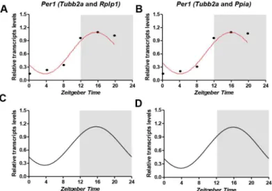
geNorm (Tubb2a/Rplp1) and NormFinder (Tubb2a/Ppia) as internal controls. The statistical analysis ofPer1diurnal expression was performed by Acrophase and CircWave softwares. We observed that with both normalization procedures, aPer1transcript levels oscillate in a rhyth-mic pattern, peaking at approximately ZT16 and with amplitude nearly of 0.47 (Fig 2).
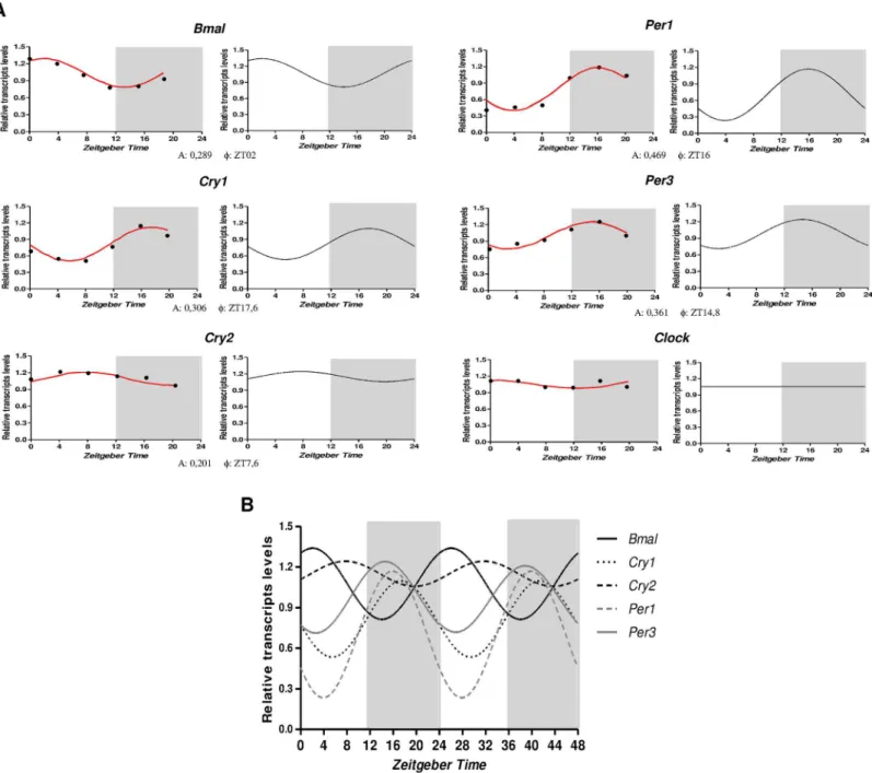
Following, we used Tubb2a/Rplp1as normalizers to evaluate, systematically, the temporal profiling ofBmal1,Per1,Per3,Cry1,Cry2andClockmRNA levels in the hippocampus of rats sacrificed every 4 hours during a 24-h period.Fig 3illustrate the temporal organization and phase relationship of the clock genes analyzed. Using both Acrophase and CircWave softwares, we observed that with exception ofClock, all genes showed a rhythmic pattern of expression, beingPer1andCry2those with the highest (0.469) and lowest (0.201) amplitude, respectively (Fig 3A). The comparison of the rhythms of each gene showed thatBmal1(F= ZT2) is in anti-phase withPer1(F= ZT16),Per3(F= ZT14.8) andCry1(F= ZT17.6), whereasBmal1peak is approximately 5 hours before acrophase ofCry2(F= ZT7.6) (Fig 3B).
Effects of diurnal variation on differential gene expression analysis in
hippocampus of epileptic rats
To investigate if the diurnal expression could be a source of variability on differential gene expression analysis in hippocampus of epileptic rats, we compared expression levels of the clock genes between epileptic rats sacrificed at ZT8 or ZT12 with naive rats sacrificed at different
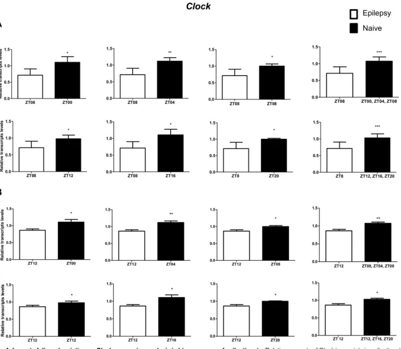
Zeit-geber times. In relation to ZT8 epileptic rats,clocktranscripts were significantly decreased in the
hippocampus of epileptic rats in all comparisons with the naive group (Fig 4A). However, the
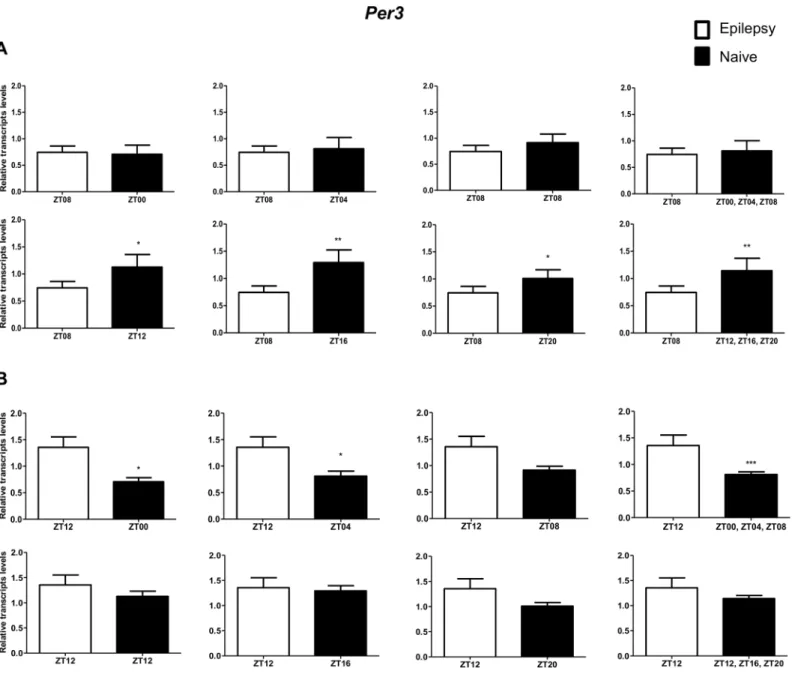
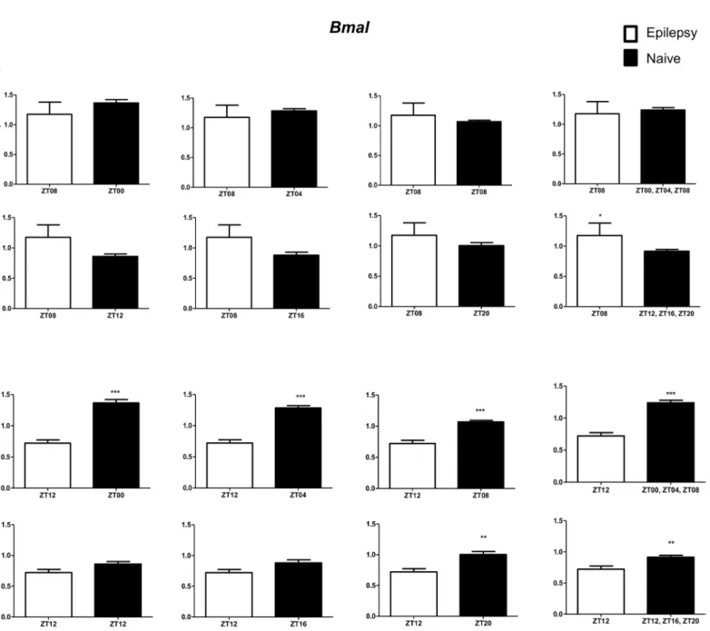
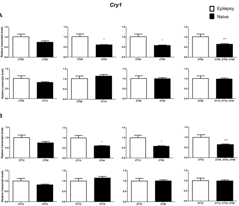
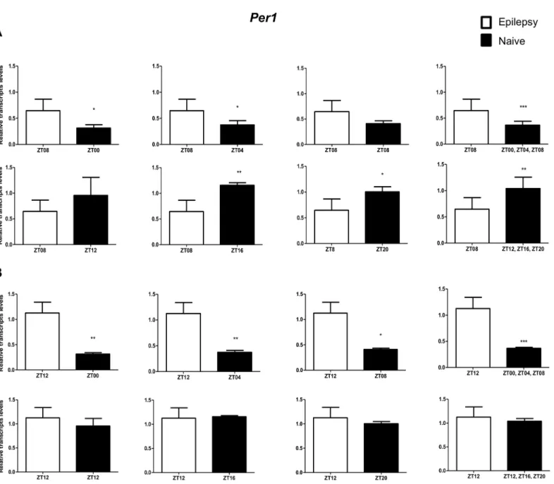
Per3was decreased only when compared with naive rats correspondent to ZT points of the dark phase (Fig 5A).Bmal1transcripts were significantly increased in epileptic rats only when com-pared with naive rats relative to the combination of dark phase ZTs (Fig 6A). Significant differ-ences inCry1(increase) andCry2(decrease) transcripts were observed only in comparisons using specifics ZTs of light phases (Figs7Aand8A). Intriguingly, Per1 transcripts levels were increased or decreased in hippocampus of epileptic rats depending on whether they were com-pared with specific ZT points of light and dark phases, respectively (Fig 9A).
Fig 2. Temporal profile ofPer1transcripts in the hippocampus based in two different normalization strategies.RT-qPCR data were normalized by best combination of genes derived by geNorm (A,C) and NormFinder (B,D) analysis (mean, n = 5). Statistical test for circadian analysis by Acrophase (A, B) and CircWave (C, D). Data are presented as mean (n = 5 rats/time point).
In relation to ZT12 epileptic rats, we also observed that the different comparisons with naive group had effects on experimental results for Bmal1, Per1, Per3, Cry1 and Cry2 (Figs4B–
9B) but not for Clock. In fact, Clock transcripts were also significantly decreased in the hippo-campus of epileptic rats in all comparisons to the naive group (Fig 4B). Per3 was increased in ZT12 only when compared with naive rats correspondent to ZT points of the dark phase (Fig 5B). Bmal1 was decreased when compared with all ZT of naive group, with exception for ZT12 and ZT16 (Fig 6B). Significant increases in Cry1 transcripts were observed only in comparisons using specifics ZTs of light phases (Fig 7B), while the CRY2 decreased was significant in
Fig 3. Temporal expression of the core clock transcripts in the hippocampus of rats.A) Relative amounts of transcripts at different ZT after
normalization toTubb2a/Rplp1. Data are presented as mean (n = 5 rats/ZT). Statistical test for circandian analysis by Acrophase (left) and CirWawe (right). B) Overlap of cosine fitting curves illustrating the phase relation of clock transcripts. For clarity reasons, data are doubleblotted againstZeitgeber time(ZT).
doi:10.1371/journal.pone.0141121.g003
comparisons using ZT of light phases and specifics ZT of dark phase (Fig 8B). Per1 transcripts were significantly increased only in comparisons with ZT of light phases (Fig 9B).
Discussion
Studies that aim to measure transcript levels, particularly using RT-qPCR, must use proper ref-erence genes to avoid erroneous conclusions [5,38,40]. To the best of our knowledge, this is the first study that investigated, systematically, suitable reference genes for circadian analysis in hippocampus of Wistar rats. The 8 candidate reference genes were ranked for expression
Fig 4. Impact of diurnal variation onClockexpression analysis in hippocampus of epileptic rats.Relative amounts ofClocktranscripts in epileptic rats ZT08 (A) and ZT12 (B) after normalization toTubb2a/Rplp1. Significant differences were evaluated using Unpaired Student’s t-test comparing results between epileptic and each ZT of naive group.*p<0.05,**p<0.01 and***p<0.001. Data are presented as mean+SEM (n = 4 rats in each epileptic group and 5 rats/time point in naive).
stability in the hippocampus of rats sacrificed at six different ZT of a 12:12 light-dark cycle. The slight differences in geNorm and NormFinder ranking probably were due to specific math-ematical algorithms used by the software. The pairwise variation value (V2/3) below of cutoff (0.15) indicated that normalization of target genes with a combination of the two best genes is sufficient. This is especially important considering that the most of circadian studies perform normalization using a single reference gene [28,32,41–43]. Thus, the best combination of refer-ence genes indicated wasTubb2a/Rplp1andTubb2a/Ppiaby geNorm and NormFinder soft-wares, respectively. In fact, when we used these combinations of genes separately to normalize thePer1transcripts amounts in hippocampus of rats at different ZT, we observed a similar
Fig 5. Impact of diurnal variation onPer3expression analysis in hippocampus of epileptic rats.Relative amounts ofPer3, transcripts in epileptic rats ZT08 (A) and ZT12 (B) after normalization toTubb2a/Rplp1. Significant differences were evaluated using Unpaired Student’s t-test comparing results between epileptic and each ZT of naive group.*p<0.05,**p<0.01 and***p<0.001. Data are presented as mean+SEM (n = 5 (ZT8) and 4 (ZT12) rats in epileptic group and n = 5 rats/time point in naive).
doi:10.1371/journal.pone.0141121.g005
diurnal oscillation pattern of gene expression (Fig 2). In the literature, we found reported reli-able reference genes for circadian studies in retina, adrenal glands, liver and leukocytes [44–
46]. However, since reference genes suitable for one tissue or species/strain should not auto-matically be used for normalization in another, our data are unique and provide a useful refer-ence for researchers that search for stable referrefer-ence genes in hippocampus of Wistar rats.
Following, we usedTubb2a/Rplp1as normalizers to analyze the temporal variation of core clock transcript levels in the hippocampus of rats sacrificed at different times thoroughly a 24h period. Supporting previous reports, we observed thatPer1,Per3,Cry1,Cry2andBmal1are expressed in an oscillating manner in this structure (Fig 3). Anti-phasic patterns ofBmal1 Fig 6. Impact of diurnal variation onBmalexpression analysis in hippocampus of epileptic rats.Relative amounts ofBmal, transcripts in epileptic rats ZT08 (A) and ZT12 (B) after normalization toTubb2a/Rplp1. Significant differences were evaluated using Unpaired Student’s t-test comparing results between epileptic and each ZT of naive group.*p<0.05,**p<0.01 and***p<0.001. Data are presented as mean+SEM (n = 5 (ZT8) and 4 (ZT12) rats in epileptic group and n = 5 rats/time point in naive).
compared withPer1,Per3andCry1are consistently observed in the SCN [47–49], which can be explained by the actions of these genes products in transcriptional/translational feedback loops of core clock mechanism. However, Jilg et al. [27] observed that mRNAs of clock genes
Cry1,Cry2andBmal1(but not ofPer1) are simultaneously elevated in the hippocampus during
late night, suggesting a common mechanism of transcriptional regulation of these genes in hip-pocampus. Regardless theClockexpression, we were unable to show significant alterations over a 24h period (Fig 3). Although a non-rhythmicClockexpression had already been reported in the SCN and some peripheral oscillators [42], our findings contrast with previous studies showing diurnal rhythms forClockexpression in hippocampus of rats [27,32,42,50].
Fig 7. Impact of diurnal variation onCry1expression analysis in hippocampus of epileptic rats.Relative amounts ofCry1, transcripts in epileptic rats ZT08 (A) and ZT12 (B) after normalization toTubb2a/Rplp1. Significant differences were evaluated using Unpaired Student’s t-test comparing results between epileptic and each ZT of naive group.*p<0.05,**p<0.01 and***p<0.001. Data are presented as mean+SEM (n = 5 (ZT8) and 4 (ZT12) rats in epileptic group and n = 5 rats/time point in naive).
doi:10.1371/journal.pone.0141121.g007
This divergent result could be explained by differences in experimental design, including spe-cies/strain, photoperiod or statistical software for circadian analysis.
Although we still do not know much about the functional significance of the rhythm’s clock genes expression in hippocampus, it is probably associated to a local temporal program that control morphological and physiological circadian changes in this structure. In fact, hippocam-pal diurnal rhythms are observed in dendritic patterning and spine density of neuron [51,52], neurogenesis [53] and long-term potentiation [50], which must be implicated in the rhythm of hippocampal-dependent learning, memory formation and aging [54–59].
Fig 8. Impact of diurnal variation onCry2expression analysis in hippocampus of epileptic rats.Relative amounts ofCry2, transcripts in epileptic rats ZT08 (A) and ZT12 (B) after normalization toTubb2a/Rplp1. Significant differences were evaluated using Unpaired Student’s t-test comparing results between epileptic and each ZT of naive group.*p<0.05,**p<0.01 and***p<0.001. Data are presented as mean+SEM (n = 5 (ZT8) and 4 (ZT12) rats in epileptic group and n = 5 rats/time point in naive).
Effects of diurnal variation on differential gene expression analysis in
hippocampus of epileptic rats
The assessment of stochastic variation of transcript abundance by analyzing multiple samples from the same tissue/individual highlights a number of potential biological and technical fac-tors that interfere in gene expression [10,60]. These studies revealed that the gene pathways and functions most closely associated with high baseline variance reflects functions that are sensitive to environmental cues, such as circadian rhythm [61]. However, the impact of these variations on differential gene expression studies in general and, particularly, in epilepsy has
Fig 9. Impact of diurnal variation onPer1expression analysis in hippocampus of epileptic rats.Relative amounts ofPer1, transcripts in epileptic rats ZT08 (A) and ZT12 (B) after normalization toTubb2a/Rplp1. Significant differences were evaluated using Unpaired Student’s t-test comparing results between epileptic and each ZT of naive group.*p<0.05,**p<0.01 and***p<0.001. Data are presented as mean+SEM (n = 4 rats in each epileptic group and 5 rats/time point in naive).
doi:10.1371/journal.pone.0141121.g009
been underscored. Here, we verified that diurnal expression is an important source of variabil-ity on differential gene expression analysis in hippocampus of epileptic rats. In fact, we show that differences at ZT could have effects on experimental results forBmal1,Per1,Per3,Cry1
andCry2(Figs5–9) but not forClock(Fig 4). Indeed, only the clock gene did not confirm a diurnal pattern of expression in hippocampus of naive rats. This is of great relevance consider-ing that the components of the core clock mechanism regulate circadian rhythms in genome-wide mRNA expression. In fact, although there is no estimate for the fraction of the hippocam-pal transcriptome that shows circadian variation, a total of 2,927 genes were identified as circa-dian regulated in mouse prefrontal cortex [62]. Thus, it may be hypothesized that part of the controversies in differential expression studies of epileptogenic process is due to differences in the time of sampling. Interesting, some of the genes with divergent expression results for epi-leptogenic process, such asIl-1β[63–65],Npy[66,67] andCox-2[68,69], have been reported presenting a rhythmic expression in different organs [70–72]. Our data highlight the impor-tance of avoiding genes with circadian expression which are out of phase when comparing experimental and control groups. Moreover, since the circadian phase difference may also affect other processes underlying epileptogenesis, such as neurogenesis, inflammation and neu-rodegeneration [53,73], we strongly recommend to take the time of day in consideration when designing all types of experiments.
Regarding the differential expression of core clock genes, we observed that only theClock
transcript abundance presented a consistent alteration (decrease) in epileptic rats (ZT8 or ZT12) for all comparisons with the naive group, suggesting that a subexpression ofClockmay be involved in epileptogenicity, possibly due to its role in gene expression regulation [74,75].
Clock/Clockmutants display in midbrain ventral tegmental area a decreased expression of the 1
subunit of the GABAA receptor and voltage-gated potassium channel (KCNQ2); and have increased levels of theGLUR1subunit of the AMPA glutamate receptor, which could contrib-ute to the observed increase in neuronal excitability of epileptic brains [75]. Interestingly, the same pattern of these gene expression alterations is seen in the hippocampus of different mod-els of epilepsy, and may enhance epileptogenicity [76–80].
The interpretation of functional significance ofBmal1,Per1,Per3,Cry1andCry2altered expression here observed in epileptic rats is jeopardized because the results change depending on the ZT of both naive and epileptic rats in the comparison. To the best of our knowledge, two reported studies investigated whether the core clock genes expression (Bmal1andPer1, only) is modulated in epilepsy. Gerstner el al. [81] showed that seizure activity itself does not influence hippocampalBMAL1expression, despite ofBMAL1gene regulates baseline threshold for seizures. Eun et al. [82] observed thatPer1expression is induced in hippocampus of rats submitted to two different seizure induction protocols. In agreement, we also observed that
Per1expression is increased in epileptic rats (ZT8 and ZT12) while theBmal1is unchanged in hippocampus of epileptic rats (ZT8, only) when compared with naive rats correspondent to ZT points of the light phase. However, future studies examining diurnal profile gene expression in epilepsy coupled to functional assays will be important to disentangle the involvement of clock genes on generation or maintenance of seizures.
suitability reference genes for circadian studies in hippocampus of rats and suggested that a subexpression ofClockmay be involved in epileptogenicity.
Author Contributions
Conceived and designed the experiments: DLGG EASS. Performed the experiments: EASS TEBSM HCM. Analyzed the data: DLGG EASS TEBSM JPL NGC MLPL HCM. Contributed reagents/materials/analysis tools: DLGG NGC MLPL. Wrote the paper: DLGG EASS TEBSM JPL NGC MLPL.
References
1. Pitkänen A, Lukasiuk K. Molecular and cellular basis of epileptogenesis in symptomatic epilepsy. Epi-lepsy Behav. Elsevier Inc.; 2009; 14:16–25. doi:10.1016/j.yebeh.2008.09.023PMID:18835369 2. Curia G, Lucchi C, Vinet J, Gualtieri F, Marinelli C, Torsello a, et al. Pathophysiogenesis of mesial
tem-poral lobe epilepsy: is prevention of damage antiepileptogenic? Curr Med Chem. 2014; 21:663–88. Available:http://www.ncbi.nlm.nih.gov/pubmed/24251566PMID:24251566
3. Brooks-Kayal AR, Raol YH, Russek SJ. Alteration of Epileptogenesis Genes. Neurotherapeutics. 2009; 6:312–318. doi:10.1016/j.nurt.2009.01.019PMID:19332325
4. Romcy-Pereira RN, Gitaí DLG, Gitaí LLG, Leite JP, Garcia-Cairasco N, Paçó-Larson ML. Genes e epi-lepsia II: expressão gênica diferencial. Rev Assoc Med Bras. 2008; 54:461–466. doi: 10.1590/S0104-42302008000500022PMID:18989569
5. Bustin SA, Benes V, Nolan T, Pfaffl MW. Quantitative real-time RT-PCR—a perspective. J Mol Endocri-nol. 2005; 34:597–601. doi:10.1677/jme.1.01755PMID:15956331
6. Ginzinger DG. Gene quantification using real-time quantitative PCR: an emerging technology hits the mainstream. Exp Hematol. 2002; 30:503–12. Available:http://www.ncbi.nlm.nih.gov/pubmed/ 12063017PMID:12063017
7. Cikos S, Koppel J. Transformation of real-time PCR fluorescence data to target gene quantity. Anal Bio-chem. 2009; 384:1–10. doi:10.1016/j.ab.2008.08.031PMID:18812161
8. Bustin SA. Quantification of mRNA using real-time reverse transcription PCR (RT-PCR): trends and problems. J Mol Endocrinol. 2002; 29:23–39. Available:http://www.ncbi.nlm.nih.gov/pubmed/ 12200227PMID:12200227
9. Bustin SA, Nolan T. Pitfalls of quantitative real-time reverse-transcription polymerase chain reaction. J Biomol Tech. 2004; 15:155–66. Available:http://www.pubmedcentral.nih.gov/articlerender.fcgi?artid= 2291693&tool=pmcentrez&rendertype=abstractPMID:15331581
10. Corton JC, Bushel PR, Fostel J, O'Lone RB. Sources of variance in baseline gene expression in the rodent liver. Mutat Res—Genet Toxicol Environ Mutagen. Elsevier B.V.; 2012; 746:104–112. doi: 10.1016/j.mrgentox.2011.12.017
11. Reppert SM, Weaver DR. Coordination of circadian timing in mammals. Nature. 2002; 418:935–41. doi: 10.1038/nature00965PMID:12198538
12. Sukumaran S, Almon RR, DuBois DC, Jusko WJ. Circadian rhythms in gene expression: Relationship to physiology, disease, drug disposition and drug action. Adv Drug Deliv Rev. 2010; 62:904–17. doi: 10.1016/j.addr.2010.05.009PMID:20542067
13. Welsh DK, Takahashi JS, Kay SA. Suprachiasmatic nucleus: cell autonomy and network properties. Annu Rev Physiol. 2010; 72:551–77. doi:10.1146/annurev-physiol-021909-135919PMID:20148688 14. Colwell CS. Linking neural activity and molecular oscillations in the SCN. Nat Rev Neurosci. 2011;
12:553–69. doi:10.1038/nrn3086PMID:21886186
15. Smarr BL, Jennings KJ, Driscoll JR, Kriegsfeld LJ. A time to remember: the role of circadian clocks in learning and memory. Behav Neurosci. 2014; 128:283–303. doi:10.1037/a0035963PMID:24708297 16. Reppert SM, Weaver DR. Molecular analysis of mammalian circadian rhythms. Annu Rev Physiol.
2001; 63:647–76. doi:10.1146/annurev.physiol.63.1.647PMID:11181971
17. Ko CH, Takahashi JS. Molecular components of the mammalian circadian clock. Hum Mol Genet. 2006; 15 Spec No:R271–7. PMID:16987893
18. Welsh DK, Logothetis DE, Meister M, Reppert SM. Individual neurons dissociated from rat suprachias-matic nucleus express independently phased circadian firing rhythms. Neuron. 1995; 14:697–706. Available:http://www.ncbi.nlm.nih.gov/pubmed/7718233PMID:7718233
19. Panda S, Antoch MP, Miller BH, Su AI, Schook AB, Straume M, et al. Coordinated transcription of key pathways in the mouse by the circadian clock. Cell. 2002; 109:307–20. Available:http://www.ncbi.nlm. nih.gov/pubmed/12015981PMID:12015981
20. Akhtar RA, Reddy AB, Maywood ES, Clayton JD, King VM, Smith AG, et al. Circadian cycling of the mouse liver transcriptome, as revealed by cDNA microarray, is driven by the suprachiasmatic nucleus. Curr Biol. 2002; 12:540–50. Available:http://www.ncbi.nlm.nih.gov/pubmed/11937022PMID: 11937022
21. Kucho K, Okamoto K, Tsuchiya Y, Nomura S, Nango M, Kanehisa M, et al. Global analysis of circadian expression in the cyanobacterium Synechocystis sp. strain PCC 6803. J Bacteriol. 2005; 187:2190–9. doi:10.1128/JB.187.6.2190–2199.2005PMID:15743968
22. McDonald MJ, Rosbash M. Microarray analysis and organization of circadian gene expression in Dro-sophila. Cell. 2001; 107:567–78. Available:http://www.ncbi.nlm.nih.gov/pubmed/11733057PMID: 11733057
23. Covington MF, Maloof JN, Straume M, Kay SA, Harmer SL. Global transcriptome analysis reveals cir-cadian regulation of key pathways in plant growth and development. Genome Biol. 2008; 9:R130. doi: 10.1186/gb-2008-9-8-r130PMID:18710561
24. Wakamatsu H, Yoshinobu Y, Aida R, Moriya T, Akiyama M, Shibata S. Restricted-feeding-induced anticipatory activity rhythm is associated with a phase-shift of the expression of mPer1 and mPer2 mRNA in the cerebral cortex and hippocampus but not in the suprachiasmatic nucleus of mice. Eur J Neurosci. 2001; 13:1190–6. Available:http://www.ncbi.nlm.nih.gov/pubmed/11285016PMID: 11285016
25. Wang X, Wang Y, Xin H, Liu Y, Wang Y, Zheng H, et al. Altered expression of circadian clock gene, mPer1, in mouse brain and kidney under morphine dependence and withdrawal. J Circadian Rhythms. 2006; 4:9. doi:10.1186/1740-3391-4-9PMID:16925815
26. Wyse CA, Coogan AN. Impact of aging on diurnal expression patterns of CLOCK and BMAL1 in the mouse brain. Brain Res. 2010; 1337:21–31. doi:10.1016/j.brainres.2010.03.113PMID:20382135 27. Jilg A, Lesny S, Peruzki N, Schwegler H, Selbach O, Dehghani F, et al. Temporal dynamics of mouse
hippocampal clock gene expression support memory processing. Hippocampus. 2010; 20:377–388. doi:10.1002/hipo.20637PMID:19437502
28. Kondratova Dubrovsky Y.V., Antoch M.P.& Kondratov R.V. a a. Circadian clock proteins control adap-tation to novel environment and memeory formation. Aging (Albany NY). 2010; 2:285–297.
29. Vosko AM, Hagenauer MH, Hummer DL, Lee TM. Period gene expression in the diurnal degu (Octodon degus) differs from the nocturnal laboratory rat (Rattus norvegicus). Am J Physiol Regul Integr Comp Physiol. 2009; 296:R353–R361. doi:10.1152/ajpregu.90392.2008PMID:19036829
30. O’Callaghan EK, Anderson ST, Moynagh PN, Coogan AN. Long-Lasting Effects of Sepsis on Circadian Rhythms in the Mouse. PLoS One. 2012; 7:1–14. doi:10.1371/journal.pone.0047087
31. Harbour VL, Weigl Y, Robinson B, Amir S. Phase Differences in Expression of Circadian Clock Genes in the Central Nucleus of the Amygdala, Dentate Gyrus, and Suprachiasmatic Nucleus in the Rat. 2014;9:1–9. doi:10.1371/journal.pone.0103309
32. Boone DR, Sell SL, Micci MA, Crookshanks JM, Parsley M, Uchida T, et al. Traumatic Brain Injury-Induced Dysregulation of the Circadian Clock. PLoS One. 2012; 7.
33. Curia G, Longo D, Biagini G, Jones RSG, Avoli M. The pilocarpine model of temporal lobe epilepsy. J Neurosci Methods. 2008; 172:143–57. doi:10.1016/j.jneumeth.2008.04.019PMID:18550176 34. Turski WA, Cavalheiro EA, Schwarz M, Czuczwar SJ, Kleinrok Z, Turski L. Limbic seizures produced
by pilocarpine in rats: behavioural, electroencephalographic and neuropathological study. Behav Brain Res. 1983; 9:315–35. Available:http://www.ncbi.nlm.nih.gov/pubmed/6639740PMID:6639740 35. Liu Z, Nagao T, Desjardins GC, Gloor P, Avoli M. Quantitative evaluation of neuronal loss in the dorsal
hippocampus in rats with long-term pilocarpine seizures. Epilepsy Res. 1994; 17:237–47. Available: http://www.ncbi.nlm.nih.gov/pubmed/8013446PMID:8013446
36. Clifford DB, Olney JW, Maniotis A, Collins RC, Zorumski CF. The functional anatomy and pathology of lithium-pilocarpine and high-dose pilocarpine seizures. Neuroscience. 1987; 23:953–68. Available: http://www.ncbi.nlm.nih.gov/pubmed/3437996PMID:3437996
37. Racine RJ. Modification of seizure activity by electrical stimulation. II. Motor seizure. Electroencepha-logr Clin Neurophysiol. 1972; 32:281–94. Available:http://www.ncbi.nlm.nih.gov/pubmed/4110397 PMID:4110397
39. Livak KJ, Schmittgen TD. Analysis of relative gene expression data using real-time quantitative PCR and the 2(-Delta Delta C(T)) Method. Methods. 2001; 25:402–8. doi:10.1006/meth.2001.1262PMID: 11846609
40. De Araújo MA, Marques TEBS, Taniele-Silva J, Souza FMDA, De Andrade TG, Garcia-Cairasco N, et al. Identification of endogenous reference genes for the analysis of microRNA expression in the hip-pocampus of the pilocarpine-induced model of mesial temporal lobe epilepsy. PLoS One. 2014; 9. 41. Golini RS, Delgado SM, Navigatore Fonzo LS, Ponce IT, Lacoste MG, Anzulovich AC. Daily patterns of
clock and cognition-related factors are modified in the hippocampus of vitamin A-deficient rats. Hippo-campus. 2012; 22:1720–32. doi:10.1002/hipo.22007PMID:22434687
42. Fonken LK, Aubrecht TG, Meléndez-Fernández OH, Weil ZM, Nelson RJ. Dim light at night disrupts molecular circadian rhythms and increases body weight. J Biol Rhythms. 2013; 28:262–71. doi:10. 1177/0748730413493862PMID:23929553
43. Vilches N, Spichiger C, Mendez N, Abarzua-Catalan L, Galdames H a., Hazlerigg DG, et al. Gestational chronodisruption impairs hippocampal expression of NMDA receptor subunits Grin1b/Grin3a and spa-tial memory in the adult offspring. PLoS One. 2014; 9:17–19. doi:10.1371/journal.pone.0091313 44. Kamphuis W, Cailotto C, Dijk F, Bergen A, Buijs RM. Circadian expression of clock genes and
clock-controlled genes in the rat retina. Biochem Biophys Res Commun. 2005; 330:18–26. doi:10.1016/j. bbrc.2005.02.118PMID:15781226
45. Kosir R, Acimovic J, Golicnik M, Perse M, Majdic G, Fink M, et al. Determination of reference genes for circadian studies in different tissues and mouse strains. BMC Mol Biol. 2010; 11:60. doi: 10.1186/1471-2199-11-60PMID:20712867
46. De Siqueira Figueredo D, Gitaí DLG, de Andrade TG. Daily variations in the expression of miR-16 and miR-181a in human leukocytes. Blood Cells, Mol Dis. 2015; doi:10.1016/j.bcmd.2015.01.004 47. Johnston JD, Ebling FJP, Hazlerigg DG. Photoperiod regulates multiple gene expression in the
supra-chiasmatic nuclei and pars tuberalis of the Siberian hamster (Phodopus sungorus). Eur J Neurosci. 2005; 21:2967–74. doi:10.1111/j.1460-9568.2005.04148.xPMID:15978008
48. Oishi K, Sakamoto K, Okada T, Nagase T, Ishida N. Antiphase circadian expression between BMAL1 and period homologue mRNA in the suprachiasmatic nucleus and peripheral tissues of rats. Biochem Biophys Res Commun. 1998; 253:199–203. doi:10.1006/bbrc.1998.9779PMID:9878515
49. Shearman LP, Sriram S, Weaver DR, Maywood ES, Chaves I, Zheng B, et al. Interacting molecular loops in the mammalian circadian clock. Science. 2000; 288:1013–9. Available:http://www.ncbi.nlm. nih.gov/pubmed/10807566PMID:10807566
50. O’Callaghan EK, Anderson ST, Moynagh PN, Coogan AN. Long-lasting effects of sepsis on circadian rhythms in the mouse. PLoS One. 2012; 7:e47087. doi:10.1371/journal.pone.0047087PMID: 23071720
51. Ikeno T, Weil ZM, Nelson RJ. Photoperiod affects the diurnal rhythm of hippocampal neuronal morphol-ogy of Siberian hamsters. Chronobiol Int. 2013; 30:1089–100. doi:10.3109/07420528.2013.800090 PMID:23879697
52. Ikeno T, Weil ZM, Nelson RJ. Timing of light pulses and photoperiod on the diurnal rhythm of hippocam-pal neuronal morphology of Siberian hamsters. Neuroscience. 2014; 270:69–75. doi:10.1016/j. neuroscience.2014.04.002PMID:24726983
53. Tamai S, Sanada K, Fukada Y. Time-of-day-dependent enhancement of adult neurogenesis in the hip-pocampus. PLoS One. 2008; 3:e3835. doi:10.1371/journal.pone.0003835PMID:19048107
54. Chaudhury D, Wang LM, Colwell CS. Circadian regulation of hippocampal long-term potentiation. J Biol Rhythms. 2005; 20:225–36. doi:10.1177/0748730405276352PMID:15851529
55. Ruby NF, Hwang CE, Wessells C, Fernandez F, Zhang P, Sapolsky R, et al. Hippocampal-dependent learning requires a functional circadian system. Proc Natl Acad Sci U S A. 2008; 105:15593–8. doi:10. 1073/pnas.0808259105PMID:18832172
56. Takahashi Y, Sawa K, Okada T. The diurnal variation of performance of the novel location recognition task in male rats. Behav Brain Res. 2013; 256:488–93. doi:10.1016/j.bbr.2013.08.040PMID: 24008072
57. Rawashdeh O, Jilg A, Jedlicka P, Slawska J, Thomas L, Saade A, et al. PERIOD1 coordinates hippo-campal rhythms and memory processing with daytime. Hippocampus. 2014; 24:712–23. doi:10.1002/ hipo.22262PMID:24550127
58. Kondratova A a., Kondratov R V. The circadian clock and pathology of the ageing brain. Nat Rev Neu-rosci. 2012; 13:325–335. doi:10.1038/nrn3208PMID:22395806
59. Ikeno T, Nelson RJ. Acute melatonin treatment alters dendritic morphology and circadian clock gene expression in the hippocampus of Siberian Hamsters. Hippocampus. 2015; 25:142–8. doi:10.1002/ hipo.22358PMID:25160468
60. Vedell PT, Svenson KL, Churchill G a. Stochastic variation of transcript abundance in C57BL/6J mice. BMC Genomics. BioMed Central Ltd; 2011; 12:167. doi:10.1186/1471-2164-12-167PMID:21450099 61. Hsu PY, Harmer SL. Circadian Phase Has Profound Effects on Differential Expression Analysis. PLoS
One. 2012; 7:18–21. doi:10.1371/journal.pone.0049853
62. Yang S, Wang K, Valladares O, Hannenhalli S, Bucan M. Genome-wide expression profiling and bioin-formatics analysis of diurnally regulated genes in the mouse prefrontal cortex. Genome Biol. 2007; 8: R247. doi:10.1186/gb-2007-8-11-r247PMID:18028544
63. Gleeson LC, Ryan KJ, Griffin EW, Connor TJ, Harkin A. Theβ2-adrenoceptor agonist clenbuterol elicits neuroprotective, anti-inflammatory and neurotrophic actions in the kainic acid model of excitotoxicity. Brain Behav Immun. 2010; 24:1354–61. doi:10.1016/j.bbi.2010.06.015PMID:20599496
64. Cardoso ALC, Costa P, de Almeida LP, Simões S, Plesnila N, Culmsee C, et al. Tf-lipoplex-mediated c-Jun silencing improves neuronal survival following excitotoxic damage in vivo. J Control Release. 2010; 142:392–403. doi:10.1016/j.jconrel.2009.11.004PMID:19913061
65. Pernot F, Heinrich C, Barbier L, Peinnequin A, Carpentier P, Dhote F, et al. Inflammatory changes dur-ing epileptogenesis and spontaneous seizures in a mouse model of mesiotemporal lobe epilepsy. Epi-lepsia. 2011; 52:2315–25. doi:10.1111/j.1528-1167.2011.03273.xPMID:21955106
66. Ruiz N, Pacheco LF, Farrell B, Cox CB, Ermolinsky BS, Garrido-Sanabria ER, et al. Metabolic gene expression changes in the hippocampus of obese epileptic male rats in the pilocarpine model of tempo-ral lobe epilepsy. Brain Res. 2011; 1426:86–95. doi:10.1016/j.brainres.2011.10.006PMID:22050960 67. Wilson DN, Chung H, Elliott RC, Bremer E, George D, Koh S. Microarray analysis of postictal
transcrip-tional regulation of neuropeptides. J Mol Neurosci. 2005; 25:285–98. doi:10.1385/JMN:25:3:285 PMID:15800381
68. Sumanont Y, Murakami Y, Tohda M, Vajragupta O, Watanabe H, Matsumoto K. Effects of manganese complexes of curcumin and diacetylcurcumin on kainic acid-induced neurotoxic responses in the rat hippocampus. Biol Pharm Bull. 2007; 30:1732–9. Available:http://www.ncbi.nlm.nih.gov/pubmed/ 17827730PMID:17827730
69. Joseph SA, Lynd-Balta E, O’Banion MK, Rappold PM, Daschner J, Allen A, et al. Enhanced cyclooxy-genase-2 expression in olfactory-limbic forebrain following kainate-induced seizures. Neuroscience. 2006; 140:1051–65. doi:10.1016/j.neuroscience.2006.02.075PMID:16677768
70. Xu Y-Q, Zhang D, Jin T, Cai D-J, Wu Q, Lu Y, et al. Diurnal variation of hepatic antioxidant gene expres-sion in mice. PLoS One. 2012; 7:e44237. doi:10.1371/journal.pone.0044237PMID:22952936 71. Sherman H, Frumin I, Gutman R, Chapnik N, Lorentz A, Meylan J, et al. Long-term restricted feeding
alters circadian expression and reduces the level of inflammatory and disease markers. J Cell Mol Med. 2011; 15:2745–59. doi:10.1111/j.1582-4934.2010.01160.xPMID:20731750
72. Wiater MF, Mukherjee S, Li A-J, Dinh TT, Rooney EM, Simasko SM, et al. Circadian integration of sleep-wake and feeding requires NPY receptor-expressing neurons in the mediobasal hypothalamus. Am J Physiol Regul Integr Comp Physiol. 2011; 301:R1569–83. doi:10.1152/ajpregu.00168.2011 PMID:21880863
73. Bouchard-Cannon P, Mendoza-Viveros L, Yuen A, Kærn M, Cheng HYM. The Circadian Molecular Clock Regulates Adult Hippocampal Neurogenesis by Controlling the Timing of Cell-Cycle Entry and Exit. Cell Rep. The Authors; 2013; 5:961–973. doi:10.1016/j.celrep.2013.10.037PMID:24268780 74. Doi M, Hirayama J, Sassone-Corsi P. Circadian Regulator CLOCK Is a Histone Acetyltransferase. Cell.
2006; 125:497–508. doi:10.1016/j.cell.2006.03.033PMID:16678094
75. McClung C a, Sidiropoulou K, Vitaterna M, Takahashi JS, White FJ, Cooper DC, et al. Regulation of dopaminergic transmission and cocaine reward by the Clock gene. Proc Natl Acad Sci U S A. 2005; 102:9377–9381. doi:10.1073/pnas.0503584102PMID:15967985
76. Drexel M, Kirchmair E, Sperk G. Changes in the expression of GABAA receptor subunit mRNAs in parahippocampal areas after kainic acid induced seizures. Front Neural Circuits. 2013; 7:142. doi:10. 3389/fncir.2013.00142PMID:24065890
77. Watanabe H, Nagata E, Kosakai A, Nakamura M, Yokoyama M, Tanaka K, et al. Disruption of the epi-lepsy KCNQ2 gene results in neural hyperexcitability. J Neurochem. 2000; 75:28–33. Available:http:// www.ncbi.nlm.nih.gov/pubmed/10854243PMID:10854243
78. Soldovieri MV, Castaldo P, Iodice L, Miceli F, Barrese V, Bellini G, et al. Decreased subunit stability as a novel mechanism for potassium current impairment by a KCNQ2 C terminus mutation causing benign familial neonatal convulsions. J Biol Chem. 2006; 281:418–28. doi:10.1074/jbc.M510980200PMID: 16260777
80. Steiger JL, Russek SJ. GABAA receptors: building the bridge between subunit mRNAs, their promot-ers, and cognate transcription factors. Pharmacol Ther. 2004; 101:259–81. doi:10.1016/j.pharmthera. 2003.12.002PMID:15031002
81. Gerstner JR, Smith GG, Lenz O, Perron IJ, Buono RJ, Maio R Di. BMAL1 controls the diurnal rhythm and set point for electrical seizure threshold in mice. 2014;8:1–7. doi:10.3389/fnsys.2014.00121 82. Eun B, Kim HJ, Kim SY, Kim TW, Hong ST, Choi KM, et al. Induction of Per1 expression following an
experimentally induced epilepsy in the mouse hippocampus. Neurosci Lett. Elsevier Ireland Ltd; 2011; 498:110–113. doi:10.1016/j.neulet.2011.03.039