Combinations of
β
-Lactam or Aminoglycoside
Antibiotics with Plectasin Are Synergistic
against Methicillin-Sensitive and
Methicillin-Resistant
Staphylococcus aureus
Yanmin Hu1*, Alexander Liu2, James Vaudrey1, Brigita Vaiciunaite1, Christiana Moigboi3, Sharla M. McTavish3, Angela Kearns3, Anthony Coates1
1Institute for Infection and Immunity, St George’s, University of London, London, United Kingdom, 2University of Oxford Centre for Clinical Magnetic Resonance Research, University of Oxford, Oxford, United Kingdom,3Antimicrobial Resistance and Healthcare Associated Infections Reference Unit, Public Health England, Colindale, London, United Kingdom
*ymhu@sgul.ac.uk
Abstract
Bacterial infections remain the leading killer worldwide which is worsened by the continuous emergence of antibiotic resistance. In particular, methicillin-sensitive (MSSA) and methicil-lin-resistantStaphylococcus aureus(MRSA) are prevalent and the latter can be difficult to treat. The traditional strategy of novel therapeutic drug development inevitably leads to emergence of resistant strains, rendering the new drugs ineffective. Therefore, rejuvenating the therapeutic potentials of existing antibiotics offers an attractive novel strategy. Plectasin, a defensin antimicrobial peptide, potentiates the activities of other antibiotics such as
β-lactams, aminoglycosides and glycopeptides against MSSA and MRSA. We performedin vitroandin vivoinvestigations to test against genetically diverse clinical isolates of MSSA (n = 101) and MRSA (n = 115). Minimum inhibitory concentrations (MIC) were determined by the broth microdilution method. The effects of combining plectasin withβ-lactams, ami-noglycosides and glycopeptides were examined using the chequerboard method and time kill curves. A murine neutropenic thigh model and a murine peritoneal infection model were used to test the effect of combinationin vivo. Determined by factional inhibitory concentra-tion index (FICI), plectasin in combinaconcentra-tion with aminoglycosides (gentamicin, neomycin or amikacin) displayed synergistic effects in 76-78% of MSSA and MRSA. A similar synergistic response was observed when plectasin was combined withβ-lactams (penicillin, amoxicillin or flucloxacillin) in 87–89% of MSSA and MRSA. Interestingly, no such interaction was ob-served when plectasin was paired with vancomycin. Time kill analysis also demonstrated significant synergistic activities when plectasin was combined with amoxicillin, gentamicin or neomycin. In the murine models, plectasin at doses as low as 8 mg/kg augmented the ac-tivities of amoxicillin and gentamicin in successful treatment of MSSA and MRSA infections. We demonstrated that plectasin strongly rejuvenates the therapeutic potencies of existing a11111
OPEN ACCESS
Citation:Hu Y, Liu A, Vaudrey J, Vaiciunaite B, Moigboi C, McTavish SM, et al. (2015) Combinations ofβ-Lactam or Aminoglycoside Antibiotics with Plectasin Are Synergistic against Methicillin-Sensitive and Methicillin-ResistantStaphylococcus aureus. PLoS ONE 10(2): e0117664. doi:10.1371/journal. pone.0117664
Academic Editor:Oliver Schildgen, Kliniken der Stadt Köln gGmbH, GERMANY
Received:September 1, 2014
Accepted:December 30, 2014
Published:February 18, 2015
Copyright:© 2015 Hu et al. This is an open access article distributed under the terms of theCreative Commons Attribution License, which permits unrestricted use, distribution, and reproduction in any medium, provided the original author and source are credited.
Data Availability Statement:All relevant data are within the paper and its Supporting Information files.
antibioticsin vitroandin vivo. This is a novel strategy that can have major clinical implica-tions in our fight against bacterial infecimplica-tions.
Introduction
Bacterial infection remains a leading killer worldwide [1] and antibiotic resistance continues to
plague the effective control of this pandemic health problem. Currently, resistance to marketed antibiotics is prevalent and most of the existing classes are now redundant for an increasing
number of bacterial species [2,3] and the number of new antibiotics reaching the market
annual-ly is unable to keep up with the development of resistance [4].Staphylococcus aureusis one of
the commonest pathogens in both hospital and community acquired infections.
Methicillin-sensitive (MSSA) and methicillin-resistantS. aureus(MRSA) cause significant morbidity and
mortality and are associated with an array of life-threatening infections including surgical site
infections, bacteremia, pneumonia and catheter-associated infections [5,6]. There is a very
limit-ed antimicrobial armamentarium to treat infections causlimit-ed by MRSA, of which vancomycin and
linezolid are the major agents but resistance to these agents is emerging [7,8]. It may be argued
that the traditional strategy of antibiotic discovery is flawed because rapid bacterial selection in-evitably leads to resistance development within only a few years post market release. Moreover, the drug discovery process itself is arduous and costly, meaning the emergence of a large group of effective antibiotics within a short time period is almost impossible. Therefore, a different ap-proach is needed to replenish our armamentarium against resistant bacteria and the most
prom-ising strategy is to restore the therapeutic potencies of existing antibiotics [9–11].
Weakening the cell envelope and increasing cellular permeability are two effective
therapeu-tic strategies to enhance the effectiveness of some antibiotherapeu-tics [12–14]. This is exploited by
com-pounds that target the bacterial cell membrane or cell wall [12–14]. In particular, peptides have
been shown to be potential enhancers to augment the activities of other antibiotics [15–17].
Plectasin, a defensin derived fromPseudoplectania nigrella, strongly synergises with other
anti-biotics [16]. Plectasin alone possesses antimicrobial activity and is bactericidal against many
Gram-positive bacteria including drug resistant Streptococci and Staphylococci [18]. It acts by
binding to the pyrophosphate moiety of lipid II in the bacterial cell wall, thus leading to the
rapid killing of the target bacteria [19]. A newer version of plectasin, NZ2114, has shown a
strong therapeutic efficacy againstS. aureusandStreptococcus pneumoniaein various different
animal infection models [20–22]. Furthermore, plectasin is stable in serum, exhibits a long
half-lifein vivoand is non-toxic in both cell culture and animals [18,23]. The pharmacokinetic
and pharmacodynamic property of plectasinin vitroandin vivomakes it an ideal adjuvant
compound to enhance the activity of other clinically used antibiotics.
In this study, we aimed to test thein vitroandin vivoactivity of plectasin in combination
with a number of antibiotics, namely penicillins (penicillin, amoxicillin, and flucloxacillin), aminoglycosides (gentamicin, neomycin and amikacin) and a glycopeptide (vancomycin) against clinical isolates of MSSA and MRSA.
Results
In vitro
susceptibility of plectasin against MSSA and MRSA
The MIC of plectasin NZ2114 was determined against 101 MSSA and 115 MRSA. As shown in
Table 1, the MIC range for plectasin was 0.5 to 4 mg/L for MSSA and 0.5 to 8 mg/L for MRSA.
The MIC50and MIC90of the isolates were 2 and 4 mg/L, respectively.
no role in study design, data collection and analysis, decision to publish, or preparation of the manuscript.
Competing Interests:This work was funded with support from the European Commission under grant agreement no: 278998, BacAttack. This
Chequerboard analysis of combination effects
The activity of plectasin in combination with (i) beta-lactam antibiotics (penicillin, amoxicillin and flucloxacillin), (ii) aminoglycosides (gentamicin, neomycin and amikacin) or (iii) vanco-mycin was determined using the broth microdilution chequerboard assay against the same test panel of MSSA and MRSA strains. The values of the FIC index for these combinations are
shown inTable 2. The combination of plectasin with theβ-lactams tested demonstrated
syner-gistic activities for 87–89% of test isolates. Notably, significant reductions inβ-lactam MICs
were observed, including the majority of the MRSAs tested (S1andS2Tables). Similarly, the
combination of plectasin with the aminoglycosides tested showed synergistic activities against
76–78% of test isolates. The MIC of the aminoglycosides in the test strains was significantly
re-duced with the presence of plectasin (S1andS2Tables). In addition, a 4- to 32-fold reduction
in the plectasin MIC was observed with both antibiotic classes (data not shown). In contrast, there was no interaction between plectasin and vancomycin. No antagonism was observed among any of the combinations tested.
Spa
typing of MSSA and MRSA
Spatyping data showed the MSSAs were more heterogeneous genotypically than the MRSAs
studied, belonging to 16 and 6 different MLST-CCs respectively (Table 3). The majority of
MRSAs (109; 94.8%) were representatives of the EMRSA-15 and-16 lineages (CC22 and CC30) which have remained the dominant HA-MRSA clones seen in the UK for over two decades.
Time kill analysis of the combinations against MSSA and MRSA
The synergistic activities of plectasin with amoxicillin, gentamicin and neomycin were exam-ined with a time kill assay against log-phase MSSA and MRSA with 5 of each MSSA and
MRSA which showed FIC index<0.5 for each drug combination. A range of different
concen-trations for the antibiotics and plectasin was determined according to the chequerboard results.
Table 1. Minimal inhibitory concentration of plectasin against clinical isolates of MSSA and MRSA.
MICa(mg/L)
Bacterial strains Number of isolates MIC range MIC50 MIC90
MSSA 101 0.5–4 2 4
MRSA 115 0.5–8 2 4
a MIC50and MIC9050% and 90% indicate that MIC values at which 50 and 90% of the isolates were inhibited, respectively.
doi:10.1371/journal.pone.0117664.t001
Table 2. Combination actives of plectasin with ß-lactams, aminoglycosides and vancomycin.
Total no. (%) of strains detected when plectasin combined with
Strains Combination activity FICI amoxicillin penicillin flucloxacillin gentamicin neomycin amikacin vancomycin
MSSA synergy 0.5 90 (89.1%) 89 (88.1%) 88 (87.1%) 79 (78.2%) 76 (75.3%) 77 (76.2%) 0
no interaction 0.56–1 11 (10.9%) 12 (11.9%) 13 (12.9%) 22 (21.8%) 25 (24.7%) 24 (23.8%) 101 (100%)
antagonism >4 0 0 0 0 0 0 0
MRSA synergy 0.5 101 (87.8%) 102 (88.7%) 102 (88.7%) 89 (77.4%) 88 (76.5%) 88 (76.5%) 0
no interaction 0.56–1 14 (12.2%) 13 (11.3%) 13 (11.3%) 26 (22.6%) 27 (23.5%) 27 (23.5%) 115 (100%)
antagonism >4 0 0 0 0 0 0 0
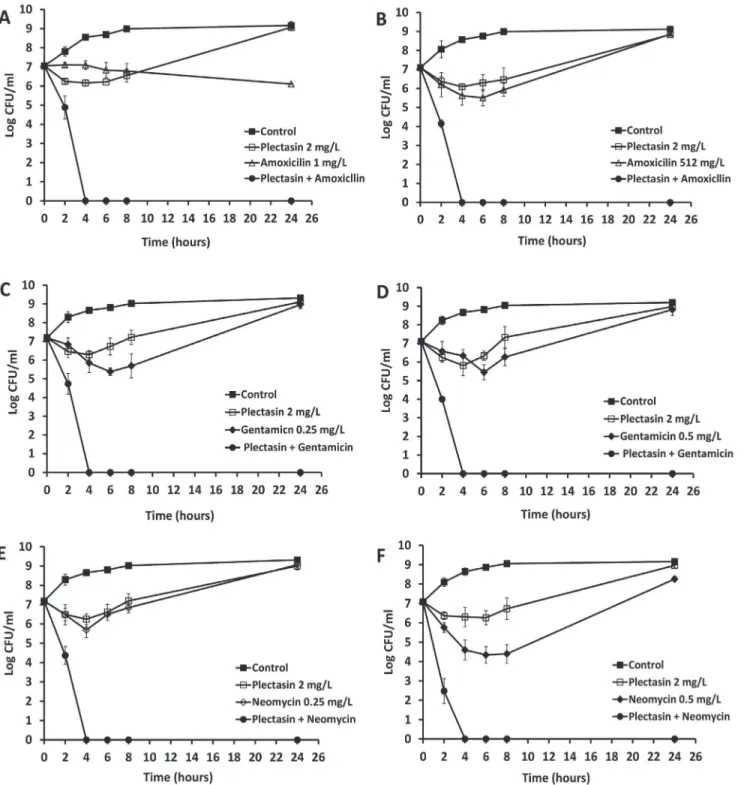
These concentrations were used for each treatment, either with a single agent or in combina-tion. The combinations with most effective synergistic activity against log-phase bacteria are
shown. InFig. 1, plectasin at the MIC value of 2 mg/L killed the bacteria initially, but regrowth
was observed after 4 hours of incubation. Amoxicillin at a concentration of 1 mg/L (MIC) in-hibited the MSSA growth (A) but had poor efficacy against MRSA at the MIC concentration 512 mg/L (B). However, in combination with plectasin, the initial inoculum was reduced by 100% at about 4 hours post treatment for both MSSA and MRSA (A and B). Gentamicin (C and D) and neomycin (E and F) at a concentration of 0.25 or 0.5 mg/L (MIC) showed bacte-ricidal activities initially against MSSA and MRSA, respectively, followed by a regrowth reach-ing a peak at 24 hours. The regrowth may be attributable to drug degradation or
microorganism adaptation [24,25]. However, in combination with plectasin, there was a 100%
reduction in viable counts at 4 hours against both MSSA and MRSA for all combinations test-ed. The CFU counts remained at zero for 24 hours and no regrowth was observed at 48 hours (data not shown). The time kill assay demonstrated that there was a significant synergistic ac-tivity between plectasin and amoxicillin, gentamicin or neomycin.
In vivo
combination activities of plectasin with amoxicillin and gentamicin
Thein vivoactivities of plectasin combination with amoxicillin and gentamicin were studied using two murine infection models. In the well-established neutropenic mouse thigh infection
Table 3.Spatypes of MSSA and MRSA isolates.
Strains MLST-CC (inferred)
Spa types (n)
MSSA 1 t127 (4), t189 (1), t273 (1), t1778 (1), t14364 (1) 5 t179 (1), t242 (1), t442 (1), t502 (1), t1305 (1), t14367 (1)
7 t091 (1)
8 t008 (5), t024 (3), t118 (1), t334 (1), t2067 (1), t14363 (1) 12 t160 (1), t213 (1), t771 (1), t888 (1), t2133 (1)
15 t084 (3), t328 (1), t346 (1), t393 (1), t491 (1)
20 t164 (1)
22 t005 (3), t032 (3), t709 (1), t891 (1), t1370 (1), t6642 (3), t10276 (1) 25 t1149 (2), t2313 (1)
30 t012 (2), t018 (3), t019 (1), t021 (5), t089 (1), t136 (1), t318 (4), t817 (1), t14366 (2)
45 t015 (4), t026 (1), t040 (1), t073 (1), t563 (1), t848 (1), t2195 (1), t4982 (1), t6243 (1), t14365 (1)
59 t216 (1)
80 t044 (1)
97 t267 (7)
101 t056 (1)
121 t171 (1), t645 (1)
MRSA 1 t2478 (1)
5 t002 (1), t311 (1)
8 t008 (1), t037 (1)
22 t020 (3), t022 (4), t025 (1), t032 (55), t379 (2), t578 (1), t611 (1), t756 (1), t1249 (2), t1370 (1), t1467 (1), t2006 (1), t3178 (2), t12788 (1)
30 t018 (29), t021 (1), t253 (3)
101 t280 (1)
Fig 1. Time-kill analysis showing the effects of plectation in combination with amoxicillin, gentamicin and neomycin against log phase MSSA and MRSA.The peptide and antibiotics alone or each combined with plectasin were added to the log-phase cultures and CFU counts were carried out at different time points. Combination of plectasin (2 mg/L) and amoxicillin (1 mg/L or 512 mg/L) against MSSA (A) and MRSA (B). Combination of plectasin (2 mg/L) and gentamicin (0.25 and 0.5 mg/L) against MRSA (C) and MRSA (D). Combination of plectasin (2 mg/L) and neomycin (0.25 and 0.5 mg/L) against MSSA (E) and MRSA (F). Results shown are the mean of three independent experiments.
model, combination of plectasin with either amoxicillin or gentamicin was tested against one
MSSA and an MRSA. These strains showed FIC indices of<0.5 and significant synergistic
ac-tivities in time kill curve for each of the drug combinations. Both test isolates had a plectasin MIC of 2 mg/L and gentamicin MIC of 1 mg/L; the MIC of amoxicillin for the MSSA and the MRSA was 0.5 mg/L and 512 mg/L, respectively. A range of different doses (mg/kg) of both an-tibiotic and plectasin was used. Treatment was performed singly or in combination after infec-tion. The data for the combinations most effective in killing the bacteria in mouse thigh muscle
or peritoneal cavity are presented. As shown inFig. 2A, 2B, 2C and 2D, plectasin at 8 mg/kg
re-duced the bacterial counts in the thigh muscles and showed 2.15 log kill at 8 hour post
treat-ment for MSSA (Fig. 2A and 2C) and 2.26 log kill for MRSA (Fig. 2B and 2D). Amoxicillin at
20 mg/kg killed 1.38 log of MSSA over an 8 hour period (Fig. 2A) and failed to control the
growth of the MRSA (Fig. 2B). Plectasin in combination with amoxicillin significantly
in-creased the kill rate against the MSSA compared to the single drug treatment (P<0.02
com-pared with plectasin or amoxicillin alone, n = 6) and reached 2.89 log kill at 8 hours (Fig. 2A).
Similarly, combination of plectasin and amoxicillin showed significant reduction of 2.83 log of CFU counts in the mouse thigh muscles with MRSA infection compared to the single drug
treatment (Fig. 2B. P<0.01 compared with plectasin or P<0.0001 with amoxicillin alone, n =
6). Gentamicin at 5 mg/kg showed bactericidal activity against both the MSSA and MRSA
strains in the model (Fig. 2C and 2D) which reduced 2.79 and 2.9 log CFU counts at 8 hours,
respectively. There was significant reduction of the CFU counts when gentamicin was
com-bined with plectasin showing 3.57 and 3.82 log kill at 8 hours for both MSSA (P<0.002
com-pared with plectasin or P<0.01 with gentamicin alone, n = 6) and MRSA (P<0.001 compared
with plectasin or P<0.02 with gentamicin alone, n = 6), respectively. Mice in both control and
amoxicillin treated MRSA infection groups developed clinical signs such as leg swelling at 8 hours of post treatment. There were no clinical signs for other treatment groups with amoxicil-lin, gentamicin, neomycin or plectasin singly and in combination with plectasin.
In the murine peritoneal infection model, combination of plectasin with amoxicillin or gen-tamicin was tested against the same MSSA and MRSA strains. At 5 hours after treatment,
plec-tasin at 8 mg/kg killed 1.3 log of the MSSA (Fig. 2E and 2G) and 1.44 log MRSA (Fig. 2F and
2H). Amoxicillin at 20 mg/kg reduced about half a log of CFU counts against the MSSA
(Fig. 2E) and showed no activity against the MRSA strain (Fig. 2F). Combination of plectasin
and amoxicillin exhibited 2.13 and 2.26 log kill against the MSSA (Fig. 2E) and MRSA strains
(Fig. 2F), respectively. The difference between the combination and the single drug was
signifi-cant for both MSSA (p<0.02 compared with plectasin or P<0.01 with amoxicillin alone,
n = 4) and MRSA (P<0.02 compared with plectasin or P<0.0001 with amoxicillin alone,
n = 4). As seen inFig. 2G and 2H, gentamicin alone killed 1.26 log of the MSSA and 1.6 log of
the MRSA at 5 hours. In combination with plectasin, 2.13 and 2.54 log kill was observed for
both the MSSA (P<0.02 compared with plectasin or P<0.02 with gentamicin alone, n = 4)
and MRSA strains (P<0.02 compared with plectasin or P<0.02 with gentamicin alone, n = 4),
respectively. In both untreated control groups and the MRSA infected group treated with amoxicillin, all animals in each group developed clinical signs such as raised fur and mild im-mobility at 5 hours after infection. The animals in other treatment groups showed no discom-fort with normal and heathy behaviors.
Investigation of membrane potential of plectasin in combination with
amoxicillin, gentamicin or neomycin
Fig 2. Effects of plectasin in combination with amoxicillin and gentamicin against MSSA and MRSA in a murine thigh infection model and in a mouse peritoneal infection model.In the murine thigh model, mice were infected with a strain of MSSA (t021) and a strain of MRSA (t032). Treatment was initiated 2 hours after infection with plectasin and amoxicillin against MSSA (A) and MRSA (B) and with plectasin and gentamicin against MSSA (C) and MRSA (D). Viability of the bacterial cells was determined from 6 mice for each group at 2, 4, 6 and 8 hours after treatment. In the mouse peritoneal infection model, mice were infected with the same strain of MSSA or MRSA. One hour after infection, treatment was initiated with plectasin and amoxicillin for MSSA (E) and for MRSA (F) and with plectasin and gentamicin for MSSA (G) and for MRSA (H). Bacterial counts in the peritoneal cavity were determined from 4 mice for each group at 2 and 5 hours post-treatment. The data have been repeated once.
or MRSA strain used in the animal models were treated with the fluorescent probe DiSC3(5) which accumulates in bacterial cells and self-quenches its own fluorescence. Plectasin was added to the DiSC3(5) treated cultures at final concentrations starting from 64 mg/L to 0 mg/ L. For the MSSA strain, no increase in fluorescent release was observed when the concentration
of plectasin at 64 mg/L (S1A Fig.). Similarly, there was no release of fluorescence after
treat-ment with amoxicillin, gentamicin or neomycin singly at concentrations from 64 to 2 mg/L,
re-spectively (S1B, S1C and S1D Fig.) compared to the positive control (S1E Fig.). The
cytoplasmic membrane potential was also examined when plectasin at 64 mg/L was combined with amoxicillin, gentamicin or neomycin. Combination of plectasin with amoxicillin,
neomy-cin or gentamineomy-cin showed no impact on the fluorescence release (S1F, S1G and S1H Fig.).
Simi-lar profiles of membrane potential were observed for the MRSA strain after treatment with plectasin and the antibiotics individually or in combination (data not shown).
Discussion
Plectasin NZ2114 is a promising synergistic agent. In our study, we found that there were sig-nificant synergistic activities when plectasin was combined with amoxicillin, penicillin,
fluclox-acillin, gentamicin, neomycin or amikacinin vitroandin vivo.
The panel of isolates tested represented a random collection from a range of clinical sites, the majority of which were associated with bacteraemia. Molecular analyses confirmed these
isolates were genotypically diverse and included representatives of the dominant lineages ofS.
aureusrecovered from humans. Plectasin NZ2114 showed significant antibacterial activity
against 216 clinical isolates of both MSSA and MRSA with an MIC50and MIC90of 2 and 4 mg/
L, respectively. On examination of the 216 clinical strains using chequerboard array analysis, synergistic activity was observed when plectasin was combined with penicillin, amoxicillin or flucloxacillin for more than 86% of both MSSA and MRSA strains. Combination of plectasin with gentamicin, neomycin and amikacin showed synergistic effects for more than 75% of each group of the strains. This positive combined effect was also confirmed using time-kill analysis which is superior to the chequerboard assay with dynamic and detailed viability measurement over time.
Bacterial resistance to antibiotics is associated with an increase in MIC to one or more anti-biotics, for example, MICs of penicillin and amoxicillin for the MRSA strains tested in this study were 256 mg/L or higher. This indicates that these antibiotics are no longer effective even at the maximum tolerated dose. Here, we clearly showed that plectasin enhances the activity of penicillin, amoxicillin and flucloxacillin against both MSSA and MRSA. In particular there was
a marked (2- to 512-fold) reduction in MIC for most of the MRSA strains (S2 Table).
Amoxi-cillin at 512 mg/L was ineffective against the MRSA strain tested (Fig. 1B), however, in
combi-nation with 2 mg/L of plectasin, a rapid bactericidal effect was noted.
Aminoglycosides are potent, broad spectrum antibiotics. However, it is well known that re-peated exposure of aminoglycosides, for example gentamicin, increases the risk of nephrotoxi-city if used systemically or ototoxinephrotoxi-city, particularly affecting the vestibulo-cochlear system.
This limits the therapeutic usage of this class of antibiotics [26,27]. In our study, we showed
that the bactericidal activities of gentamicin (one of the most commonly prescribed aminogly-cosides in clinical practice) and neomycin (one of the topical antibiotics used to clear the MRSA in nose and on skin) were significantly enhanced when combined with plectasin. The concentrations of gentamicin or neomycin which produced 100% kill of log-phase MSSA or MRSA were 2 to 4 or 2 to 8 mg/L, respectively at 2 to 4 hours (data not shown). At a lower dose of 0.25 to 0.5 mg/L, gentamicin or neomycin killed the bacterial cells initially followed by a
with plectasin, 100% kill was seen at 4 hours for both MSSA and MRSA which gave the equiva-lent potency of the drug used singly to achieve a complete kill. Furthermore, the culture re-mained sterile after 48 hours, which indicated that all the bacteria were eliminated. This is significant since therapeutic effects can be achieved with lower doses of aminoglycoside, thus can dramatically reduce its side effect profile, which is particularly important where prolonged therapy is required.
The findings from our study will undoubtedly convey important positive clinical implications. Firstly, our demonstration of the efficacy of plectasin as a powerful synergistic agent strongly sug-gests that other similar peptides will most likely be beneficial above and beyond their direct anti-microbial properties. Secondly, synergistic combinations improve the effectiveness of antibiotics in terms of faster action and lower relapse. Thirdly, highly resistant bacteria, such as MRSA, which can be difficult to treat, may now be successfully eliminated from culture with combina-tion therapy. Finally, combinacombina-tion therapy has the promising potential to reduce the therapeutic dose of antibiotics such as aminoglycosides, thus minimizing their toxic side effects.
One of the reasons for the emergence of antibiotic resistance is believed to be the poor
per-meability of bacterial cell envelope to antimicrobial agents [7,28,29]. Vancomycin resistance in
S. aureushas primarily been associated with thickening of bacterial cell wall [28]. Amikacin
re-sistance of MRSA is associated with cell wall thickening [29]. Compounds which target the cell
wall or cell membrane were found to potentiate the activities of other antibiotics [14,15,17].
Such compounds also exhibited activities against stationary-phase bacteria [30–32]. Plectasin
has no influence on cytoplasmic membrane potential or pore formation of the membrane [19]
which was confirmed in our depolarisation study. Plectasin was found to bind to the bacterial cell wall precursor lipid II, an essential cell wall composition, to form a stoichiometric complex
which resulted in the inhibition of cell wall biosynthesis [19]. This was confirmed in our study
that plectasin has no bactericidal activity against stationary-phase bacteria which terminate or
remodel their cell wall synthesis [33]. Further work is required to elucidate the precise
mecha-nism underlying the activity of plectasin. Treatment with plectasin might increase the cell wall permeability against log-phase bacteria which may accelerate other antibiotics such as gentami-cin and neomygentami-cin to accumulate in the cells. Gentamigentami-cin and neomygentami-cin inhibit bacterial
pro-tein synthesis and their bactericidal activities are concentration dependent [34]. Increased
bactericidal activities are associated with increased bacterial intracellular concentration of the
antibiotics [35,36]. The synergistic combination of plectasin and aminoglycoside antibiotics
suggested that increased levels of the antibiotics inside the bacterial cells as a result of the per-meabilizing effect of plectasin accelerate bacterial kill. A previous study demonstrated that van-comycin which permeabilised bacterial cell wall increased the intracellular concentration of
gentamicin to 186% [37] resulting rapid death of the bacteria. It was shown that [38]
strepto-mycin uptake was enhanced inEnterococcussp. when combined with penicillin or other
antibi-otics which changed cell wall permeability by inhibiting cell wall synthesis. Interestingly,
plectasin synergised withβ-lactam antibiotics such as penicillin and amoxicillin which inhibit
bacterial cell wall synthesis by predominately binding to the DD-transpeptidase and disrupt
the formation of peptidoglycan cross-links [39]. The positive interaction between plectasin and
β-lactam antibiotics might represent a double-hit on cell wall biosynthesis. Interestingly, in
contrast to previous reports [16], in our study, there was no synergy between plectasin and
van-comycin although vanvan-comycin also binds the D-alanyl-D-alanine (D-ala-D-ala) part of the
pentapeptide in Lipid II [19,40]. This difference could be due to the strains used and numbers
of the strains tested. In the study of Zhanget al[16], only two MSSA ATCC strains were tested
demonstrate the dynamic changes in reduction of CFU counts after treatment with plectasin combination. It would be prudent to conduct similar studies with strains from other geograph-ic locations such as Asia, Australia and the United States of Amergeograph-ica to demonstrate whether the plectasin combination approach can be applied globally.
The therapeutic utility of plectasin combinations with amoxicillin and gentamicin was also examined using a mouse neutropenic thigh model and a mouse peritoneal infection model. As a potential therapeutic agent, plectasin possesses many valuable properties, it is non-toxic with a maximum tolerated dose at 125 mg/kg and has an extended stability in serum and a long
half-life [16,18]. Its bactericidal activity has been reported in various animal models [20–22].
Here we demonstrate that plectasin at 8 mg/kg killed MSSA and MRSA in both mouse neutro-penic thigh model and mouse peritoneal infection model. However, the combination of plecta-sin with amoxicillin or gentamicin improved the therapeutic activities of each plecta-single agent with significant kill of MSSA and MRSA in the mouse thigh muscles and the peritoneal cavity. Most importantly, when amoxicillin was completely ineffective against MRSA, the addition of plecta-sin was able to significantly reduce bacterial counts and attenuate the severity of clinical signs in the animals. Collectively, the data show the application of plectasin-antibiotic combination
therapyin vivooffers the potential to revive the potency of conventional antibiotics, such as
amoxicillin, against both MSSA and MRSA.
In conclusion, this study has shown a number of important novel mechanistic findings which we believe will be of major clinical benefit in our fight against antibiotic resistance. We
have shown that plectasin, a novel agent, when used in combination withβ-lactams and
ami-noglycosides can successfully eliminate both MSSA and MRSAin vitroand significantly
re-duced the bacterial burdenin vivo. This combination therapy allows a lower dose of the main
treatment antibiotic to be used while maintaining therapeutic potency. Most importantly, we demonstrated that rejuvenating the efficacy of existing antibiotics against resistant bacteria can potentially be applied to a range of different antibiotic classes. This early groundwork lays the foundation for further validation in clinical trials with the aim to translate combination therapy into clinical benefits for patients.
Materials and Methods
Bacterial strains and growth conditions
Bacterial strains used in this study included MRSA strains (115 clinical isolates from St
George’s Hospital, London) and methicillin-sensitiveS. aureus(101 clinical isolates from St
George’s Hospital, London). These clinical isolates were collected from blood cultures, tissue
fluid or routine screening on skin of the patients in the South West London area. Most of these strains were isolated from cases of bacteremia. Some were isolated as organ or skin coloniza-tion. The isolates were grown in nutrient broth (Oxoid) or on blood agar and trypticase soy agar (Oxoid) plates.
Spa
typing of MSSA and MRSA clinical isolates
Spatyping was performed as described previously [41]. Multi Locus Sequence Type Clonal
Complex (MLST-CC) assignments were inferred based onspatyping data and by reference to
the spa server (http://spa.ridom.de/mlst.shtml) and MLST database (http://saureus.mlst.net).
Susceptibility tests of antibiotics against exponentially growing bacteria
Institute guidelines for broth microdilution MIC [42]. Serial two-fold dilutions of antibiotics
were prepared in triplicate followed by addition of a standard bacterial suspension of 1–5 × 10⁵
CFU/mL. After 24 hours at 37°C, the optical density (OD) readings were determined using an ELx800 absorbance microplate reader (BioTek). The MIC was determined as the lowest con-centration of an antibiotic with similar OD reading as the control (medium only). The test anti-biotics were: penicillin (powder for injection Genus Pharmaceuticals), amoxicillin (powder for injection Bowmed Limited), flucloxacillin (powder for injection Bowmed Limited) all of which
were obtained from Pharmacy of St George’s hospital, London; gentamicin, neomycin and
amikicin were obtained from Sigma UK; Plectacin (NZ2114) was supplied by Novozymes A/S Demark.
Chequerboard assays to measure combination effects of drugs against
log-phase bacteria
The chequerboard assay was used for the measurement of combination effects of plectasin with the antibiotics. Combinations of two drugs were prepared using 96 well plates using drug con-centrations starting from two fold higher than their MIC values, then serially diluted in a two-fold manner. The effects of the combinations were examined by calculating the fractional inhibitory concentration index (FICI) of each combination as follows: (MIC of drug A, tested in combination)/(MIC of drug A, tested alone) + (MIC of drug B, tested in combination)/(MIC of drug B, tested alone). The profile of the combination was defined as synergy if the FICI was
0.5, no interaction if the FICI was>0.5 but<4.0 and antagonism if the FICI was>4.0 [43].
In vitro
time kill curve tests of single drugs and combinations
Time kill curves of single drugs and drug combination were performed with a range of different
concentration containing a final inoculum of 107CFU/ml of the test isolates. At 0, 2, 4, 7 and
24 hours of incubation, viability expressed as CFU/ml was determined by plating 100μl of
seri-al dilutions onto tryptone soya agar (Oxoid) plates or blood agar plates which were incubated at 37°C for 24 hours. The CFU was counted using aCOLyte colony counter (Synbiosis) and
an-alyzed using the counter’s software.
Mouse thigh infection model
Female ICR mice (five to six weeks old, body weight 24–26g) were used (Harlan UK Ltd) for
the mouse thigh infection model [7]. The animal husbandry and animal care guidelines were
followed according to the Animals Scientific Procedures Act, 1986 (an Act of the Parliament of the United Kingdom 1986 c. 14). The study was specifically approved by the animal ethical
committee of St George’s, University of London. In order to render the mice neutropenic, two
doses of cyclophosphamide (Sigma) were injected intraperitoneally 3 days (150 mg/kg) and 1 day (100 mg/kg) before the infection of the bacteria. A previous study demonstrated that this
regimen of cyclophosphamide produced neutropenia in mice for 5 days [44]. Fifty microlitres
of the log phase culture which contained 5 ×10⁵bacteria was injected into right posterior thigh
Mouse peritoneal infection model
Female ICR mice (five to six weeks old, body weight 24–26g) were used (Harlan UK Ltd) for
the mouse peritoneal infection model [18]. The mice were infected intraperitoneally with 200 l
of an overnight culture of the test isolate. After one hour, plectasin (8 mg/kg) and amoxicillin (20 mg/kg) or gentamicin (5 mg/kg) singly or in combination was injected intravenously into the mice. A group of untreated mice was included as a control. At 2 and 5 hours after treat-ment, 4 mice in each group were sacrificed and 1 ml sterile phosphate buffered saline (PBS) was injected intraperitoneally followed by gently massaging of the abdomen. Peritoneal fluid was sampled aseptically. The fluid was diluted and CFU counts were performed.
Measurement of bacterial cytoplasmic membrane potential
The effect of drug treatment on bacterial cytoplasmic membrane potential was measured using a fluorescent assay with a membrane potential sensitive cyanine dye, DiSC3(5)
(Dipropylthia-carbocyanine), as described previously [45]. Bacterial cells from log phase cultures were washed
with PBS and resuspended with the same buffer to an optical density of 0.05 at 600 nm. The
cell suspension was incubated with 0.4μM DiSC3(5) (Sigma) until a stable (approximately
90%) reduction in fluorescence was reached as a result of DiSC3(5) uptake and quenching in the cell due to an intact membrane potential. This was followed by addition of KCl into the cell
suspension at a final concentration of 100 mM to equilibrate the intracellular and external K+
concentrations. The treated cell suspension was placed into wells of a 96 well flat bottom fluo-rescence microtitre plate (Fischer Scientific UK) followed by addition of different concentra-tions of drugs individually or in combination in triplicate. Fluorescence was monitored using a fluorescence spectrophotometer (Glomax Multi detection system, Promega) at an excitation wavelength of 622 nm and an emission wavelength of 670 nm. The background was subtracted using a control which contained only the cells and the dye.
Statistical analysis
The significance of differences between experimental groups was determined by Student’s t
test. P values<0.05 were considered significant.
Supporting Information
S1 Fig. Determination of cytoplasmic membrane potential by plectasin, amoxicillin, genta-micin and neomycin alone or plectasin in combination with each antibiotic.Log phase
MSSA culture was incubated with DiSC3(5) to a final concentration of 0.4μM until no further
quenching was detected, followed by addition of 0.1 M KCl. Plectasin, amoxicillin, gentamicin and neomycin were incubated with the cultures individually or in combination. The changes in fluorescence were monitored at various time points. A, plectasin alone. B, amoxicillin alone. C, gentamicin alone. D, neomycin alone. E, HT61 as a positive control. F, plectasin in tion with amoxicillin. G, plectasin in combination with gentamicin. H, plectasin in combina-tion with neomycin. The data were confirmed in two independent experiments.
(EPS)
S1 Table. Reduction of MICs of antibiotics in combination with plectasin against MSSA.
(DOCX)
S2 Table. Reduction of MICs of antibiotics in combination with plectasin against MRSA.
Acknowledgments
We would like to thank Dr Julie Johnson from St George’s Healthcare NHS Trust for kindly
providing the clinical isolates of MSSA and MRSA and Novozymes A/S Demark for providing plectasin NZ2114. We would also like to thank Yingjun Liu for technical support.
Author Contributions
Conceived and designed the experiments: YH AC. Performed the experiments: YH JV BV CM SM. Analyzed the data: YH AL AK. Contributed reagents/materials/analysis tools: YH AC. Wrote the paper: YH AL AC AK.
References
1. Toone EJ (2011) Bacterial infection remains a leading cause of death in both Western and developing world. Preface. Adv Enzymol Relat Areas Mol Biol 77: xi–xiii. PMID:21692365
2. Arias CA, Murray BE (2009) Antibiotic-resistant bugs in the 21st century—a clinical super-challenge. N Engl J Med 360: 439–443. doi:10.1056/NEJMp0804651PMID:19179312
3. Boucher HW, Talbot GH, Bradley JS, Edwards JE, Gilbert D, et al. (2009) Bad bugs, no drugs: no ESKAPE! An update from the Infectious Diseases Society of America. Clin Infect Dis 48: 1–12. doi:10. 1086/595011PMID:19035777
4. Coates AR, Halls G, Hu Y (2011) Novel classes of antibiotics or more of the same? Br J Pharmacol 163: 184–194. doi:10.1111/j.1476-5381.2011.01250.xPMID:21323894
5. Fry DE, Barie PS (2011) The changing face of Staphylococcus aureus: a continuing surgical challenge. Surg Infect (Larchmt) 12: 191–203. doi:10.1089/sur.2011.068PMID:21812657
6. Magret M, Lisboa T, Martin-Loeches I, Manez R, Nauwynck M, et al. (2011) Bacteremia is an indepen-dent risk factor for mortality in nosocomial pneumonia: a prospective and observational multicenter study. Crit Care 15: R62. doi:10.1186/cc10036PMID:21324159
7. Hiramatsu K (2001) Vancomycin-resistant Staphylococcus aureus: a new model of antibiotic resis-tance. Lancet Infect Dis 1: 147–155. PMID:11871491
8. Hill RL, Kearns AM, Nash J, North SE, Pike R, et al. (2010) Linezolid-resistant ST36 methicillin-resistant Staphylococcus aureus associated with prolonged linezolid treatment in two paediatric cystic fibrosis patients. J Antimicrob Chemother 65: 442–445. doi:10.1093/jac/dkp494PMID:20089543
9. Kalan L, Wright GD (2011) Antibiotic adjuvants: multicomponent anti-infective strategies. Expert Rev Mol Med 13: e5. doi:10.1017/S1462399410001766PMID:21342612
10. Aligholi M, Emaneini M, Taherikalani M, Shahsavan S, Jabalameli F, et al. (2011) Time-kill study and synergistic activity of cell-wall inhibitor antibiotics in combination with gentamicin against Enterococcus faecalis and Enterococcus faecium. Acta Microbiol Immunol Hung 58: 219–226. doi:10.1556/AMicr. 58.2011.3.5PMID:21983323
11. Gordon NC, Png K, Wareham DW (2010) Potent synergy and sustained bactericidal activity of a vanco-mycin-colistin combination versus multidrug-resistant strains of Acinetobacter baumannii. Antimicrob Agents Chemother 54: 5316–5322. doi:10.1128/AAC.00922-10PMID:20876375
12. Yoon J, Urban C, Terzian C, Mariano N, Rahal JJ (2004) In vitro double and triple synergistic activities of Polymyxin B, imipenem, and rifampin against multidrug-resistant Acinetobacter baumannii. Antimi-crob Agents Chemother 48: 753–757. PMID:14982760
13. Elemam A, Rahimian J, Doymaz M (2010) In vitro evaluation of antibiotic synergy for polymyxin B-resis-tant carbapenemase-producing Klebsiella pneumoniae. J Clin Microbiol 48: 3558–3562. doi:10.1128/ JCM.01106-10PMID:20686085
14. Hu Y, Coates AR (2012) Enhancement by novel anti-methicillin-resistant Staphylococcus aureus com-pound HT61 of the activity of neomycin, gentamicin, mupirocin and chlorhexidine: in vitro and in vivo studies. J Antimicrob Chemother. doi:10.1093/jac/dks475PMID:23264512
15. Anantharaman A, Rizvi MS, Sahal D (2010) Synergy with rifampin and kanamycin enhances potency, kill kinetics, and selectivity of de novo-designed antimicrobial peptides. Antimicrob Agents Chemother 54: 1693–1699. doi:10.1128/AAC.01231-09PMID:20176897
16. Zhang Y, Teng D, Mao R, Wang X, Xi D, et al. (2014) High expression of a plectasin-derived peptide NZ2114 in Pichia pastoris and its pharmacodynamics, postantibiotic and synergy against Staphylococ-cus aureus. Appl Microbiol Biotechnol 98: 681–694. doi:10.1007/s00253-013-4881-2PMID:
17. Giacometti A, Cirioni O, Del Prete MS, Barchiesi F, Fortuna M, et al. (2000) In vitro activities of mem-brane-active peptides alone and in combination with clinically used antimicrobial agents against Steno-trophomonas maltophilia. Antimicrob Agents Chemother 44: 1716–1719. PMID:10817738
18. Mygind PH, Fischer RL, Schnorr KM, Hansen MT, Sonksen CP, et al. (2005) Plectasin is a peptide anti-biotic with therapeutic potential from a saprophytic fungus. Nature 437: 975–980. PMID:16222292
19. Schneider T, Kruse T, Wimmer R, Wiedemann I, Sass V, et al. (2010) Plectasin, a fungal defensin, tar-gets the bacterial cell wall precursor Lipid II. Science 328: 1168–1172. doi:10.1126/science.1185723
PMID:20508130
20. Ostergaard C, Sandvang D, Frimodt-Moller N, Kristensen HH (2009) High cerebrospinal fluid (CSF) penetration and potent bactericidal activity in CSF of NZ2114, a novel plectasin variant, during experi-mental pneumococcal meningitis. Antimicrob Agents Chemother 53: 1581–1585. doi:10.1128/AAC. 01202-08PMID:19188395
21. Xiong YQ, Hady WA, Deslandes A, Rey A, Fraisse L, et al. (2011) Efficacy of NZ2114, a novel plecta-sin-derived cationic antimicrobial peptide antibiotic, in experimental endocarditis due to methicillin-re-sistant Staphylococcus aureus. Antimicrob Agents Chemother 55: 5325–5330. doi:10.1128/AAC. 00453-11PMID:21859940
22. Andes D, Craig W, Nielsen LA, Kristensen HH (2009) In vivo pharmacodynamic characterization of a novel plectasin antibiotic, NZ2114, in a murine infection model. Antimicrob Agents Chemother 53: 3003–3009. doi:10.1128/AAC.01584-08PMID:19414576
23. Brinch KS, Tulkens PM, Van Bambeke F, Frimodt-Moller N, Hoiby N, et al. (2010) Intracellular activity of the peptide antibiotic NZ2114: studies with Staphylococcus aureus and human THP-1 monocytes, and comparison with daptomycin and vancomycin. J Antimicrob Chemother 65: 1720–1724. doi:10. 1093/jac/dkq159PMID:20534628
24. Aeschlimann JR, Dresser LD, Kaatz GW, Rybak MJ (1999) Effects of NorA inhibitors on in vitro antibac-terial activities and postantibiotic effects of levofloxacin, ciprofloxacin, and norfloxacin in genetically re-lated strains of Staphylococcus aureus. Antimicrob Agents Chemother 43: 335–340. PMID:9925528
25. Chan EL, Zabransky RJ (1987) Determination of synergy by two methods with eight antimicrobial com-binations against tobramycin-susceptible and tobramycin-resistant strains of Pseudomonas. Diagn Microbiol Infect Dis 6: 157–164. PMID:3102156
26. Quiros Y, Vicente-Vicente L, Morales AI, Lopez-Novoa JM, Lopez-Hernandez FJ (2011) An integrative overview on the mechanisms underlying the renal tubular cytotoxicity of gentamicin. Toxicol Sci 119: 245–256. doi:10.1093/toxsci/kfq267PMID:20829429
27. Mazurek B, Lou X, Olze H, Haupt H, Szczepek AJ (2012) In vitro protection of auditory hair cells by sa-licylate from the gentamicin-induced but not neomycin-induced cell loss. Neurosci Lett 506: 107–110. doi:10.1016/j.neulet.2011.10.060PMID:22075224
28. Cui L, Ma X, Sato K, Okuma K, Tenover FC, et al. (2003) Cell wall thickening is a common feature of vancomycin resistance in Staphylococcus aureus. J Clin Microbiol 41: 5–14. PMID:12517819
29. Yuan W, Hu Q, Cheng H, Shang W, Liu N, et al. (2013) Cell wall thickening is associated with adaptive resistance to amikacin in methicillin-resistant Staphylococcus aureus clinical isolates. J Antimicrob Chemother 68: 1089–1096. doi:10.1093/jac/dks522PMID:23322605
30. Hu Y, Shamaei-Tousi A, Liu Y, Coates A (2010) A new approach for the discovery of antibiotics by tar-geting non-multiplying bacteria: a novel topical antibiotic for staphylococcal infections. PLoS One 5: e11818. doi:10.1371/journal.pone.0011818PMID:20676403
31. Alborn WE Jr., Allen NE, Preston DA (1991) Daptomycin disrupts membrane potential in growing Staphylococcus aureus. Antimicrob Agents Chemother 35: 2282–2287. PMID:1666494
32. Mascio CT, Alder JD, Silverman JA (2007) Bactericidal action of daptomycin against stationary-phase and nondividing Staphylococcus aureus cells. Antimicrob Agents Chemother 51: 4255–4260. PMID:
17923487
33. Lam H, Oh DC, Cava F, Takacs CN, Clardy J, et al. (2009) D-amino acids govern stationary phase cell wall remodeling in bacteria. Science 325: 1552–1555. doi:10.1126/science.1178123PMID:19762646
34. Hancock RE (1981) Aminoglycoside uptake and mode of action-with special reference to streptomycin and gentamicin. II. Effects of aminoglycosides on cells. J Antimicrob Chemother 8: 429–445. PMID:
7037727
35. Pages JM, Sandrine AF, Mahamoud A, Bolla JM, Davin-Regli A, et al. (2010) Efflux pumps of gram-negative bacteria, a new target for new molecules. Curr Top Med Chem 10: 1848–1857. PMID:
20615189
37. Cottagnoud P, Cottagnoud M, Tauber MG (2003) Vancomycin acts synergistically with gentamicin against penicillin-resistant pneumococci by increasing the intracellular penetration of gentamicin. Anti-microb Agents Chemother 47: 144–147. PMID:12499182
38. Moellering RC Jr., Weinberg AN (1971) Studies on antibiotic syngerism against enterococci. II. Effect of various antibiotics on the uptake of 14 C-labeled streptomycin by enterococci. J Clin Invest 50: 2580–2584. PMID:5001959
39. Shahid M, Sobia F, Singh A, Malik A, Khan HM, et al. (2009) Beta-lactams and beta-lactamase-inhibi-tors in current- or potential-clinical practice: a comprehensive update. Crit Rev Microbiol 35: 81–108. doi:10.1080/10408410902733979PMID:19514910
40. Reynolds PE (1989) Structure, biochemistry and mechanism of action of glycopeptide antibiotics. Eur J Clin Microbiol Infect Dis 8: 943–950. PMID:2532132
41. Harmsen D, Claus H, Witte W, Rothganger J, Claus H, et al. (2003) Typing of methicillin-resistant Staphylococcus aureus in a university hospital setting by using novel software for spa repeat determi-nation and database management. J Clin Microbiol 41: 5442–5448. PMID:14662923
42. Barry AL (1999) Methods for determining bactericidal activity of antimicrobial agents: approved guide-line. Wayne, Pa.: [ National Committee for Clinical Laboratory Standards]. xvi, 32 p. p.
43. Odds FC (2003) Synergy, antagonism, and what the chequerboard puts between them. J Antimicrob Chemother 52: 1. PMID:12805255
44. Zuluaga AF, Salazar BE, Rodriguez CA, Zapata AX, Agudelo M, et al. (2006) Neutropenia induced in outbred mice by a simplified low-dose cyclophosphamide regimen: characterization and applicability to diverse experimental models of infectious diseases. BMC Infect Dis 6: 55. PMID:16545113