AÇÃO
RESUMO
Introdução: A ressonância magnética é considerada o exame complementar mais importante para o diagnóstico de esclerose múltipla, seus diagnósticos diferenciais e avaliação da sua progressão/resposta terapêutica. No entanto, para um uso ótimo desta ferramenta na esclerose múltipla, é essencial a aplicação de um protocolo de imagem padronizado, reprodutível e comparável. Neste contexto, o Grupo de Estudos de Esclerose Múltipla e a Sociedade Portuguesa de Neurorradiologia, após discussão conjunta, designaram um comité de peritos para a criação de recomendações adaptadas à realidade nacional sobre a utilização da ressonância magnética na esclerose múltipla. Este documento corresponde à segunda parte das primeiras recomendações de consenso portuguesas sobre a utilização da ressonância magnética na esclerose múltipla na prática clínica.
Material e Métodos: O Grupo de Estudos de Esclerose Múltipla e a Sociedade Portuguesa de Neurorradiologia após discussão do tema em reuniões de âmbito nacional e de uma reunião do grupo de trabalho que teve lugar na Figueira da Foz em maio de 2017, designaram um comité de peritos que elaboraram por método de consenso protocolos padronizados sobre o uso da ressonância magnética na esclerose múltipla. O documento teve como base a melhor evidência científica e a opinião dos peritos. Posteriormente,
Consensus Recommendations of the Multiple Sclerosis
Study Group and the Portuguese Neuroradiological
Society for the Use of Magnetic Resonance Imaging in
Multiple Sclerosis in Clinical Practice: Part 2.
Recomendações e Consensos do Grupo de Estudos de
Esclerose Múltipla e da Sociedade Portuguesa de
Neurorradiologia sobre Ressonância Magnética na
Esclerose Múltipla na Prática Clínica: Parte 2.
1. Unidade Funcional de Neurorradiologia. Serviço de Imagem Médica. Centro Hospitalar e Universitário de Coimbra. Coimbra. Portugal. 2. Serviço de Neurologia. Centro Hospitalar Universitário de São João. Porto. Portugal.
3. Departamento de Neurociências e Saúde Mental. Faculdade de Medicina. Universidade do Porto. Porto. Portugal. 4. Unidade de Neurorradiologia. Serviço de Imagiologia. Unidade Local de Saúde de Matosinhos. Matosinhos. Portugal. 5. Departamento de Biomedicina. Faculdade de Medicina. Universidade do Porto. Porto. Portugal.
6. Hospital de Cascais Dr. José de Almeida. Cascais. Portugal.
7. Serviço de Neurorradiologia. Centro Hospitalar de Lisboa Ocidental. Lisboa. Portugal. 8. Conselho Português do Cérebro. Coimbra. Portugal.
9. Serviço de Neurorradiologia. Hospital da Luz. Guimarães. Portugal. 10. Serviço de Neurologia. Hospital Beatriz Ângelo. Loures. Portugal. 11. Serviço de Neurorradiologia. Hospital da Luz. Lisboa. Portugal.
Autor correspondente: Daniela Jardim Pereira. [email protected]
Recebido: 06 de dezembro de 2018 - Aceite: 07 de outubro de 2019 | Copyright © Ordem dos Médicos 2020
Daniela Jardim PEREIRA1, Pedro ABREU2,3, Ana Mafalda REIS4, Daniela SEIXAS5, Inês CARREIRO1,
Isabel CRAVO6, Joana GRAÇA7, Pedro Melo FREITAS1,8, Olga BRITO1, Solange SILVA9, José VALE10, Pedro VILELA10,11 Acta Med Port 2020 Jan;33(1):66-75 ▪ https://doi.org/10.20344/amp.11532
ABSTRACT
Introduction: Magnetic resonance imaging is recognized as the most important diagnostic test in the diagnosis of multiple sclerosis, differential diagnosis and evaluation of progression/therapeutic response. However, to make optimal use of magnetic resonance im-aging in multiple sclerosis, the use of a standard, reproducible and comparable imim-aging protocol is of uttermost importance. In this context, the Portuguese Society of Neuroradiology and the Group of Studies of Multiple Sclerosis, after a joint discussion, appointed a committee of experts to create recommendations adapted to the national reality on the use of magnetic resonance imaging in multiple sclerosis. This document represents the second part of the first Portuguese consensus recommendations on the use of magnetic reso-nance imaging in multiple sclerosis in clinical practice.
Material and Methods: The Portuguese Society of Neuroradiology and the Group of Studies of Multiple Sclerosis, after discussing the topic in national meetings and after a working group meeting held in Figueira da Foz, May 2017, appointed a committee of experts that have developed several standard protocols on the use of magnetic resonance imaging on multiple sclerosis by consensus. The docu-ment obtained was based on the best scientific evidence and expert opinion. Portuguese multiple sclerosis consultants and depart-ments of neuroradiology scrutinized and reviewed the consensus paper; comdepart-ments and suggestions were considered. Standardized strategies of magnetic resonance imaging referral in clinical practice for diagnosis and follow-up of multiple sclerosis were published in the first part of this paper.
Results: We provide magnetic resonance imaging acquisition protocols regarding multiple sclerosis diagnostic and monitoring and the information to be included in the report for application across Portuguese healthcare institutions.
Conclusion: We hope that these first Portuguese magnetic resonance imaging guidelines will contribute to optimize multiple sclerosis management and improve patient care in Portugal.
NORMAS ORIENT
AÇÃO
o documento foi enviado para escrutínio à maioria dos responsáveis de consulta de esclerose múltipla e dos departamentos de neu-rorradiologia; tendo sido considerados os seus comentários e sugestões. As estratégias padronizadas de referenciação imagiológica na prática clínica para o diagnóstico e seguimento da esclerose múltipla foram publicadas na primeira parte deste artigo.
Resultados: Neste artigo são propostos os protocolos de aquisição de ressonância magnética adequados para o diagnóstico e moni-torização da esclerose múltipla, bem como a informação a constar do relatório imagiológico, tendo em vista a sua aplicação nas várias instituições de saúde portuguesas.
Conclusão: Os autores esperam que estas primeiras orientações portuguesas sobre a utilização da ressonância magnética na escle-rose múltipla na prática clínica contribuam para otimizar a gestão desta patologia e melhorar o tratamento destes doentes em Portugal. Palavras-chave: Doenças Desmielinizantes; Esclerose Múltipla; Protocolos; Ressonância Magnética
INTRODUCTION
Since the first formal inclusion of magnetic resonance imaging (MRI) in multiple sclerosis (MS) diagnostic criteria in 2001,1 we have witnessed significant imaging advances
and widespread clinical implementation. MRI is presently the most important diagnostic test in the diagnosis of MS, differential diagnosis and to evaluate MS dissemination in space and/or time. MRI is a fundamental tool for monitor-ing therapeutic response and depictmonitor-ing t adverse effects of treatment.
MRI accuracy in detecting MS plaques and in differ-entiating these from other mimickers depends on the MR protocols and specific technical parameters. This is even more critical for monitoring response to therapy and to de-termine progression of clinically silent disease. The use of a standard, reproducible and comparable imaging protocol with satisfactory image quality between serial studies is of uttermost importance to guide the management of MS pa-tients.
The purpose of this document, based on the profession-al experience and the best scientific evidence available, is to present the first Portuguese consensus recommendations of an MS MR imaging protocol, for nationwide implementa-tion. These recommendations are aimed at making better initial diagnoses as well as reliable imaging comparisons during follow-up, in the clinical practice setting. A structured neuroimaging report for MS, using a universal language with clinical appropriateness, will also be presented.
MATERIAL AND METHODS
The Portuguese Society of Neuroradiology-forum (SPNR-forum) and the Grupo de Estudos de Esclerose
Múltipla (GEEM, the main Portuguese healthcare
profes-sionals group dedicated to MS study and treatment), sup-ported by the Portuguese Neurological Society, nominated among their respective members a group of experts, origi-nating from academic and community-based MS centers, to convey and write the first draft of a consensus, based on the best available scientific evidence and clinical expertise. The SPNR-forum initiated its activity in 2016 by revising the recently published scientific evidence, integrated with the clinical expertise and available advances in imaging technology, to define standardized MR protocols for diagno-sis and monitoring of MS in order to implement them nation-wide in imaging departments/institutions.
The SPNR-forum and the GEEM nominated a working group to develop the first Portuguese National recommen-dations for the use of the magnetic resonance imaging in multiple sclerosis in clinical practice. After several
discus-sions about the topic in national meetings and after a work-ing group meetwork-ing held in Figueira da Foz, in May 2017, a standard protocol on the use of MRI in MS was developed by consensus. The document obtained was based on the best scientific evidence and expert opinion. Subsequently, in order to generate a broader agreement and evaluation of ease of implementation, the majority of Portuguese MS consultants and departments of neuroradiology scrutinized and reviewed the consensus paper - comments and sug-gestions were considered. Timing and frequency of investi-gations, and other considerations such as MS criteria, were addressed in a separate paper.2
RESULTS
Generic practical aspects of MRI protocols implemen-tation
High-field MR imaging improves MS characterization, in-creasing lesion load quantification both on T2 weighted fluid attenuation inversion recovery (T2-FLAIR) and gadolinium-enhanced T1 weighted sequence and having a greater cor-relation with physical disability and cognitive measures.3,4
In patients with clinical isolated syndrome (CIS), the higher lesion load in 3T MRI units influenced the imaging classi-fication of dissemination in space (but not in time) on first McDonald criteria.5 Until now, it has not been proved that
the use of higher fields results in earlier diagnosis and we must consider that most Portuguese imaging centres are equipped with 1.5T scanners.
The application of advanced techniques in high-field and ultra-high-field scanners, such as susceptibility weight-ed imaging (SWI) and double-inversion recovery (DIR) may put in evidence characteristic features of multiple sclerosis plaques such as the perivenular distribution and cortical involvement, respectively, improving diagnostic specificity (see above).
The consensus recommendation is that it is mandatory to perform multiple sclerosis imaging at least in 1.5T MR unit. If the institution has a 3T MR scanner available, this may be preferably used, especially for brain imaging, but what is crucial is to guarantee that follow-up studies will be performed on the same magnetic field to allow an accurate comparison.
Regarding the spinal cord, the use of magnetic fields higher than 1.5T adds no diagnostic or prognostic value. Instead, the increase of B0 generates more Gibbs artefacts and movement artefacts, from cerebrospinal fluid (CSF) pulsation and breathing, inducing false positives. It also results in higher energy deposit and consequent higher
NORMAS ORIENT
AÇÃO
specific absorption rate, which can be partially compensat-ed by fast parallel imaging.
All brain must be covered with axial slices, which should be oriented parallel to the subcallosal line, both on 2D se-quences and 3D sese-quences reformations. Precise and con-sistent repositioning is fundamental for longitudinal evalua-tion of disease progression across time.
We recommend non-gapped slice thickness of ≤ 3 mm and in-plane spatial resolution of 1 x 1 mm for brain studies. For the spinal cord, sequences in sagittal planes should be performed with non-gapped slice thickness of ≤ 3 mm and in axial planes with non-gapped slice thickness of ≤ 5 mm.
Recommendations summary
Generic practical aspects of MRI protocols implementation
• Mandatory: Multisequence MRI must be performed at magnetic field strength of at least 1.5T or higher.
• Highly recommended: Brain MR imaging should have non-gapped slice thickness of ≤ 3 mm and in-plane spatial resolution of 1x1mm for brain studies.
• Highly recommended: Spinal cord sequences in sagittal planes should be performed with non-gapped slice thickness of ≤ 3 mm and in axial planes with non-gapped slice thickness of ≤ 5 mm.
MRI protocols for diagnosis and follow-up 1. Brain imaging protocol
Brain MRI scan is essential in order to make an accurate diagnosis of MS, as well as monitoring disease activity and treatment efficacy and/or adverse effects. However, its sen-sitivity directly depends on a standardized imaging protocol, which includes at least two T2-weighted sequences on the axial plane, a sagittal T2-FLAIR and a contrast-enhanced
T1-weighted sequence, acquired with non-gapped slice thickness of ≤ 3 mm and in-plane spatial resolution of 1 x 1 mm. Additional sequences might complement the informa-tion given by the brain MRI, namely in differential diagnosis and detection of treatment adverse effects, as discussed further. The protocol for brain MRI is summarized in Table 1.
1.1 Proton Density (PD) / T2 WI and T2-FLAIR/DF
T2 weighted imaging (WI) sequences are imperative in multiple sclerosis both for diagnosis and follow-up. This should be acquired with a non-gapped slice thickness of ≤ 3 mm and in-plane spatial resolution of 1 x 1 mm. T2 and pro-ton-density have better sensitivity for infra-tentorial lesions, while T2-FLAIR allows better detection of periventricular and juxtacortical lesions. In particular, sagittal T2-FLAIR is useful to characterize disease affecting the corpus callosum and to demonstrate the ovoid morphology of perivenular lesions (‘Dawnson fingers’). Additionally, in areas particu-larly susceptible to artefacts, such as temporal poles and posterior fossa, we must confirm the presence of a demy-elinating lesion in two T2-weighted sequences. Therefore, we recommend axial planes of conventional spin-echo or fast spin-echo T2 and proton-density (acquired with a dual echo) and/or T2-FLAIR, combined with sagittal T2-FLAIR. We highly recommend acquiring a 3D T2-FLAIR/dark fluid (DF) (1 mm3 isotropic voxel) followed by multiplanar
reconstructions on the axial plane with slice thickness of 3 mm without gap. The advantages of using a 3D T2-FLAIR include: more homogenous CSF suppression, important reduction of CSF and blood flow artefacts, and increased posterior fossa lesion detection (equal or superior to T2-weighted6). Post-processing flexibility, including longitudinal
co-registration for subtraction images and automated lesion segmentation, is an additional advantage.
We also suggest the use of coronal T2 FAT-SAT/ short tau inversion recovery (STIR) for optic nerve evaluation in case of optic neuritis clinical suspicion.
Recommendations summary
MRI protocols for diagnosis and follow-up (PD / T2 WI and T2-FLAIR/DF)
• Mandatory: It is mandatory to include an axial T2 and proton-density, combined with a sagittal T2-FLAIR/DF.
• Highly recommended: It is highly recommended to use a 3D T2-FLAIR/DF instead of sagittal and axial T2-FLAIR.
• Optional: We suggest that coronal T2 FAT-SAT / STIR should be used for optic nerve evaluation if optic neuritis is suspected.
1.2 T1 weighted and contrast-enhanced
At 1.5T it is well established that conventional 2D spin-echo sequences are more sensitive for identification of active MS lesions enhanced with gadolinium. Axial T1 spin-echo images should be acquired with a non-gapped slice thickness of ≤ 3 mm and in-plane spatial resolution
Table 1 – Protocol for brain MRI at baseline and follow up) Brain MRI
Mandatory sequences Axial T2 axial
Axial PD and/or T2-FLAIR axial Sagittal T2-FLAIR (2D or 3D) Axial T1 SE 2D + gad Optional sequences
Axial T1 SE 2D
3D T1-weighted sequences (particularly at 3T) Axial DWI
DIR SWI
Non-gapped slice thickness of ≤ 3 mm and in-plane spatial resolution of 1x1mm.
Gadolinium (single dose: 0,1 mmol/kg body weight) must be injected at least 5 minutes prior to T1 acquisition - we suggest injection before FLAIR to save time.
NORMAS ORIENT
AÇÃO
of 1 x 1 mm.
The paradigm has been changing with technical devel-opments including the wide implementation of single-slab 3D images, parallel imaging in higher field strengths, to-gether with better receiver coil arrays and gradients. The higher field strength of 3T MRI scanners improves image resolution, allowing better detection of small lesions, and increases T1 shortening effect with gadolinium and higher detection rates of MS lesions compared to conventional 2D spin-echo at 3T.7 Also, 3D-GRE is less susceptible to
pulsa-tile flow artefacts than 2D spin-echo. Other advantages of including a 3D-T1 sequence in the MS protocol are easier comparison on follow-up and possible co-registration of lon-gitudinal studies with subtraction image, atrophy measure on T1 pre-contrast study (which ideally should be the same sequence as the post-contrast) and improved classification of cortical lesions.8 Also, we can obtain high-quality images
at any plane by reformatting the generated data set. Contrast administration is mandatory whenever lesions are detected on T2-weighted sequences, at least in the ini-tial study to demonstrate dissemination in time. Gadolinium enhancing traduces breakdown of the blood-brain barrier caused by inflammatory activity, distinguishing chronic le-sions from acute new lele-sions, in which enhancement may persist from three weeks to three months.6 The
recom-mendation is a standard dose of gadolinium (single dose: 0,1mmol/kg body weight) with a minimum delay of five min-utes before acquisition.9 This time should be used to
per-form other sequences, namely T2/DP and/or T2-FLAIR, so that the total acquisition time is not extended.
In accordance with recent European Medicines Agency (EMA) recommendations,10 the use of gadolinium is now
re-stricted to macrocyclic agents (gadobutrol, gadoteric acid and gadoteridol). These restrictions followed the emer-gence of several studies proving gadolinium deposition in brain tissues (see Gulani et al11 for a recent review) after
the first description in 201412 of a relationship between
cu-mulative dose of gadolinium and hyperintensity of dentate nucleus and globus pallidus. The Food and Drug Adminis-tration (FDA)13 also states “health care professionals should
limit gadolinium-based contrast agents (GBCA) use to cir-cumstances in which additional information provided by the contrast agent is necessary, and assess the necessity of repetitive MRIs with GBCAs.” Given these recent concerns, even though no data exists proving biological or neuro-logical consequences from brain deposition of gadolinium, we should reconsider the administration of gadolinium as standard in follow up studies in multiple sclerosis. We can define disease activity by detecting new T2 lesions, al-though gadolinium administration improves sensitivity. The risk-benefit ratio in this group of patients must be cautiously evaluated. In a patient without clinical relapses or new MRI lesions for the last five years it may be reasonable to per-form follow up studies without contrast administration.14,15
However, this aspect is still a matter of debate in the radiol-ogy community.
Recommendations summary
MRI protocols for diagnosis and follow-up (T1 weighted and contrast-enhanced)
• Mandatory: It is mandatory to acquire a conventional 2D T1 spin-echo after gadolinium injection (single dose: 0,1 mmol/kg body weight) with a minimum delay of 5 minutes. Isotropic 3D T1-weighted sequences are an equivalent and valuable option in 3T scanners.
• Optional: We suggest that a conventional 2D T1 spin-echo before gadolinium injection should be obtained to facilitate MS lesion enhancement depiction.
1.3 Diffusion-weighted imaging
Axial diffusion-weighted imaging (DWI) (≤ 5 mm) is mandatory in the imaging follow up for progressive multifo-cal leukoencephalopathy (PML) surveillance, a potentially devastating complication of therapy with natalizumab. PML is an opportunistic infection due to the reactivation and replication of the John Cunningham virus (JCV). There-fore, patients with detectable JCV serum antibodies are at higher risk and should follow a PML-surveillance algorithm described in the first part of these consensus recommen-dations.2 MRI has high sensitivity in the detection of PML
lesions months before the first symptoms, and it has been shown that those patients that were asymptomatic at the time of PML diagnosis had less functional disability and higher survival.16 DWI hyperintensity is considered a very
helpful feature for diagnosing PML,17 reflecting acute
de-myelination with consequent swelling of oligodendrocytes and astrocytes.18 However, we should be aware that high
signal intensity in DWI might be absent in 40% of the pre-symptomatic patients, particularly in smaller and/or cortical lesions.19
We highly recommend the use of DWI in the core MR protocol of the first examination to exclude non-MS lesions, in particular differentiating it from acute ischemia. Indeed, most MS acute and chronic MS lesions have increased ADC values, largely due to extracellular oedema and axonal loss. However, in a subgroup of patients, we may also find hyperacute demyelinating lesions with transient diffusion re-striction.20 DWI cannot replace gadolinium-enhanced T1WI
for the distinction between acute and chronic lesions.21 Recommendations summary
MRI protocols for diagnosis and follow-up (Diffusion-weighted imaging)
• Mandatory: Is mandatory to obtain 2D axial DWI (≤5 mm) for patients with higher risk of PML • Optional: we suggest that 2D axial DWI (≤5 mm)
should be obtained in the initial MRI scan for differential diagnosis purposes.
1.4 Optional sequences
a) SWI and other susceptibility-based techniques
NORMAS ORIENT
AÇÃO
haemoglobin (SWI and T2*) made accessible in vivo the typical perivenous morphology of MS lesions, already known from histological data. This was first demonstrated on T2* at 7T22 and the same group proved further that this
perivenous appearance was predictive of demyelination vs non-MS white matter lesions.23 In order to simultaneously
highlight white matter lesions and veins, MR imaging con-trast techniques were developed combining a single image 3T-FLAIR and 7T-SWI phase data24 or T2* and FLAIR (both
at 3T) called FLAIR*.25 This last technique was recently
applied in a clinical dataset at 3T showing 100% of sen-sitivity and 80% of specificity for more than 45% of ‘vein in lesion’, while dissemination in space (DIS) criteria had 96% sensitivity and 40% specificity.26 The FLAIR* technique
uses a T2*-weighted segmented echo-planar imaging (T2*-segEPI) acquired during contrast injection (single-dose), using the paramagnetic properties of gadolinium to com-pensate for the less sensitivity to susceptibility effects at 3.0 T compared to 7.0 T.25 Furthermore, MS lesions also exhibit
a characteristic rim or nodular low signal on susceptibility-based sequences, which may help to differentiate CIS or MS from other neurological disorders.27,28
The inclusion of susceptibility-based techniques as an optional sequence on the first diagnostic may be useful for the differential diagnosis.
FLAIR* is not available in most Portuguese imaging centres, but is possible to have a perception of the “vein in lesion” by merging 3D FLAIR and SWI (Fig. 1).
b) DIR or PSIR
Cortical lesions are typical and abundant on MS, as shown by histopathological data.29 MRI techniques allow
as-sessment in vivo and characterization of grey matter pathol-ogy in MS in such a way that cortical lesions were included on 2016 MAGNIMS criteria30 and on the recently revised
McDonald criteria (2017).31 DIR is one of those sequences
that improves the detection of cortical lesions, in this case
by suppressing signals from white matter and CSF. On the other hand, DIR is susceptible to flow-related artefacts and variations on grey matter signal intensity, leading to fre-quent false positives and low interobserver concordance.8,32
Phase-sensitivity inversion recovery (PSIR) seems to improve detection and classification of intracortical lesions when combined with DIR33, even though lesions with
mini-mal extension into the white matter still remain difficult to classify, even with this combined protocol. In this context, 3D MPRAGE provides additional information to improve lesion classification,8 with the advantage of being widely
available in most manufacturers without additional cost and, consequently, being easily implemented in clinical practice. Any of these sequences may be included as optional on the MS protocol at 3T since cortical lesions can now be used to fulfil MRI criteria for DIS.31 However, they are not
considered mandatory since we must be aware that MR ca-pacity for detection of grey matter pathology is far below the gold standard of histopathology and we lack standardiza-tion of image acquisistandardiza-tion and image analysis of cortical le-sions with specific imaging criteria.34 Furthermore, DIR and
PSIR are not universally available in Portuguese imaging centres.
Recommendations summary
MRI protocols for diagnosis and follow-up (optional sequences)
• Optional: SWI and DIR may be included on the MS protocol as optional sequences.
1.5 Spinal cord imaging protocol
Spinal cord MRI imaging is prone to different types of artefacts, being technically challenging and sometimes dif-ficult to interpret not only because spinal cord has a small cross-sectional area, which is surrounded by a high amount of fat, bone, CSF and vessels, but also because spinal cord imaging is susceptible to both participant movements and
Figure 1 – (A) Susceptibility-weighted imaging (MIP). (B) Axial reconstruction of 3D T2-FLAIR. (C) Fused image (A+B). MS lesions tend to be distributed along the course of deep medullary veins, a phenomenon that can be depicted in fused SWI-FLAIR images, contributing to the differential diagnosis
NORMAS ORIENT
AÇÃO
intrinsic motion promoted by cardiac and respiratory cycles. Nevertheless, spinal cord imaging significantly adds diagnostic and prognostic value in MS, with asymptomatic lesions being detected in 42% of patients with CIS.35 The
presence of spinal cord MS lesions may also contribute to fulfil the 2017 McDonaldcriteria for space and time dissemi-nation31 and to predict conversion to clinically definite MS.21
Specific indications on the frequency and timing to perform spinal cord MRI were also stated in the first part of these guidelines2.
Protocol for spinal cord MRI is summarized in Table 2.
1.5.1 T2 weighted
Sagittal planes are the main approach in spinal cord im-aging since they allow an extensive coverage of the cervical
and/or dorsal segments with a reasonable acquisition time compared to axial planes. However, sagittal imaging is also more susceptible to artefacts that can easily lead to false positives. Conventional spin-echo or fast spin-echo T2 is considered the reference standard, always being part of the protocol. But, it is generally recognized that these con-ventional sequences lack sensitivity and specificity for MS lesions.36-38 It is mandatory to complement conventional
T2-weighted sequence with a proton-density or a STIR se-quence (Fig. 2). STIR has a higher contrast-to-noise ratio, making the lesions ‘brighter’, but it is also more affected by flow-related artefacts, frequently leading to the identification of erroneous lesions. That is, compared to proton-density, STIR has higher sensitivity but lower specificity.
More recently, an alternative to STIR in the cervical seg-ment (where flow-related artefacts become more problem-atic) is PSIR, which has an excellent lesion-to-cord contrast ratio. However, it is not as widely available in our healthcare institutions and, although it performs slightly better than STIR in cervical cord,39,40 it is far less sensitive in the dorsal
segment.40
In the axial plane, it is important to perform high-res-olution sequences (pixel size ≤ 1 mm2) due to the small
cross-sectional area of spinal cord.41,42 It is common to use
T2-weighted gradient echo sequences with short echo time in order to reduce CSF flow artefacts and acquisition time, especially in the cervical segment. Although more time-con-suming, thin-slice T2-weighted fast-spin echo sequences (2D or 3D) are also appropriate to increase detection of MS lesions, particularly in the dorsal segment. Axial T2 WI should be performed for better characterization of lesions suspected/detected on sagittal planes.
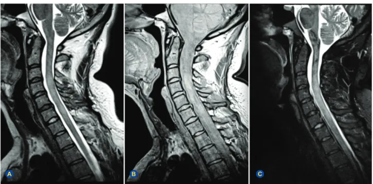
In conclusion, we can only define a spinal cord lesion if we detect a hyperintense area 1) in the sagittal plane both
Figure 2 – Sagittal cervical spinal cord images. (A) T2-weighted fast spin-echo. (B) Proton-density (acquired at dual-echo). (C) Short-tau inversion recovery (STIR). The images depict confluent cervical lesions in an MS patient extending from C2-C3 to C6. At least two T2 sequences are required to identify a demyelinating plaque at the spinal cord. Spinal lesions should also be confirmed on the axial plane.
A B C
Table 2 – Protocol for spinal cord MRI Spinal Cord MRI
Mandatory sequences Sagittal T2 SE or FSE
Sagittal PD (acquired in dual echo) or STIR Axial T2 (lesion focused)
Sagittal T1 SE + gad (if T2 lesions present) Optional sequences
Sagittal T1 SE Axial T1 SE + gad
Axial 2D or 3D T2 FSE (for all spinal cord) PSIR
Non-gapped slice thickness of ≤ 3 mm in sagittal planes and non-gapped slice thickness of ≤ 5 mm in axial planes. Gadolinium injection (single dose: 0,1 mmol/kg body weight) should be done preferably in a “one-stop-strategy”.
NORMAS ORIENT
AÇÃO
on T2 and another appropriate sequence (STIR, DP, PSIR) or 2) if we identify it in two T2 weighted planes.31
Recommendations summary
Spinal cord MRI imaging (T2 weighted)
• Mandatory: It is mandatory to obtain at least two T2 weighted sequences in the sagittal plane, and they must include a conventional spin-echo or fast spin-spin-echo T2 plus proton-density-weighted (acquired with a dual-echo) or short-tau inversion recovery sequences (STIR). • Mandatory: If spinal cord lesions are detected
on sagittal plane sequences, it is mandatory to include a focussed axial T2-weighted sequence. 1.5.2 T1 weighted
Only a small number of spinal cord lesions enhances after gadolinium administration compared with the brain (four to ten times more common) and are usually related to new clinical symptoms.21,41 We recommended acquiring
a sagittal contrast-enhanced T1-weighted spin-echo, when T2 lesions are present, if possible in the same session as brain MRI. This will save time and reduce the number of gadolinium administrations and its potential adverse ef-fects in these chronic patients (as discussed in 2.1.2). Axial contrast-enhanced T1-weighted spin-echo acquisition is op-tional.
MS abnormalities are rarely seen on spontaneous sagit-tal T1-weighted imaging, adding no significant value to the standard protocol regarding spinal cord evaluation.42 In
or-der to facilitate the lesion enhancement depiction, the ac-quisition of a conventional (sagittal) 2D T1 spin-echo before gadolinium injection may be useful.
Recommendations summary
Spinal cord MRI imaging (T1 weighted)
• Mandatory: It is mandatory to obtain a sagittal contrast-enhanced T1-weighted spin-echo when spinal cord MS T2 lesions are identified on spinal imaging.
• Highly recommended: It is highly recommended to follow the “one-stop-strategy”: include a contrast-enhanced T1-weighted spin-echo when brain MS T2 lesions are identified in brain imaging.
• Optional: We suggest that a conventional sagittal 2D T1 spin-echo should be obtained before gadolinium injection to facilitate MS lesion enhancement depiction.
2. Advanced techniques
In the past years, the great advance in acquisition and analysis of non-conventional MR imaging encouraged the publication of several MS studies using those advanced techniques to better characterize both the pathophysiology at tissular/microscopical level and the prognosis in a more individualized manner.
Magnetization transfer imaging (MTI), based on the ex-change of magnetization between pools of bound and free-protons,43 provides quantitative metrics sensitive to
neuro-degenerative microstructural changes on MS, in contrast to conventional techniques that predominantly reflect the inflammatory aspect of the disease.44 This method is easy
to implement in the clinical setting, but it lacks specificity, widely changing with biophysical parameters and between scanners.43,44
Myelin-water imaging (MWI) is a multi-echo T2 re-laxation technique that assesses water trapped in myelin bilayers. It quantifies the myelin content, with strong his-topathological correlations,45 which was shown to be
het-erogeneously reduced in different MS-lesion type and even in normal-appearing white matter (NAWM).46 There are
sev-eral potential confounding factors that may influence this quantitative data and difficult MWI clinical implementation,47
even though recent advances allowed shorter acquisition times and whole-brain coverage.43
The integrity of white matter tracts can be assessed by diffusion tensor imaging (DTI) and seems to be linked with cognitive impairment and progression of physical disabil-ity.43
MR spectroscopy may also contribute to the assess-ment of axonal damage, with NAA decreases consistently reported, and to study grey matter pathology, nowadays recognized as significant in MS48. Both have unsolved
tech-nical issues that compromise reproducibility and translation to clinical practice.
Perfusion, both arterial spin labelling (ASL) and dynamic susceptibility contrast (DSC), has produced contradictory results43 and has no place in routine imaging evaluation of
MS patients (except in case of tumefactive demyelinating lesions, in which perfusion can be useful for differential di-agnosis with neoplasms).49
Functional MRI (fMRI) has proved to be an interesting tool to assess adaptive cortical changes/reorganization that may limit the clinical impact of structural injury.50 fMRI
ap-plications are presently limited to group studies, in research or, eventually, clinical trials, and does not have a role in clini-cal practice.
In conclusion, despite being theoretically appealing, most MR advanced techniques are technically complex, time-consuming and difficult to implement outside the re-search framework.
Imaging postprocessing methods, specially automated methods, to measure brain atrophy progression (longitudi-nal volumetric studies) or to perform automatic lesion count (including subtraction images and automatic and semiauto-matic segmentation) may be included only as an aid to our imaging evaluation in clinical practice, taking into account that they are not formally approved yet and still have some limitations. In addition, neuroradiologists must have the ex-pertise and the hospitals/imaging centres need to provide access to the tools demanded for this type of evaluation.
NORMAS ORIENT
AÇÃO
Recommendations summary Advanced Techniques
• Not recommended: MR advanced techniques (e.g. MTI, MWI, fMRI, DTI, spectroscopy or perfusion) lack standardization in acquisition, postprocessing and interpretation, not being recommended for routine clinical use.
• Optional: Automated postprocessing methods for brain atrophy quantification and automatic lesion count / lesion load quantification may be used if available.
3. Structured Neuroimaging Report
A concise and accurate structured neuroimaging report is warranted as specified in Table 3.
For this goal, the examination request must contain all the fundamental clinical information, such as a brief clinical history (type and duration of symptoms), the patient thera-peutic (discrimination of the administered drugs, such as corticosteroids or MS-disease modifying therapy) and clini-cal diagnostic hypothesis. If the patient performed previous MR imaging, the neuroradiologist must be informed and have access to the images and report of that examination. We recommend patients to have copies of their own stud-ies in a standard readable format (DICOM), particularly if it is likely that they will perform the follow-up examinations in different imaging centres. A standard neuroimaging report should be adopted and divided into technical description, imaging reading and interpretation, with a final summary of the main imaging findings.
Recommendations summary Structured Neuroimaging Report
• Mandatory: We recommend the neuroimaging report to be divided into technical description, imaging reading and interpretation with a final summary of the main imaging findings (see Table 3).
CONCLUSION
In these first Portuguese MRI consensus recommenda-tions, we provide standard imaging protocols adapted to the Portuguese reality, based on the most recent scientific evidence and on our own practical experience. The harmo-nisation of MRI protocols throughout imaging centres will allow a better diagnostic acuity and precise follow-up of the disease. Due to the great technical advances in MRI and in MS knowledge, these guidelines must be reviewed periodi-cally.
OBSERVATIONS
The Grupo de Estudos de Esclerose Múltipla (GEEM, the main Portuguese healthcare professionals group dedi-cated to MS study and treatment, supported by the Por-tuguese Neurological Society) experts group for the first
Portuguese Consensus Recommendations for the Use of the Magnetic Resonance Imaging in Multiple Sclerosis in Clinical Practice is composed by: Pedro Abreu, Rui Pedro-sa, Maria José Sá, João Cerqueira, Lívia SouPedro-sa, Ana Mar-tins da Silva, Joaquim Pinheiro, João de Sá, Sónia Batista, Rita Moiron Simões, José Vale.
ACKNOWLEDGEMENTS
We would like to thank Rita Gouveia Nunes (Institute for Systems and Robotics/IST) and Rafael Simões (B2QUANT Lda) for their contribution to this work.
COMPETING INTERESTS
The authors have declared that no competing interests exist.
FUNDING SOURCES
This work had an investigational grant from Roche Far-macêutica Química LDA: EPAM129844-G.
Table 3 – Guidelines for the neuroimaging report Structured neuroimaging report
1. Technique
• Magnetic field strength;
• Anatomic coverage (brain or spinal cord and which segment); • MR sequences and planes acquired (including thickness); • Gadolinium-based agent and dose;
• Availability and date of a previous brain and/or spinal MR exam for comparison.
2. Imaging findings
• Number (count if ≥ 3 mm) and anatomical distribution of T2 lesions, specifying if juxtacortical/cortical, periventricular, infratentorial or in spinal cord;
• Subjective evaluation of lesion load (mild, moderate, severe); • Number and anatomical distribution of gadolinium-enhancing T1 lesions and type of enhancement (ring, solid, concentric, etc.);
• Atrophy characterization with the use of validated clinical imaging scales, such as global cortical atrophy (GCA) scale. The qualitative impression of the initial atrophy and/or atrophy progression should be included;
• Incidental/non-MS related findings and its clinical significance ; • Follow up: new T2 lesions, gadolinium -enhancing T1 lesions and increased size of previously detected MS plaques. 3. Conclusion
• Interpret if findings are typical, atypical or not consistent with MS and, in this case, provide differential diagnosis;
• Indicate if MR criteria of DIS and dissemination in time (DIT) are fulfilled according to the 2017 MS McDonald criteria1;
• Follow-up: conclude if there are imaging signs of new silent lesions or active plaques and identify potential therapeutic adverse effects (particularly, PML-IRIS).
1DIS is defined by one or more T2-hyperintense lesions in two or more of these four areas:
periventricular, cortical/juxtacortical, infratentorial and spinal cord. DIT is demonstrated by: new T2 and/or gadolinium-enhancing lesion(s) on follow up MRI with reference to a baseline scan OR simultaneous presence of gadolinium-enhancing and non-enhancing lesions at any time.
NORMAS ORIENT
AÇÃO
REFERENCES
1. Mcdonald W, Compston A, Edan G, Goodkin D, Hartung HP, Lublin FD, et al. Recommended diagnostic criteria for multiple sclerosis: guidelines from the International Panel on the diagnosis of multiple sclerosis. Ann Neurol. 2001;50:121-7.
2. Abreu P, Pedrosa R, Sá MJ, Cerqueira J, Sousa L, Silva AM, et al. Recomendações e consensos do Grupo de Estudos de Esclerose Múltipla e da Sociedade Portuguesa de Neurorradiologia sobre ressonância magnética na esclerose múltipla na prática clínica: parte 1. Acta Med Port. 2018;31:281-9.
3. Wattjes MP, Barkhof F. High field MRI in the diagnosis of multiple sclerosis: high field-high yield? Neuroradiology. 2009;51:279-92. 4. Stankiewicz JM, Glanz BI, Healy BC, Arora A, Neema M, Benedict R, et
al. Brain MRI lesion load at 1.5T and 3T versus clinical status in multiple aclerosis. J Neuroimaging. 2011;21:e50-6.
5. Wattjes MP, Harzheim M, Kuhl CK, Gieseke J, Schmidt S, Klotz L, et al. Does high-field MR imaging have an influence on the classification of patients with clinically isolated syndromes according to current diagnostic MR imaging criteria for multiple sclerosis? Am J Neuroradiol. 2006;27:1794-8.
6. Traboulsee A, Simon JH, Stone L, Fisher E, Jones DE, Malhotra A, et al. Revised recommendations of the CMSC Task Force for a standardized MRI protocol and clinical guidelines for the diagnosis and follow-up of multiple sclerosis. Am J Neuroradiol. 2015;37:394-401.
7. Crombé A, Saranathan M, Ruet A, Durieux M, Roquefeuil E, Ouallet JC, et al. MS lesions are better detected with 3D T1 gradient-echo than with 2D T1 spin-echo gadolinium-enhanced imaging at 3T. Am J Neuroradiol. 2015;36:501-7.
8. Nelson F, Poonawalla A, Hou P, Wolinsky JS, Narayana P. 3D MPRAGE improves classification of cortical lesions in multiple sclerosis. Mult Scler. 2008;14:1214-9.
9. Uysal E, Erturk SM, Yildirim H, Seleker F, Basak M. Sensitivity of immediate and delayed gadolinium-enhanced MRI after injection of 0.5 M and 1.0 M gadolinium chelates for detecting multiple sclerosis lesions. Am J Roentgenol. 2007;188:697-702.
10. ema.europa.eu - EMA’s final opinion confirms restrictions on use of linear gadolinium agents in body scans. Recommendations conclude EMA’s scientific review of gadolinium deposition. [accesseed 2018 Jan 7]. Available from: http://www.ema.europa. eu/ema/index.jsp?curl=pages/medicines/human/referrals/ Gad%0Aoliniumcontaining_contrast_agents/human_referral_ prac_000056.jsp&mid=WC0b01ac05805%0Ac516f.
11. Gulani V, Calamante F, Shellock FG, Kanal E, Reeder SB. Gadolinium deposition in the brain: summary of evidence and recommendations. Lancet Neurol. 2017;16:564-70.
12. Kanda T, Ishii K, Kawaguchi H, Kitajima K, Takenaka D. High signal intensity in the dentate nucleus and globus pallidus on unenhanced T1-weighted MR images: relationship with increasing cumulative dose of a gadolinium-based contrast material. Radiology. 2014;270:834-41. 13. fda.gov. FDA Drug Safety Communication: FDA identifies no harmful
effects to date with brain retention of gadolinium-based contrast agents for MRIs; review to continue. [accessed 2018 Jan 7]. Available from: https://www.fda.gov/Drugs/DrugSafety/ucm559007.htm.
14. Vågberg M, Axelsson M, Birgander R, Burman J, Cananau C, Forslin Y et al. Guidelines for the use of magnetic resonance imaging in diagnosing and monitoring the treatment of multiple sclerosis: recommendations of the Swedish Multiple Sclerosis Association and the Swedish Neuroradiological Society. Acta Neurol Scand. 2017;135:17-24. 15. Karimian-Jazi K, Wildemann B, Diem R, Schwarz D, Hielscher T, Wick
W, et al. Gd contrast administration is dispensable in patients with MS without new T2 lesions on follow-up MRI. Neurol Neuroimmunol Neuroinflamm. 2018;5:e480.
16. Dong-Si T, Richman S, Wattjes MP, Wenten M, Gheuens S, Philip J, et al. Outcome and survival of asymptomatic PML in natalizumab-treated MS patients. Ann Clin Transl Neurol. 2014;1:755-64.
17. Popescu V, Battaglini M, Hoogstrate WS, Verfaillie SC, Sluimer IC, van Schijndel RA, et al. Optimizing parameter choice for FSL-brain extraction tool (BET) on 3D T1 images in multiple sclerosis. Neuroimage. 2012;61:1484-94.
18. Bergui M, Bradac GB, Oguz KK, Boghi A, Geda C, Gatti G, et al. Progressive multifocal leukoencephalopathy: diffusion-weighted imaging and pathological correlations. Neuroradiology. 2004;46:22-5. 19. Wattjes MP, Vennegoor A, Steenwijk MD, de Vos M, Kilestein J, van
Oosten B, et al. MRI pattern in asymptomatic natalizumab-associated PML. J Neurol Neurosurg Psychiatry. 2014;86:793-8.
20. Eisele P, Szabo K, Griebe M, Rossmanith C, Forster A, Hennerici M, et al. Reduced diffusion in a subset of acute MS lesions: a serial multiparametric MRI study. Am J Neuroradiol. 2012;33:1369-73. 21. Rovira À, Wattjes MP, Tintoré M, Tur C, Yousry T, Sormani M, et al.
Evidence-based guidelines: MAGNIMS consensus guidelines on the use of MRI in multiple sclerosis—clinical implementation in the diagnostic process. Nat Rev Neurol. 2015;11:471-82.
22. Tallantyre EC, Brookes MJ, Dixon JE, Morgan PS, Evangelou N, Morris PG. Demonstrating the perivascular distribution of MS lesions in vivo with 7-Tesla MRI. Neurology. 2008;70:2076-8.
23. Tallantyre EC, Dixon JE, Donaldson I, Owens T, Morgan PS, Morris PG, et al. Ultra-high-field imaging distinguishes MS lesions from asymptomatic white matter lesions. Neurology. 2011;76:534-9. 24. Grabner G, Dal-Bianco A, Schernthaner M, Vass K, Lassmann H,
Trattnig S. Analysis of multiple sclerosis lesions using a fusion of 3.0 T FLAIR and 7.0 T SWI phase: FLAIR SWI. J Magn Reson Imaging. 2011;33:543-9.
25. Sati P, George IC, Shea CD, Gaitán MI, Reich DS. FLAIR*: A combined MR contrast technique for visualizing white matter lesions and parenchymal veins. Radiology. 2012;265:926-32.
26. Campion T, Smith RJ, Altmann DR, Brito GC, Turner BP, Evanson J, et al. FLAIR* to visualize veins in white matter lesions: a new tool for the diagnosis of multiple sclerosis? Eur Radiol. 2017:4257-63.
27. Wuerfel J, Sinnecker T, Ringelstein EB, Jarius S, Schwindt W, Niendorf T, et al. Lesion morphology at 7 Tesla MRI differentiates Susac syndrome from multiple sclerosis. Mult Scler. 2012;18:1592-9.
28. Kelly JE, Mar S, D’Angelo G, Zhou G, Rajderkar D, Benzinger TL. Susceptibility-weighted imaging helps to discriminate pediatric multiple sclerosis from acute disseminated encephalomyelitis. Pediatr Neurol. 2015;52:36-41.
29. Sinnecker T, Dörr J, Pfueller CF, Harms L, Ruprecht K, Jarius S, et al. Distinct lesion morphology at 7-T MRI differentiates neuromyelitis optica from multiple sclerosis. Neurology. 2012;79:708-14.
30. Vercellino M, Plano F, Votta B, Mutani R, Giordana MT, Cavalla P. Grey matter pathology in multiple sclerosis. Lancet Neurol. 2008;7:841-51. 31. Filippi M, Rocca MA, Ciccarelli O, De Stefano N, Kappos L, Rovira A,
et al. MRI criteria for the diagnosis of multiple sclerosis: MAGNIMS consensus guidelines. Lancet Neurol. 2016;15:292-303.
32. Thompson AJ, Banwell BL, Barkhof F, Carrol W, Coetzee T, Comi G, et al. Diagnosis of multiple sclerosis: 2017 revisions of the McDonald criteria. Lancet Neurol. 2018;17:162-73.
33. Nelson F, Poonawalla AH, Hou P, Huang F, Wolinsky JS, Narayana PA. Improved identification of intracortical lesions in multiple sclerosis with phase-sensitive inversion recovery in combination with fast double inversion recovery MR imaging. Am J Neuroradiol. 2007;28:1645-9. 34. Wattjes MP, Steenwijk MD, Stangel M. MRI in the diagnosis and
monitoring of multiple sclerosis: an update. Clin Neuroradiol. 2015;25:157-65.
35. Brex PA, O’Riordan JI, Miszkiel KA, Moseley IF, Thompson AJ, Plant GT, et al. Multisequence MRI in clinically isolated syndromes and the early development of MS. Neurology. 1999;53:1184-90.
36. Dietemann JL, Thibaut-Menard A, Warter JM, Neugroschl C, Tranchant C, Gillis C, et al. MRI in multiple sclerosis of the spinal cord: Evaluation of fast short-tan inversion-recovery and spin-echo sequences. Neuroradiology. 2000;42:810-3.
37. Philpott C, Brotchie P. Comparison of MRI sequences for evaluation of multiple sclerosis of the cervical spinal cord at 3 T. Eur J Radiol. 2011;80:780-5.
38. Kearney H, Miller DH, Ciccarelli O. Spinal cord MRI in multiple sclerosis-diagnostic, prognostic and clinical value. Nat Rev Neurol. 2015;11:327-38.
39. Poonawalla AH, Hou P, Nelson F, Wolinsky J, Narayana P. Cervical spinal cord lesions in multiple sclerosis: T1-weighted inversion-recovery MR imaging with phase-sensitive reconstruction. Radiology. 2008;246:258-64.
40. 40. Alcaide-Leon P, Pauranik A, Alshafai L, Rawal S, Oh J, Montanera W et al. Comparison of Sagittal FSE T2, STIR, and T1-Weighted Phase-Sensitive Inversion Recovery in the Detection of Spinal Cord Lesions in MS at 3T. Am J Neuroradiol. 2016;37:970-5.
41. Lycklama G, Thompson A, Filippi M, Miller D, Polman C, Fazekas F, et al. Spinal-cord MRI in multiple sclerosis. Lancet Neurol. 2003;2:555-62. 42. Bot JC, Barkhof F. Spinal-cord MRI in multiple sclerosis: conventional and nonconventional MR techniques. Neuroimaging Clin N Am. 2009;19:81-99.
NORMAS ORIENT
AÇÃO
43. Enzinger C, Barkhof F, Ciccarelli O, Filippi M, Kappos L, Rocca M, et al. Nonconventional MRI and microstructural cerebral changes in multiple sclerosis. Nat Rev Neurol. 2015;11:676-86.
44. Lema A, Bishop C, Malik O, Mattoscio M, Ali R, Nicholas R, et al. A comparison of magnetization transfer methods to assess brain and cervical cord microstructure in multiple sclerosis. J Neuroimaging. 2017:27:221-6.
45. Laule C, Leung E, Li DK, Traboulsee AL, Paty DW, MacKay AL, et al. Myelin water imaging in multiple sclerosis: quantitative correlations with histopathology. Mult Scler. 2006;12:747-53.
46. Faizy TD, Thaler C, Kumar D, Sedlacik J, Broocks G, Grosser M, et
al. Heterogeneity of multiple sclerosis lesions in multislice myelin water imaging. PLoS One. 2016;11:1-13.
47. MacKay AL, Laule C. Magnetic resonance of myelin water: an in vivo marker for myelin. Brain Plast. 2016;2:71-91.
48. De Stefano N, Filippi M. MR spectroscopy in multiple sclerosis. J Neuroimaging. 2007;1:31S-5.
49. Cha S, Pierce S, Knopp EA, Johnson G, Yang C, Ton A, et al. Dynamic contrast-enhanced T2*-weighted MR imaging of tumefactive demyelinating lesions. Am J Neuroradiol. 2001;22:1109-16.
50. Filippi M, Rocca MA. Present and future of fMRI in multiple sclerosis. Expert Rev Neurother. 2013;13:S27-31.