with Conventional Antibiotics against Methicillin
Resistant
Staphylococcus aureus
Madhuri Singh1, Ravisekhar Gadepalli2, Benu Dhawan2, Kasturi Mukhopadhyay1*
1School of Environmental Sciences, Jawaharlal Nehru University, New Delhi, India,2Department of Microbiology, All India Institute of Medical Sciences, New Delhi, India
Abstract
Our previous studies revealed that alpha-melanocyte stimulating hormone (a-MSH) is strongly active againstStaphylococcus aureus(S. aureus) including methicillin resistantS. aureus(MRSA). Killing due to a-MSH occurred by perturbation of the bacterial membrane. In the present study, we investigated thein vitrosynergistic potential of a-MSH with five selected conventional antibiotics viz., oxacillin (OX), ciprofloxacin (CF), tetracycline (TC), gentamicin (GM) and rifampicin (RF) against a clinical MRSA strain which carried a type III staphylococcal cassette chromosomemec(SCCmec) element and belonged to the sequence type (ST) 239. The strain was found to be highly resistant to OX (minimum inhibitory concentration (MIC) = 1024mg/ml) as well as to other selected antimicrobial agents includinga-MSH. The possibility of the existence of intracellular target sites ofa-MSH was evaluated by examining the DNA, RNA and protein synthesis pathways. We observed a synergistic potential ofa-MSH with GM, CF and TC. Remarkably, the supplementation ofa-MSH with GM, CF and TC resulted in$64-, 8- and 4-fold reductions in their minimum bactericidal concentrations (MBCs), respectively. Apart from membrane perturbation, in this study we found that a-MSH inhibited ,53% and ,47% DNA and protein synthesis, respectively, but not RNA synthesis. Thus, the mechanistic analogy betweena-MSH and CF or GM or TC appears to be the reason for the observed synergy between them. In contrast,a-MSH did not act synergistically with RF which may be due to its inability to inhibit RNA synthesis (,10%). Nevertheless, the combination ofa-MSH with RF and OX showed an enhanced killing by,45% and,70%, respectively, perhaps due to the membrane disrupting properties ofa-MSH. The synergistic activity ofa-MSH with antibiotics is encouraging, and promises to restore the lost potency of discarded antibiotics.
Citation:Singh M, Gadepalli R, Dhawan B, Mukhopadhyay K (2013) Combination of Alpha-Melanocyte Stimulating Hormone with Conventional Antibiotics against Methicillin ResistantStaphylococcus aureus. PLoS ONE 8(9): e73815. doi:10.1371/journal.pone.0073815
Editor:John Conly, University of Calgary, Canada
ReceivedJanuary 30, 2013;AcceptedJuly 24, 2013;PublishedSeptember 9, 2013
Copyright:ß2013 Singh et al. This is an open-access article distributed under the terms of the Creative Commons Attribution License, which permits unrestricted use, distribution, and reproduction in any medium, provided the original author and source are credited.
Funding:This work is supported by the grants from Indian Council of Medical Research, India and Department of Biotechnology, India. The funders had no role in study design, data collection and analysis, decision to publish, or preparation of the manuscript.
Competing Interests:The authors have declared that no competing interests exist. * E-mail: [email protected]
Introduction
Infections due to methicillin resistant Staphylococcus aureus
(MRSA) are becoming untreatable and putting tremendous pressure on healthcare systems [1–3]. MRSA is the most common cause of infection at a variety of sites including skin and soft tissues, respiratory tract, bloodstream, and prosthetic devices [4]. Beta lactam antibiotics, the wonder drugs of the past, are now no longer effective against these resistant bacteria. Likewise, promising new antibiotics of various classes, recently approved by the Food and Drug Administration (FDA), including linezolid (an oxazolidinone) and daptomycin (a cyclic lipopeptide), could not significantly improve the outcomes of infections caused by MRSA [5,6]. In fact, emergence of resistance to these new classes of antibiotics may hinder the use of these drugs in the future. With this existing scenario, an effective solution may be the development of combination therapies involving antimicrobial agents with differ-ent mechanisms of inhibitory action [7,8]. For example, pairing of vancomycin with rifampin or gentamicin has been often successful in the treatment of endocarditis caused by MRSA [9]. The potential benefits of combination therapy over single therapy include, reduction in the dose of toxic antibiotics, decreased resistance development, and broader antibacterial activity [10,11].
In the last three decades, host defense peptides (HDPs), which are key components of innate immunity in a vast array of organisms, have drawn a lot of attention as potential therapeutic agents [12– 14]. These peptides are modulators of the immune system and evoke effector mechanisms to rapidly stop pathogen proliferation [15]. Moreover, they work synergistically with other HDPs in the host environment [16]. Because of their unique mechanism of targeting microbes and their role in triggering host immunity, HDPs exhibit immense potential to act synergistically with conventional antibiotics. Thus, the pairing of HDPs with other conventional antibiotics or with other HDPs has received greater priority over the pairing of antibiotics alone [17–20].
the lost antibacterial efficacy of conventional antibiotics. For this purpose, we evaluated the in vitro combination effect of a-MSH with five different antibiotics viz., (oxacillin (OX), gentamicin (GM), rifampicin (RF), tetracycline (TC), and ciprofloxacin (CF)) against a clinical MRSA strain which was resistant to all these five antibiotics at high concentrations. We observed a dramatic increment in the staphylocidal effect of each antibiotic even at lower doses when combined with an ineffective dose of a-MSH
in vitro. These results are very encouraging as combininga-MSH with currently discarded traditional antibiotics might be useful for the treatment of fatal infections due to resistant microorganisms.
Materials and Methods
Materials
All the antibiotics viz., oxacillin sodium salt, ciprofloxacin hydrochloride, tetracycline hydrochloride, rifampicin, gentamicin sulfate, synthetic a-MSH were purchased from Sigma-Aldrich (USA). Brain heart infusion (BHI), cation-adjusted Muller Hinton broth (CAMHB), and agar were purchased from HiMedia (India). Scintillation cocktail ‘O’ in toluene was bought from Spectrochem (Mumbai, India). Tricarboxylic acid, MTT (3-(4,5-Dimethylthia-zol-2yl)-2,5-diphenyle tetrazolium bromide), trypan blue, tri-tonX2100 and dimethyl sulfoxide (DMSO) were purchased from Fisher Scientific (Germany). [methyl-3H] thymidine, [4,5-3H]
leucine and [5-3H] uridine were purchased from the Board of Radiation and Isotope Technology (BRIT, India). The purity ofa -MSH was.97% and its concentration was determined spectro-photometrically (UV-2450 UV-VIS spectrophotometer, Shi-madzu). Stocks of all the antibiotics anda-MSH were stored at 4uC.
Bacterial Strains
The clinical strain of S. aureus was isolated from a patient admitted to the All India Institute of Medical Sciences (AIIMS), New Delhi, India with skin and soft tissue infection (SSTI). Ethical approval was obtained from the Institute Ethics Committee, AIIMS, New Delhi, India.S. aureuswas identified using standard biochemical tests using multiplex PCR. Cefoxitin disk diffusion method and mecA PCR was used to characterize the strain as MRSA [24]. Additionally, one prototype ATCC MRSA 33591 and ATCC MSSA 29213 were used to perform quality control as recommended by the Clinical Laboratory Standard Institute (CLSI) [25]. Strains were stored at280uC in 15% glycerol until sub-cultured onto BHI agar plate followed by secondary culture in BHI broth. The mid log phase grown cells (OD600 nm= 0.5) were
used for all the experiments. The cell suspension of desired density was prepared in 10 mM sodium potassium phosphate buffer containing 150 mM NaCl (PBS).
Molecular Characterization of the Clinical MRSA Strain The clinical MRSA isolate was processed for staphylococcal cassette chromosomemec(SCCmec) typing and multilocus sequence typing (MLST) as previously described [24,26,27]. The sequences of the seven housekeeping genes (arcC,aroE,glpF,gmk,pta,tpi, and
yqiL) obtained were compared with the sequences at the MLST website (http://www.mlst.net) to determine the sequence type (ST).
Minimum Inhibitory Concentration (MIC)
MIC values of all the antibiotics, i.e., OX, RF, GM, CF, and TC were determined by broth microdilution assay following CLSI guidelines using CAMHB [25]. The assay was repeated on three days independently, and a constant MIC value was found. MIC
values of OX .2mg/ml [28], GM .0.25mg/ml [29], CF
.0.25mg/ml [30], TC.0.25mg/ml [31], and RF.0.015mg/ ml [28] were considered resistant. MIC value ofa-MSH could not be determined as MHB/BHI medium tends to reduce its antibacterial activity. The same inhibitory effect of MHB medium for another cationic antimicrobial peptide, i.e., LL237 has also previously been reported by Turner et al., [32]. Nevertheless, they had found that the peptide was very active when they performed the killing assays by viable colony count method using PBS buffer [32].
Minimum Bactericidal Concentration (MBC)
MBC was determined by the killing assay using 105 to 106CFU/ml of mid log culture as described elsewhere [22,23,32,33]. Briefly, the cells were incubated with each of the selected antibiotic (2mg/ml to 2048mg/ml) anda-MSH (2mg/ml to 160mg/ml) individually in PBS. The mixture was incubated at 37uC for 2 h and the aliquots were 10 -fold diluted two times serially in PBS (to reduce the drug carry over and to allow accurate colony count) followed by plating on BHI agar plate in triplicate. Viable colonies were counted after overnight incubation and percentage of killing of treated sample was determined with respect to the untreated samples (control). The concentrations at which $50%, and $90% of cells were killed, were defined as MBC50and MBC90,respectively. MBC values of OX.0.5mg/ml
[28], GM.6.5mg/ml [34], CF.2mg/ml [35], TC.3mg/ml [36], and RF.0.125mg/ml [37] were considered resistant.
Synergism Studies
For determining synergism, both the pairing agents (antibiotic and a-MSH) were administered at doses which did not show significant bacterial killing ($80% cell survival) when used alone. The methodology was similar to the killing assay protocol as described above. Briefly, the cells (105 to 106CFU/ml) were treated with ineffective doses of each antibiotic alone ranging from 2 to 32mg/ml and in presence and absence ofa-MSH (8mg/ml). For example, to test synergism between a-MSH and OX, cells were treated with OX alone in the concentration range of 2 to 32mg/ml and in the presence of 8mg/ml ofa-MSH. An MBC50
and MBC90value was calculated for each antibiotic alone and in
the presence ofa-MSH. A$4-fold reduction in MBC90value in
the presence of a-MSH was considered a synergistic relation betweena-MSH and that antibiotic [28].
Impact ofa-MSH on Bacterial DNA, RNA and Protein Synthesis
To determine the effect ofa-MSH on bacterial macromolecule synthesis, whole cell labeling was done with radioactive precursors of DNA, RNA and protein as described elsewhere [38,39]. In brief,,108CFU/ml ofS. aureusATCC 29213 cells were labeled
confirm that the chosen doses ofa-MSH were not bactericidal, killing kinetics using the 2 and 10mg/ml of a-MSH against
,108CFU/ml of S. aureus ATCC 29213 were performed using
duplicate samples prepared separately, without radioactive precursors. Three independent experiments were done in each case.
Hemolytic Activity ofa-MSH
Fresh 5 ml aliquots of blood were collected from Swiss albino mouse in the presence of 2 mg/ml ethylenediaminetetraacetic acid (EDTA) and centrifuged at 400 rpm for 10 min and the red blood cells (RBCs) pellet was collected. The use of animals was approved by the Institutional Animal Ethics Committee of Jawaharlal Nehru University (IAEC-JNU), New Delhi, India. The pellet was washed 3 times in 10 mM PBS buffer, pH 7.4, and re-suspended in 4-fold volumes of the same buffer. 5ml of this RBC solution was added to 995ml of PBS buffer containing serially diluteda-MSH (100 pg/ ml to 100mg/ml). The RBC anda-MSH mixture was incubated at 37uC for 1 h or 18 h and centrifuged. To the 200ml of supernatant 800ml of PBS buffer was added and absorbance was measured at 413 nm using UV-VIS Spectrophotometer (Shi-madzu, Japan). To ensure 100% (positive control) and 0% (negative control) hemolysis, 0.1% tritonX2100 (TX100) and PBS buffer were added, respectively, instead of a-MSH and absorbance at 413 nm was measured. Hemolysis due to the test peptide was determined as described elsewhere [40], % Hemo-lysis = 1006(absorbance in peptide – absorbance in
PBS)/(absor-bance in TX100 - absorPBS)/(absor-bance in PBS). The assay was done in duplicate.
Cytotoxicity Due toa-MSH by MTT Assay
The efficacy of the peptide to induce cell death in a 3T3 mouse fibroblast cell line was determined by MTT assay as described elsewhere [41]. This colorimetric assay is based upon the reduction of yellow tetrazolium dye, MTT, to an insoluble purple colored formazan product by the enzyme succinate dehydrogenase found in metabolically active cells. After treatment with MTT, the cells are solubilized in the presence of organic solvent (i.e., DMSO), and the released, solubilized formazan product is measured spectrophotometrically at 570 nm. Since reduction of MTT can only happen in metabolically active cells, the intensity of formazan is directly proportional to the cell viability. Briefly, a monolayer of 1 day old 3T3 cells (105cells/ml predetermined by trypan blue staining) were grown in DMEM medium supple-mented with 10% fetal bovine serum on 24 well plates placed in a 5% CO2 incubator. The cells were washed and re-suspended in
PBS buffer and then treated witha-MSH (0.2mg/ml to 20mg/ml) for 2 h. Untreated cells in the presence of PBS buffer alone were taken as a negative control (100% intact cells). To the mixture, 20ml of 5 mg/ml MTT solution was added and incubated in the dark at 37uC for 4.5 h, followed by addition of 200ml DMSO and further incubation for 1 h. After incubation, 100ml of this mixture was removed and added to 900ml of double distilled H2O and
absorbance was measured at 570 nm against a background (100ml DMSO+900ml ddH2O). The percentage cytotoxicity
was calculated as described elsewhere [41], % of Cytotoxici-ty = 1006(absorbance of control - absorbance of sample)/(absor-bance of control). The above assay was done in triplicate.
Statistical Analysis
All data was compiled by using mean 6 standard deviation calculated for three independent replicates using Microsoft office excel. Difference in mean values among % survival data-sets for various antibiotics alone and in the presence ofa-MSH and %
survival data-sets for different concentrations of the same antimicrobial agent were measured by one-way Analysis of Variance (ANOVA) using Minitab statistical software [22,23], and p values#0.05 were considered significant.
Results
Genotypic Characterization of the Clinical MRSA Strain The clinical MRSA strain carried a type III cassette, and MLST profiling classified this strain as ST239. The strain is close to UK EMRSA-1 (NCTC11931), and the Brazilian/Hungarian clone (ST239-SCCmecIII) with allelic profile of 2-3-1-1-4-4-3 based on the DNA sequence of seven housekeeping genes. The predomi-nance of the ST239 clone has been reported previously among Indian nosocomial MRSA strains [24,42].
Susceptibility Profiles ofS. aureusstrains to the Selected Antibiotics by Microdilution Assay
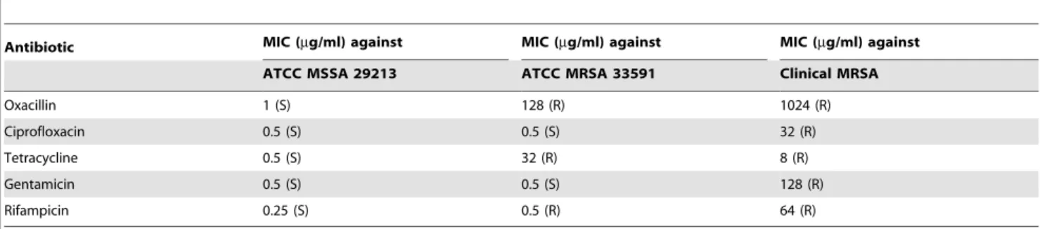
MIC values of the selected antibiotics CF, GM, TC, RF and OX against the clinical MRSA isolate as well as ATCC MRSA 33591 and ATCC MSSA 29213 were determined and are presented in Table 1. The ATCC MRSA 33591 strain was resistant to OX (MIC = 128mg/ml), RF (MIC = 0.5mg/ml), and TC (MIC = 32mg/ml), while it was susceptible to both GM and CF. However, the clinical MRSA isolate showed very high MIC values for all antibiotics tested indicating its resistance to both bacteriostatic (TC) and bactericidal (OX, GM, CF, RF) groups of drugs. For example, the MIC values for OX and GM were 1024mg/ml and 128mg/ml, respectively, which are 512-fold greater than the values in susceptible bacteria [43]. The rationale behind choosing the clinical MRSA over prototype ATCC MRSA 33591 for the synergistic study was its high resistance towards all the tested antibiotics.
Susceptibility Profile of Clinical MRSA to the Selected Antibiotics and toa-MSH by the Killing Assay
The killing activity of all selected antibiotics (at concentrations ranging from 64 to 2048mg/ml) anda-MSH (2 to 160mg/ml) was assessed against the clinical MRSA isolate. Taking into account the varying action time of the different chosen antibiotics, the antibiotic treatment was done for 2 h to allow sufficient time for both the slow-acting (i.e., OX) and fast-acting antibiotics (i.e., CF). Fig. 1a shows the percentage survival of bacteria treated with each selected antibiotic alone. Similarly, Fig. 1b shows the percentage survival ofS. aureuscells treated witha-MSH alone for 2 h. The concentrations at which an antibiotic showed 90% and 50% bacterial killing were taken as the MBC90 and MBC50 value,
respectively, of that antibiotic. These values were determined from the killing curves of the tested antibiotics (Fig. 1a), and are summarized in Table 2. It is apparent from Fig. 1a and Table 2 that the clinical MRSA strain was highly resistant to all the selected antibiotics. The MBC90 values of CF, TC and RF
were 128mg/ml, 128mg/ml and 512mg/ml, respectively (Table 2). However, in case of OX and GM, MBC90 could not
be achieved even at 2048mg/ml. Similarly, as shown in Fig. 1b, the strain was poorly susceptible to a-MSH as no killing was observed up to 8mg/ml ofa-MSH, and only 71.763.3% killing was obtained at 160mg/ml ofa-MSH (p,0.001 when multiple data-set of % survival were compared among different concen-trations ofa-MSH). It is pertinent to note here that in our previous reports we observed .95% killing by a-MSH against S. aureus
Synergistic Interaction of a-MSH with Selected Conventional Antibiotics
The synergistic potential of a-MSH with the 5 different antibiotics was determined by the killing assay. The data with each antibiotic alone, and in the presence of 8mg/ml ofa-MSH is presented in Fig. 2. A remarkable increase in the staphylocidal activity of each antibiotic was observed when combined witha
-MSH. Of the 5 antibiotics tested, GM (Fig. 2a), TC (Fig. 2b) and CF (Fig. 2c) demonstrated.90% killing in presence ofa-MSH, whereas, the killing activity of the above antibiotics was ,20% when they were administered alone in the same range of concentrations. For example, 2 to 32mg/ml of GM alone showed only 13.764.9% to 16.8611.5% bacterial killing; while at the same concentrations it showed 80.166.2% to 91.061.2% killing when used in combination with 8mg/ml of a-MSH (Fig. 2a). Similarly, 2 to 32mg/ml of OX alone showed 3.763.4% to 4.565.8% killing (Fig. 2d), whereas, in the presence of 8mg/mla -MSH, it killed 45.764.0% to 77.162.9% cells (Fig. 2d). The combination of RF and a-MSH, however, could only achieve 5660.2% killing (Fig. 2e). The change in bactericidal activity of antibiotics in the presence ofa-MSH was statistically significant (p,0.05 when compared among data-sets of different doses of each antibiotic alone and in the presence ofa-MSH). The MBC50
and MBC90values of all antibiotics were also determined in the
presence ofa-MSH from the above mentioned killing data (Fig. 2) and are summarized in Table 2. A synergistic relation was obtained whena-MSH was paired with GM, CF and TC since a
$4-fold reduction in the MBC90value was observed for all three
antibiotics. For instance, the MBC90 value of GM alone was
.2048mg/ml, while in combination witha-MSH it was 32mg/ ml, thus achieving a 64-fold reduction (Table 2). Similarly, TC showed 32-fold and 4-fold reductions in MBC50 and MBC90
value, respectively, and CF showed 32-fold and 8-fold reductions in MBC50and MBC90 value, respectively, in the presence ofa
-MSH. Although there was a huge reduction in MBC50values of
OX (512-fold) and RF (64-fold) in the presence ofa-MSH, 90% killing could not be achieved even when they were combined with a-MSH.
Impact ofa-MSH on Bacterial DNA, RNA and Protein Synthesis
To examine the effect of sub-lethal doses of a-MSH (2 and 10mg/ml) on macromolecular synthesis ofS. aureusATCC 29213 cells, incorporation of radioactive precursor [methyl-3H] thymi-dine, [5-3H] uridine and [4,5-3H] leucine into DNA, RNA and protein, respectively, was observed over a period of 2 h and percent radioactivity of these precursors is presented in Fig. 3a–c. Additionally, ciprofloxacin, rifampicin and tetracycline treated samples (2mg/ml each) were included as positive controls of DNA, RNA and protein synthesis inhibition, respectively [44]. As can be seen from Fig. 3a and Fig. 3c, a reduction in the incorporation of [methyl-3H] thymidine and [4,5-3H] leucine was observed in the a-MSH treated samples. For example, [methyl-3H] thymidine Table 1.Minimum Inhibitory concentration (MIC) values of selected antibiotics againstS. aureusstrains as determined by broth microdilution assay following CLSI guidelines [25].
Antibiotic MIC (mg/ml) against MIC (mg/ml) against MIC (mg/ml) against
ATCC MSSA 29213 ATCC MRSA 33591 Clinical MRSA
Oxacillin 1 (S) 128 (R) 1024 (R)
Ciprofloxacin 0.5 (S) 0.5 (S) 32 (R)
Tetracycline 0.5 (S) 32 (R) 8 (R)
Gentamicin 0.5 (S) 0.5 (S) 128 (R)
Rifampicin 0.25 (S) 0.5 (R) 64 (R)
Note: (S) Strain susceptible to tested antibiotic. (R) Strain resistant to tested antibiotic. doi:10.1371/journal.pone.0073815.t001
Figure 1. Determining bactericidal concentrations of each tested antibacterial agent. (a) Killing curves showing bactericidal activity of five different tested antibiotics against clinical MRSA isolate. Concentrations of each antibiotic were selected from 0 to 2048mg/ml. Symbols; Oxacillin (red diamond), Gentamicin (green square), Rifampicin (yellow triangle), Ciprofloxacin (pink square) and Tetracycline (orange circle). (b) Bactericidal activity ofa-MSH (2–160mg/ml) against clinical MRSA isolate. The killing assay was done in triplicate and repeated on three different occasions. *p value#0.001, **p value#0.01, ***p value #0.05.
radioactivity showed a reduction from 100% (untreated control) to 77.666.8% and 67.068.5% when treated with 2 and 10mg/ml of a-MSH, respectively, for 30 min. It further reduced to 53.265.9% and 50.8611.6% after 2 h of treatment (Fig. 3a). Likewise, a 1864.5% and 34.562.1% decrease in the incorporation of [4,5-3H] leucine was observed in samples treated with 2 and 10mg/ml ofa-MSH, respectively, for 30 min (Fig. 3c), and was further decreased by 40.966.3% and 4761.4%, after 2 h treatment with a-MSH (Fig. 3c). The radioactive labeling assay indicated that there was inhibition of both DNA and protein synthesis in the bacterial cells on exposure to sub-lethal concentrations of a-MSH. However, less than a 10% decrease in the incorporation of [5-3H] uridine was observed in thea-MSH treated samples compared to the untreated controls, suggesting thata-MSH had only a marginal effect on RNA synthesis (,10%) (Fig. 3b).
To confirm that the chosen doses of a-MSH were not bactericidal, killing kinetics using the 2 and 10mg/ml ofa-MSH against,108CFU/ml ofS. aureusATCC 29213 were performed
simultaneously, and the killing results are shown in Fig. 3d. The data showed that only,12% and 4066.9% killing was observed
in cells treated with 2mg/ml and 10mg/ml of a-MSH, respectively, over a period of 2 h.
Effect ofa-MSH Toxicity on Mouse Red Blood Cells (RBCs) and Fibroblast Cell Lines
The hemolysis of mouse RBCs exposed to a range ofa-MSH concentrations (100 pg/ml to 100mg/ml) was examined (Fig. 4a). As can be seen from this figure, less than 10% hemolysis was observed after 1 h treatment witha-MSH even at a concentration of 100mg/ml, and only,3% further increase in hemolysis was
observed when exposure time was increased to 18 h. Fibroblast Table 2.Minimum bactericidal concentration (MBC50and MBC90) values of selected antimicrobial agents when used alone and in
the presence of 8mg/ml ofa-MSH against clinical MRSA strain.
Antibiotic
MBC50/MBC90(mg/ml) of antibiotic alone
MBC50/MBC90(mg/ml) of antibiotic in the presence ofa-MSH
Fold reduction in MBC values (MBC50/MBC90) of antibiotic when combined witha-MSH
Oxacillin 2048/.2048 4/.32 512/NA*
Ciprofloxacin 64/128 2/16 32/8
Tetracycline 64/128 2/32 32/4
Gentamicin 2048/.2048 2/32 1024/.64
Rifampicin .256/512 8/.32 .32/NA*
Note: Fold reduction = (MBC50of antibiotic alone/MBC50in presence ofa-MSH).
NA*means MBC90not achieved.
doi:10.1371/journal.pone.0073815.t002
Figure 2. Killing curves of each of antibiotic alone and in presence of a-MSH against clinical MRSA isolate. (a) Gentamicin, (b)
Tetracycline, (c) Ciprofloxacin, (d) Oxacillin, and (e) Rifampicin. Symbols; antibiotic alone (diamond) and antibiotic+a-MSH (8mg/ml) (square). Experiments were repeated on three independent days. p value#0.05 (when multiple comparisons were done among % survival data-sets of different concentrations of same antibiotic with and withouta-MSH).
cell cytotoxicity due to a-MSH exposure was measured using MTT assay (Fig. 4b). The survival of cells was 100% upon exposure to 2mg/ml of a-MSH, and a mere 9.8% loss in cell viability occurred on exposure to 20mg/ml of a-MSH. Taken together, it is evident that a-MSH has negligible hemolytic and cytotoxic effects at concentrations well above the dose required for its antibacterial effect.
Discussion
We face a grave health risk due to the failure of existing antibiotics in treating multidrug resistant strains ofS. aureusboth in the hospital and community settings [45,46,47]. Enormous efforts
have been made worldwide to develop novel sustainable antibac-terial agents. Although HDPs have the potential to be developed as a new class of therapeutics [48], their toxicity at the required doses is a major drawback. To address this problem, among the different strategies, the combination of HDPs with conventional antibiotics is receiving wide attention [49,50].
In our previously published studies, we demonstrated the strong antibacterial activity of a neuropeptide,a-MSH, against various strains ofS. aureus, including the MRSA strains [21,22]. We further proved thata-MSH caused membrane damage leading to leakage of cellular content and depolarization, and eventual cell-killing. In the present study, we explored thein vitrosynergistic potential ofa -MSH with GM, CF, TC, OX, and RF against a clinical isolate of
Figure 3. Impact of sub-lethal doses ofa-MSH on DNA, RNA and protein synthesis ofS. aureusATCC 29213.(a) % of radioactivity of
thymidine in untreated control (red), treated with 2mg/mla-MSH (green), 10mg/mla-MSH (purple) and 2mg/ml of ciprofloxacin (blue); (b) % of radioactivity of uridine in control (red), treated with 2mg/mla-MSH (green), 10mg/mla-MSH (purple) and 2mg/ml of rifampicin (blue); (c) % of radioactivity of leucine in control (red), treated with 2mg/mla-MSH (green), 10mg/mla-MSH (purple) and 2mg/ml of tetracycline (blue); (d) killing kinetics of 2mg/ml ofa-MSH (diamond), and 10mg/ml ofa-MSH (square) against,108CFU/ml ofS. aureusATCC 29213. Experiments were done in duplicate and repeated on three independent days. *p value#0.001, **p value#0.01, ***p value#0.05.
S. aureusidentified as MRSA (ST239-SCCmecIII). The ST239 has been identified as a major cause of MRSA infections in Asian healthcare settings, including India [24,42,51]. The antibiotics were chosen because they belonged to different classes of antimicrobial agents and their modes of action are quite different from one another. For example, GM and TC primarily act on protein synthesis whereas CF targets DNA replication [44,52]. RF inhibits DNA-dependent RNA polymerase activity in bacterial cells [53] and OX belongs to theb-lactam family, interferes with bacterial cell wall synthesis by attachment with penicillin binding protein [44].
Our present study strongly suggests thata-MSH acts synergis-tically with GM, CF and TC. For example, 2 h incubation with 32mg/ml of GM, CF and TC showed 17%, 14% and 45% killing of S. aureus cells, respectively. When used in combination with 8mg/mla-MSH, killing activity increased to 91%, 93% and 90%, respectively (Fig. 2a, b, & c). The fold reduction in MBC90values
of GM, CF and TC was.64, 8, and 4, respectively, when used in combination with a-MSH. This suggests that 90% killing was achieved with a much lower dose of these antibiotics when used in combination with a-MSH. Synergy was robust with the combi-nation of GM anda-MSH with a.64-fold reduction in MBC90
(Table 2). GM is effective against staphylococcal infection but exhibits dose-limiting toxicities [54]. Since the addition ofa-MSH with GM lowered the required dose of the antibiotic, it could be an alternative and effective treatment for staphylococcal infections. The addition of a-MSH to OX substantially increased the bacterial killing (Fig. 2d). However, .90% bactericidal activity
could not be achieved using this combination. This may be attributed to the fact that the strain used was highly resistant to OX (MIC = 1024mg/ml) and the maximum dose of OX tested in the study was 32mg/ml. Synergy may be observed upon using a higher dose of OX and increasing the incubation time. The combination ofa-MSH and RF was only additive (Fig. 2e) as only
,45% increase in killing of RF was obtained when combined with
a-MSH.
We next sought to further delineate the mechanism of action of a-MSH on S. aureus cells. We have previously shown that membrane permeabilization was a major mechanism of staphy-locidal action of a-MSH. However, other targets could not be ruled out [22]. This was because a time lag was observed between bacterial cell death (occurring within 15 minutes of peptide exposure) and substantial membrane damage (occurring between 30–120 min after a-MSH exposure) [23]. In an attempt to understand whether membrane damage due toa-MSH exposure was the lone cause of cell death, or whether like other HDPs (LL237 and human a-defensin) [55], a-MSH also caused pleiotropic intracellular effects, we evaluated the impact of a -MSH on DNA, RNA and protein synthesis. Radioactive whole-cell labeling assays showed 53% and 47% reduction in the incorporation of thymidine and leucine into DNA and protein, respectively, in the presence of sub-inhibitory doses ofa-MSH. In contrast, only a marginal (,10%) reduction in the incorporation of uridine into RNA (Fig. 3b.) was observed in a-MSH treated cells. Taken together, these observations indicate that besides membrane damaging properties;a-MSH possesses the capability of hampering DNA replication and protein synthesis ofS. aureus
ATCC 29213, directly or indirectly, with little effect on RNA synthesis.
These observations have important implications in understand-ing the synergy observed between the antibiotics and the peptide. It has been reported that the membrane permeabilizing activity of HDPs can increase the uptake of antibiotics in the resistant S. aureus strains, thereby decreasing the effective antibiotic dose [14,54–57]. As observed by others [56,57], the ability ofa-MSH to increase membrane permeability may have facilitated the entry of all the antibiotics studied here, thus increasing their efficacy in bacterial killing. The more pronounced synergistic activity in the case of CF, GM, and TC may be due to a mechanistic analogy between these antibiotics and a-MSH. For instance, CF targets DNA replication and GM and TC target protein synthesis to cause their antibacterial action. This study showed the diminishing effect of a-MSH on DNA and protein synthesis. The common killing mechanism (either inhibition of protein synthesis or inhibition of DNA synthesis) along with other known (like membrane damaging ability of the peptide) or unknown mechanisms make the combination of GM or TC or CF witha-MSH synergistic.
The combination of OX witha-MSH was also very promising. OX primarily acts on the staphylococcal cell wall and a-MSH causes rapid changes in cell membrane permeability. Despite their different targets, each agent may complement the effect of the other, leading to substantial increase in bacterial cell death. On the contrary, a moderate increase in the effect of RF and a-MSH combination may be due to the absence of a common mechanism of bactericidal action. This pair appeared to act additively rather than synergistically. As already pointed out, the membrane permeabilizing property ofa-MSH probably helps RF in entering the cells and hence an increase in antibacterial activity of RF was obtained in presence ofa-MSH.
The lack of any appreciable mammalian cell cytotoxiciy and hemolytic activity (Fig. 4) as a result of a-MSH exposure is important since it further enhances its possible medical
applica-Figure 4. Cellular toxicity due toa-MSH.(a) Hemolytic effect of
a-MSH (100 pg/ml to 100mg/ml) on mice RBCs after 1 h (diamond) and 18 h (square) of incubation, (b) cytotoxic effect ofa-MSH (0.2mg/ml to 20mg/ml) on the mouse fibroblast cell lines. Each assay was done in triplicate on two different days.
tions. Our previous studies had already suggested thata-MSH was a prospective candidate to be developed as an anti-staphylococcal agent. The findings of the present study, using a highly resistant staphylococcal strain, have further broadened the therapeutic potential ofa-MSH.
We would also like to add a word of caution here. Mechanisms of resistance inS. aureus are multifactorial and vary significantly from strain to strain. Emergence of varied clonal complexes among MRSA strains indicates its extraordinary ability to adapt and develop resistance [45,47]. Therefore, more clinical strains from different genetic backgrounds need to be studied beforea -MSH may be used against all strains ofS. aureus. Detailedin vivo
studies will also be required before this combination therapy moves from the bench to the bedside. Nevertheless, these results raise interesting possibilities for future studies.
In summary, we have shown for the first time thata-MSH acts synergistically and additively with various classes of conventional antibiotics by drastically reducing the required dose of the antibiotics as well as the peptide itself. In the long run,
combination therapy involving antibiotics and antimicrobial peptides may perhaps be a viable strategy to improve the efficacy of treatment in a cost-effective manner.
Acknowledgments
We thank Prof. Sudha Bhattacharya (SES, JNU, New Delhi, India), Dr. Sneha Sudha Komath (SLS, JNU, New Delhi, India) for critical reading of the manuscript. We thank Dr. Ilora Ghosh (SES, JNU, New Delhi, India) for providing lab facilities and 3T3 mouse fibroblast cell line for conducting MTT assay.
Part of this work was presented at 10thASM Biodefense and Emerging Disease Research Meeting, Washington DC, 26-to 29 February 2012.
Author Contributions
Conceived and designed the experiments: KM MS. Performed the experiments: MS RG. Analyzed the data: KM MS RG BD. Contributed reagents/materials/analysis tools: KM. Wrote the paper: KM MS BD.
References
1. Tiwari HK, Sapkota D, Sen MR (2008) High prevalence of multidrug-resistant MRSA in a tertiary care hospital of northern India. Infect Drug Resist 1: 57–61. 2. Otter JA, French GL (2008) The emergence of community-associated methicillin-resistantStaphylococcus aureusat a London teaching hospital, 2000– 2006. Clin Microbiol Infect 14: 670–676.
3. Klevens RM, Morrison MA, Nadle J, Petit S, Gershman K, et al. (2007) Invasive methicillin-resistantStaphylococcus aureusinfections in the United States. JAMA 298: 1763–1771.
4. Fry DE, Barie PS (2011) The changing face ofStaphylococcus aureus: a continuing surgical challenge. Surg Infect 12: 191–203.
5. Wong A, Reddy SP, Smyth DS, Aguero-Rosenfeld ME, Sakoulas G, et al. (2010) Polyphyletic emergence of linezolid-resistant staphylococci in the United States. Antimicrob Agents Chemother 54: 742–748.
6. Yang SJ, Xiong YQ, Dunman PM, Schrenzel J, Franc¸ois P, et al. (2009) Regulation of mprF in daptomycin-nonsusceptibleStaphylococcus aureusstrains. Antimicrob Agents Chemother 53: 2636–2637.
7. Jawetz E (1968) The use of combinations of antimicrobial drugs. Annu Rev Pharmacol 8: 151–170.
8. Krogstad DJ, Moellering RC, Jr. (1980) Combinations of antibiotics; mechanisms of interaction against bacteria. In: V Lorian editor. Antibiotics in Laboratory Medicine. Baltimore. The Williams & Wilkins Co. 300–309. 9. Moreillon P, Que YA (2004) Infective endocarditis. Lancet 363: 139–149. 10. Entenza JM, Veloso TR, Vouillamoz J, Giddey M, Majcherczyk P, et al. (2011)
In vivo synergism of ceftobiprole and vancomycin against experimental endocarditis due to vancomycin-intermediateStaphylococcus aureus. Antimicrob Agents Chemother 55: 3977–3984.
11. Credito K, Lin G, Appelbaum PC (2007) Activity of daptomycin alone and in combination with rifampin and gentamicin againstStaphylococcus aureusassessed by time-kill methodology. Antimicrob Agents Chemother 51: 1504–1507. 12. Zasloff M (2002) Antimicrobial peptides of multicellular organisms. Nature 415:
389–395.
13. Catania A, Colombo G, Rossi C, Carlin A, Sordi A, et al. (2006) Antimicrobial properties ofa-MSH and related synthetic melanocortins. Scientific World Journal 6: 1241–1246.
14. Hancock REW, Patrzykat A (2002) Clinical development of cationic antimicrobial peptides: from natural to novel antibiotics. Curr Drug Targets Infect Disord 2: 79–83.
15. Yount NY, Yeaman MR (2012) Emerging themes and therapeutic prospects for anti-infective peptides. Annu Rev Pharmacol Toxicol 52: 337–360.
16. Chen X, Niyonsaba F, Ushio H, Okuda D, Nagaoka I, et al. (2005) Synergistic effect of antibacterial agents human b-defensins, cathelicidin LL237 and lysozyme againstStaphylococcus aureusandEscherichia coli. J Dermatol Sci 40: 123– 132.
17. van der Linden DS, Short D, Dittmann A, Yu PL (2009) Synergistic effects of ovine-derived cathelicidins and other antimicrobials againstEscherichia coliO157: H7 andStaphylococcus aureus1056 MRSA. Biotechnol Lett 31: 1265–1267. 18. Park Y, Park SN, Park SC, Shin SO, Kim JY, et al. (2006) Synergism of leu-Lys
antimicrobial peptides and chloramphenicol against bacterial cells. Biochim Biophys Acta 1764: 24–32.
19. Pankey GA, Ashcraft DS (2011) Detection of synergy using the combination of polymyxin B with either meropenem or rifampin against carbapenemase-producingKlebsiella pneumonia. Diagn Microbiol Infect Dis 70: 561–564. 20. Rishi P, Preet S, Bharrhan S, Verma I (2011)In vitroandin vivosynergistic effects
of cryptidin 2 and ampicilin againstSalmonella. Antimicrob Agents Chemother 55: 4176–4182.
21. Shireen T, Singh M, Dhawan B, Mukhopadhyay K (2012) Characterization of cell membrane parameters of clinical isolates ofStaphylococcus aureuswith varied susceptibility to Alpha-melanocyte stimulating hormone. Peptides 37: 334–339. 22. Madhuri, Shireen T, Venugopal SK, Ghosh D, Gadepalli R, et al. (2009)In vitro antimicrobial activity of alpha-melanocyte stimulating hormone against major human pathogenStaphylococcus aureus. Peptides 30: 1627–35.
23. Singh M, Mukhopadhyay K (2011) C-terminal amino acids of alpha-melanocyte stimulating hormone are requisite for its antibacterial activity against Staphylococcus aureus.Antimicrob Agents Chemother 55: 1920–1929.
24. Gadepalli R, Dhawan B, Kapil A, Sreenivas V, Jais M, et al. (2009) Clinical and molecular characteristics of nosocomial methicillin-resistantStaphylococcus aureus and soft tissue skin isolates from three Indian hospitals. J Hosp Infect 73: 253– 263.
25. Clinical and Laboratory Standards Institute (2009) Performance standards for antimicrobial susceptibility testing; Nineteenth informational supplement. CLSI Document M100-S19. CLSI, Wayne, PA.
26. Oliveira DC, de Lencastre H (2002) Multiplex PCR strategy for rapid identification of structural types and variants of themecelement in methicillin-resistantStaphylococcus aureus. Antimicrob Agents Chemother 46: 2155–2161. 27. Enright MC, Day NP, Davies CE, Peacock SJ, Spratt BG (2000) Multilocus
sequence typing for characterization of resistant and methicillin-susceptible clones ofStaphylococcus aureus. J Clin Microbiol 38: 1008–1015. 28. Traczewski MM, Goldmann DA, Murphy P (1983)In vitroactivity of rifampin in
combination with oxacillin against Staphylococcus aureus. Antimicrob Agents Chemother 23: 571–576.
29. Schafer JA, Hovde LB, Rotschafer JC (2006) Consistent rates of kill of Staphylococcus aureusby gentamicin over a 6-fold clinical concentration range in an in vitropharmacodynamic model (IVPDM). J Antimicrob Chemother 58: 108– 111.
30. Sanfilippo CM, Hesje CK, Haas W, Morris TW (2011) Topoisomerase mutations that are associated with high-level resistance to earlier fluoroquino-lones in Staphylococcus aureus have less effect on the antibacterial activity of besifloxacin. Chemotherapy 57: 363–371.
31. Kronvall G, Karlsson I, Walder M, Sorberg M, Nilsson LE (2006) Epidemiological MIC cut-off values for tigecycline calculated from Etest MIC values using normalized resistance interpretation. J Antimicrob Chemother 57: 498–505.
32. Turner J, Cho Y, Dinh NY, Waring AJ, Lehrer RI (1998) Activities of LL237, a cathelin-associated antimicrobial peptide of human neutrophils. Antimicrob Agents Chemother 42: 2206–2214.
33. Javadpour MM, Juban MM, Lo WC, Bishop SM, Alberty JB, et al. (1996) De novo antimicrobial peptides with low mammalian cell toxicity. J Med Chem 39: 3107–3113.
34. Pogwizd SM, Lerner SA (1976)In vitroactivity of gentamicin, amikacin, and netilmicin alone and in combination with carbenicillin againstSerratia marcescens. Antimicrob Agents Chemother 10: 878–884.
35. Cohen MA, Huband MD, Yoder SL, Gage JW, Roland GE (1998) Bacterial eradication by clinafloxacin, CI2990, and ciprofloxacin employing MBC test, in vitrotime–kill andin vivotime–kill studies. J Antimicrob Chemother 41: 605– 614.
36. Minuth JN, Holmes TM, Musher DM (1974) Activity of tetracycline, doxycycline, and minocycline against methicillin -susceptible and -resistant staphylococci. Antimicrob Agents Chemother 6: 411–414.
38. Yenugu S, Hamil KG, Radhakrishnan Y, French FS, Hall SH (2004) The androgen-regulated epididymal sperm-binding protein, humanb-Defensin 118 (DEFB118) (Formerly ESC42), is an antimicrobialb-defensin. Endocrinology 145: 3165–3173.
39. Menzel TM, Tischer M, Franc¸ois P, Nickel J, Schrenzel J, et al. (2011) Mode-of-action studies of the novel bisquaternary bisnaphthalimide MT02 against Staphylococcus aureus.Antimicrob Agents Chemother 55: 311–320.
40. Chou HT, Wen HW, Kuo TY, Lin CC, Chen WJ (2010) Interaction of cationic antimicrobial peptides with phospholipid vesicles and their antibacterial activity. Peptides 31: 1811–1820.
41. Khanavi M, Gheidarloo R, Sadati N, Ardekani MR, Nabavi SM, et al. (2010) Cytotoxicity of fucosterol containing fraction of marine algae against breast and colon carcinoma cell line. Biol Res 43: 31–37.
42. Arakere G, Nadig S, Ito T, Ma XX, Hiramatsu K (2009) A novel type-III staphylococcal cassette chromosomemec(SCCmec) variant among Indian isolates of methicillin-resistantStaphylococcus aureus. FEMS Microbiol Lett 292: 141–148. 43. Turnidge J, Paterson DL (2007) Setting and revising antibacterial susceptibility
breakpoints. Clin Microbiol Rev 20: 391–408.
44. Tenover FC (2006) Mechanisms of antimicrobial resistance in bacteria. The Am J Med 119: S3–10.
45. Wu K, Zhang K, McClure J, Zhang J, Schrenzel J, et al. (2013) A correlative analysis of epidemiologic and molecular characteristics of methicillin-resistant Staphylococcus aureus clones from diverse geographic locations with virulence measured by aCaenorhabditis eleganshost model. Eur J Clin Microbiol Infect Dis 32: 33–42.
46. Boucher HW, Corey GR (2008) Epidemiology of methicillin-resistant Staphylo-coccus aureus.Clin Infect Dis 46: S344–S349.
47. Chambers HF, DeLeo FR (2009) Waves of resistance:Staphylococcus aureusin the antibiotic era. Nat Rev Microbiol 7: 629–641.
48. Rice LB (2008) Federal funding for the study of antimicrobial resistance in nosocomial pathogens: no ESKAPE. J Infect Dis 197: 1079–1081.
49. Leszczyn´ska K, Namiot A, Janmey PA, Bucki R (2010) Modulation of exogenous antibiotic activity by host cathelicidin LL237. Acta Pathologica, Microbiologica et Immunologica Scandinavica 118: 830–836.
50. Sakoulas G, Bayer AS, Pogliano J, Tsuji BT, Yang SJ, (2012) Ampicillin enhances daptomycin- and cationic host defense peptide-mediated killing of ampicillin- and vancomycin-resistant Enterococcus faecium. Antimicrob Agents Chemother 56: 838–844.
51. Smyth DS, McDougal LK, Gran FW, Manoharan A, Enright MC, et al. (2009) Population structure of a hybrid clonal group of methicillin-resistant Staphylo-coccus aureusST239-MRSA-III. PLoS One 5: e8582.
52. Drlica K, Zhao X (1997) DNA gyrase, topoisomerase IV, and the 4-quinolones. Microbiol Mol Bio Rev 61: 377–392.
53. Campbell EA, Korzheva N, Mustaev A, Murakami K, Nair S, et al. (2001) Structural mechanism for rifampicin inhibition of bacterial RNA polymerase. Cell 104: 901–912.
54. Lopez-Novoa JM, Quiros Y, Vicente L, Morales AI, Lopez-Hernandez F (2011) New insights into the mechanism of aminoglycoside nephrotoxicity: an integrative point of view. Kidney Int 79: 33–45.
55. Xiong YQ, Yeaman MR, Bayer AS (1999)In vitroantibacterial activities of platelet microbicidal protein and neutrophil defensin againstStaphylococcus aureus are influenced by antibiotics differing in mechanism of action. Antimicrob Agents Chemother 43: 1111–1117.
56. Yenugu S, Narmadha G (2010) The human male reproductive tract antimicrobial peptides of HE2 family exhibit potent synergy with standard antibiotics. J Pept Sci 16: 337–341.