Cephalometric evaluation of adult anterior open bite
non-extraction treatment with Invisalign
Shuka Moshiri1, Eustáquio A. Araújo1, Julie F. McCray1, Guilherme Thiesen1, Ki Beom Kim1
Objective: The purpose of this study was to evaluate, by means of cephalometric appraisal, the vertical effects of non-extraction treat-ment of adult anterior open bite with clear aligners (Invisalign system, Align Technology, Santa Clara, CA, USA). Methods: Lateral cephalograms of 30 adult patients with anterior open bite treated using Invisalign (22 females, 8 males; mean age at start of treat-ment: 28 years and 10 months; mean anterior open bite at start of treattreat-ment: 1.8 mm) were analyzed. Pre- and post-treatment cephalograms were traced to compare the following vertical measurements: SN to maxillary occlusal plane (SN-MxOP), SN to mandibular occlusal plane (SN-MnOP), mandibular plane to mandibular occlusal plane (MP-MnOP), SN to mandibular plane (SN-MP), SN to palatal plane (SN-PP), SN to gonion-gnathion plane (SN-GoGn), upper 1 tip to palatal plane (U1-PP), lower 1 tip to mandibular plane (L1-MP), mesiobuccal cusp of upper 6 to palatal plane (U6-PP), mesiobuccal cusp of lower 6 to mandibular plane (L6-MP), lower anterior facial height (LAFH), and overbite (OB). Paired t-tests and descriptive statistics were utilized to analyze the data and assess any significant changes resulting from treatment. Results: Statistically significant differences were found in overall treatment changes for SN-MxOP, SN-MnOP, MP-MnOP, SN-MP, SN-GoGn, L1-MP, L6-MP, LAFH, and OB. Conclusions: The Invisalign system is a viable therapeutic modality for non-extraction treatment of adult anterior mild open bites. Bite closure was mainly achieved by a combination of counterclockwise rotation of the mandibular plane, lower molar intrusion and lower incisor extrusion.
Keywords: Open bite. Orthodontics. Orthodontic appliances.
1 Saint Louis University, Department of Orthodontics (Saint Louis, USA).
Contact address: Ki Beom Kim
Department of Orthodontics, Saint Louis University, 3320 Rutger Street Saint Louis, MO, 63104 – E-mail: [email protected]
Submitted: October 26, 2016 - Revised and accepted: April 18, 2017
DOI: https://doi.org/10.1590/2177-6709.22.5.030-038.oar
How to cite: Moshiri S, Araújo EA, McCray JF, Thiesen G, Kim KB. Cephalo-metric evaluation of adult anterior open bite non-extraction treatment with Invis-align. Dental Press J Orthod. 2017 Sept-Oct;22(5):30-8.
DOI: https://doi.org/10.1590/2177-6709.22.5.030-038.oar
» The authors report no commercial, proprietary or financial interest in the products or companies described in this article.
Objetivo: o objetivo desse estudo foi realizar uma avaliação cefalométrica dos efeitos verticais do tratamento sem extração de mor-didas abertas anteriores em adultos com o uso de alinhadores (sistema Invisalign, Align Technology, Santa Clara, CA, EUA). Méto-dos: foram analisados cefalogramas laterais de 30 pacientes adultos com mordida aberta anterior, tratados com o sistema Invisalign (22 do sexo feminino, 8 do sexo masculino, com idade média ao início do tratamento de 28 anos e 10 meses, e mordida aberta anterior média igual a 1,8mm). Os cefalogramas pré- e pós-tratamento foram traçados e as seguintes medidas verticais foram avaliadas: SN ao plano oclusal maxilar (SN-MxOP), SN ao plano oclusal mandibular (SN-MnOP), plano mandibular ao plano oclusal mandibular (MP-MnOP), SN ao plano mandibular (SN-MP), SN ao plano palatino (SN-PP), SN ao plano gônio-gnátio (SN-GoGn), incisivo central superior ao plano palatino (U1-PP), incisivo central inferior ao plano mandibular (L1-MP), cúspide mesiovestibular do molar superior ao plano palatino (U6-PP), cúspide mesiovestibular do molar inferior ao plano mandibular (L6-MP), altura facial anterior inferior (AFAI) e sobremordida (OB). Testes t pareados e estatística descritiva foram utilizados para analisar os dados e as alterações significativas resultantes do tratamento. Resultados: foram encontradas diferenças estatisticamente significativas durante o tratamento para SN-MxOP, SN-MnOP, MP-MnOP, SN-MP, SN-GoGn, L1-MP, L6-MP, AFAI e OB. Conclusões: o sistema Invisalign é uma modalidade terapêutica viável para o tratamento sem extração de mordidas abertas anteriores em pacientes adultos. O fechamento da mordida foi obtido principalmente por uma combinação de rotação do plano mandibular no sentido anti-horário, intrusão molar inferior e extrusão do incisivo inferior.
INTRODUCTION
Open bites pose as one of the more challenging dentofacial deformities in the orthodontic world, as they tend to defy treatment.1-3 Indeed, many re-searchers contend that vertical discrepancies are more difficult to manage than those in the antero-posterior dimension.4,5 The complexity of this par-ticular bite stems from both the mechanics needed to treat it and the efforts to combat its high relapse tendency. Due to lack of anterior contact, anterior open bites can lead to excessive wear of the posteri-or dentition, as the patient lacks anteriposteri-or disclusion. Impairments with mastication and speech, in addi-tion to dissatisfacaddi-tion with the esthetics of an open bite, can negatively impact patients on a psychologi-cal and emotional level.6
The etiology of anterior open bites is complex and multifaceted. It may develop from either oral habits, excessive growth of lymphatic tissues, tongue position, or a genetic predisposition. While growing patients may be treated with interceptive orthodon-tic appliances, treatment of adult patients presents a more complex picture once growth has ceased and habit-related sequela assume permanence.7,8
A meta-analysis on the long-term stability of treat-ment of anterior open bites found that both surgical and non-surgical correction had success rates greater than 75% (with an 82% mean stability value for pa-tients surgically treated and 75% for papa-tients treated only with orthodontics).9 This indicates that nonsur-gical orthodontics has nearly equal long-term stabil-ity outcomes, while being a less invasive and more economical option for the patient. Non-surgical adult treatment of anterior open bites involves either ex-trusion of the anterior segment10 or, less commonly, intrusion of over-extruded posterior segments.10-12 The rising popularity of adult orthodontics, and lack of guaranteed stability with both fixed appliance therapy and surgery, has generated impetus to dis-cover more effective treatment modalities for anterior open bites. One alternative practitioners have turned towards is that of clear aligner therapy.
Upon arrival to the market, Invisalign was pro-moted as an esthetic alternative to fixed appliances.13 Initially, it was indicated for low complexity cases, without skeletal discrepancies, mainly involving mild crowding. Since its inception, the appliance has
un-dergone several alterations to improve its ability to achieve proper alignment and occlusion. The Invis-align system has rapidly evolved and incorporated fea-tures ostensibly have enabled it to treat more complex malocclusions.
Although the literature examining orthodontic treatment with Invisalign is limited, a few investiga-tors have demonstrated its successful management of mild anterior open bites.14-16 The appliance is purport-ed to have a bite block effect and to maintain vertical control, two traits that make it a possible treatment alternative for open bite cases. Unfortunately, the few published studies are case reports that do not ad-equately evaluate the appliance’s capacity to maintain vertical control, a parameter that is often worsened by the extrusive effects of fixed appliance therapy.
Thus, the purpose of this study was to evaluate the vertical effects of non-extraction, adult anterior open bite treatment with the Invisalign system. It would be beneficial to understand Invisalign’s influence on this dimension in order to understand the appliance’s capacity for vertical control.
MATERIAL AND METHODS Sample
Institutional review board approval was obtained before the study (protocol #25918). Initially, the sample size calculation was made with Epi Info® 7 software (CDC, Atlanta, GA, USA), using the fol-lowing parameters2: an average and standard devia-tion for the vertical posidevia-tion of the lower incisor at pre-treatment of 38.26 ± 2.93 mm and at post-treat-ment of 40.97 ± 2.74 mm. Using a 90% power and 5% significance level, a sample size of 30 subjects would be sufficient.
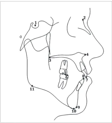
Figure 1 - Anatomical landmarks: 1) Sella; 2) Nasion; 3) Posterior nasal spine;
4) Anterior nasal spine; 5) U6 mesiobuccal cusp; 6) L6 mesiobuccal cusp;
7) U1 incisor tip; 8) L1 incisor tip; 9) Gnathion; 10) Menton; 11) Inferior gonion. crowding exceeding 6 mm either in the maxillary or
in the mandibular dental arch. Twenty-four sets of records were obtained from practice A, four sets of records were obtained from practice B, and two sets of records were obtained from practice C. These pa-tients were randomly selected between anterior open bite cases that were finished between 2011 and 2015.
The patient selection criteria were as follows: » Patients were all non-growing at the outset of treatment, determined via the cervical vertebral maturation technique.
» No vertical overlap between the upper and lower incisors, with edge-to-edge canines deemed acceptable.
» No extractions of permanent teeth were per-formed during treatment.
» No orthognathic surgery was performed as a part of treatment.
» The patients were treated exclusively with Invisalign and anteroposterior elastics during treat-ment, if necessary.
» Pre- and post-treatment lateral cephalograms were available for each patient.
Care was taken to ensure that all private practitio-ners had at least elite provider status, indicating that they treat, at minimum, up to one hundred Invisalign cases per year.
The overall goal of each treatment was to achieve overbite reduction in order to attain vertical overlap, or positive overbite, of the maxillary and mandibular incisors.
Data collection
Pre-treatment and post-treatment digital and ana-log lateral cephaana-lograms were collected, scanned, and traced for each patient digitally in the Dolphin Imag-ing 11.8 Premium software (Dolphin ImagImag-ing Sys-tems LLC, Chatsworth, CA, USA). Since the lateral cephalograms were obtained from distinct practices, the magnification rate was corrected using this soft-ware. Thirty-three hard tissue landmarks were iden-tified and traced, in addition to two reference land-marks on each radiograph (Fig 1). The mandibular and maxillary occlusal planes were manually traced and all occlusal plane measurements were manually measured. Six linear and six angular measurements were performed (Table 1, Figs 2 and 3).
Statistical methods
The data in this study was analyzed via IBM SPSS Statistics 23.0 statistical analysis software (SPSS Inc., Chicago, IL, USA). Descriptive statistics and paired
RESULTS
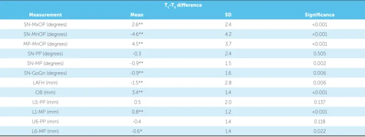
Average treatment time was 21 months (ranging from 11 to 34 months). Descriptive statistics (Ta-ble 2) and paired t-tests (Table 3) used to analyze the data revealed that nine of the twelve variables mea-sured were statistically significant in overall treatment change. Statistically significant (p < 0.01) changes were found in SN-MxOP, SN-MnOP, MP-MnOP, SN-MP, SN-GoGn, LAFH, overbite, and L1-MP.
Statistically significant (p < 0.05) changes were also observed in L6-MP. But SN-PP, U1-PP, and U6-PP did not undergo any statistically significant changes.
Chronbach’s alpha tests for intra-examiner re-liability was above 0.80 for all variables except for overbite (Cronbach’s alpha= 0.63). With regard to accuracy of measurements, overall, the original and repeated measurements were at an adequate level of reliability (Table 4).
Table 1 - Measurements definitions.
Measurement Deinition
SN-MxOP Angle formed by SN and the maxillary occlusal plane (plane drawn through the mesiobuccal cusp tip of the maxillary molar to the upper central incisor tip)
SN-MnOP Angle formed by SN and the mandibular occlusal plane (plane drawn through the mesiobuccal cusp tip of the mandibular molar to the lower central incisor tip)
MP-MnOP Angle formed by mandibular plane (inferior gonion to menton) and the mandibular occlusal plane
SN-PP Angle formed by SN to palatal plane (ANS to PNS)
SN-MP Angle formed by SN to mandibular plane (inferior gonion to menton)
SN-GoGn Angle formed by SN and the plane drawn through the points inferior gonion and gnathion
U1-PP The millimetric distance between U1 tip and the palatal plane (ANS to PNS)
L1-GoGn The millimetric distance between L1 tip and the plane drawn through inferior gonion-gnathion
U6-PP The millimetric distance between the mesiobuccal cusp tip of the maxillary molar and the palatal plane (ANS to PNS)
L6-GoGn The millimetric distance between the mesiobuccal cusp tip of the mandibular molar and the plane drawn through inferior gonion-gnathion
LAFH The millimetric distance between ANS and menton
OB The vertical millimetric distance from U1 tip to L1 tip
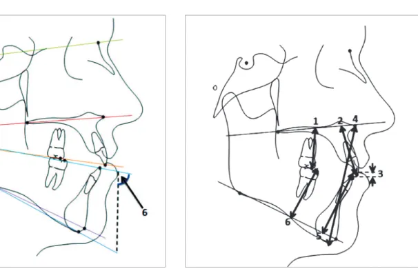
Figure 2 - Cephalometric angular measurements: 1) SN-GoGn; 2) SN-MP;
3) SN-MxOP; 4) SN-MnOP; 5) SN-PP; 6) MP-MnOP.
Figure 3 - Cephalometric linear measurements: 1) U6-PP; 2) U1-PP; 3) OB;
Table 2 - Descriptive statistics at T1 and T2.
Table 3 - Treatment changes.
Table 4 - Reliability statistics.
* denotes changes are significant at p < 0.05. ** denotes changes are significant at p < 0.01.
Measurement Pre-treatment (T1) Post-treatment (T2)
Mean SD Mean SD
SN-MxOP (degrees) 18.0 5.0 20.6 5.4
SN-MnOP (degrees) 20.7 5.5 16.2 5.6
MP-MnOP (degrees) 20.2 5.1 24.7 4.6
SN-PP (degrees) 7.8 4.2 7.5 4.7
SN-MP (degrees) 40.8 7.2 39.9 6.9
SN-GoGn (degrees) 37.6 7.1 36.7 6.9
LAFH (mm) 74.3 5.3 72.8 5.2
OB (mm) -1.8 1.2 1.5 0.9
U1-PP (mm) 30.7 2.8 31.2 2.6
L1-MP (mm) 38.3 2.8 39.1 3.1
U6-PP (mm) 25.4 2.2 25.0 2.3
L6-MP (mm) 31.3 2.5 30.7 2.4
T1-T2 diference
Measurement Mean SD Signiicance
SN-MxOP (degrees) 2.6** 2.4 <0.001
SN-MnOP (degrees) -4.6** 4.2 <0.001
MP-MnOP (degrees) 4.5** 3.7 <0.001
SN-PP (degrees) -0.3 2.4 0.505
SN-MP (degrees) -0.9** 1.5 0.002
SN-GoGn (degrees) -0.9** 1.6 0.006
LAFH (mm) -1.5** 2.8 0.006
OB (mm) 3.4** 1.4 <0.001
U1-PP (mm) 0.5 2.0 0.137
L1-MP (mm) 0.8** 1.2 <0.001
U6-PP (mm) -0.4 1.4 0.118
L6-MP (mm) -0.6* 1.4 0.022
Variable Cronbach’s alpha
SN-MxOP 0.958
SN-MnOP 0.864
MP-MnOP 0.965
SN-PP 0.950
SN-MP 0.983
SN-GoGn 0.987
LAFH 0.993
OB 0.629
U1-PP 0.837
L1-MP 0.923
U6-PP 0.946
L6-MP 0.825
DISCUSSION
incisal overlap, which is usually obtained at the ex-pense of adverse sequela. Fixed appliance therapy has a tendency to worsen the vertical dimension in open bite patients, who more often present as hyperdiver-gent, long-faced individuals.6,17,21,22
Non-surgical options for correction of anteri-or open bites in adults are limited. It is commonly thought that extractions enable a bite-closing effect by allowing protraction into the extraction space, thereby decreasing the palatomandibular wedge. The literature regarding extraction treatment does not support this idea. In actuality, protraction dur-ing space closure may cause occlusal movement of the posterior segments, which essentially cancels out the so-called “wedge effect”.23,24
Post-treatment increases in lower anterior facial height have been observed in non-extraction treat-ment as well.25 Anterior segments must be extruded via elastics, or posterior segments intruded, to achieve bite closure with fixed appliances. The multiloop edgewise archwire (MEAW) technique employs ante-rior elastics to achieve positive overbite, to correct the cant of the occlusal planes, and to address the mesial inclination of posterior teeth.26 Although the tech-nique has proven capable of attaining bite closure, it is mainly via anterior extrusion.27 Previous cephalo-metric evaluation of the technique revealed insignifi-cant changes in lower anterior face height and in the mandibular plane angle during treatment.27,28
Bite closure via extrusion of anterior teeth may not be indicated for all adult patients presenting with anterior open bites. Even more, extrusion of the max-illary incisors is deemed unstable.29 Some investiga-tors believe that maxillary incisor extrusion in adult patients may compromise the periodontal structures, lead to root resorption and ultimately jeopardize smile esthetics.10 Without the use of skeletal anchor-age devices, true molar intrusion is very difficult to be achieved in adult patients using fixed appliances.30
Angular measurement changes
In this study, the decision to split the occlusal plane angles was based on the report by Nahoum7 that it would be inaccurate to use the same plane in open and deep bite cases and; instead, he deined two separate planes: maxillary MxOP) and mandibular (SN--MnOP) occlusal planes. SN-MxOP showed a
statis-tically significant mean increase of 2.6o. Conversely, Sn-MnOP significantly decreased by a mean of 4.6o, indicating again that the bite was closed either by in-cisor extrusion and/or molar intrusion. MP-MnOP increased significantly by a mean of 4.5o; this finding indicates a decreased steepening of the mandibular occlusal plane due to bite closure. A statistically sig-nificant mean decrease of 0.9o in the SN-MP angle and mean decrease of 0.9o in the SN-GoGn angle was observed, which we can attribute to counterclock-wise rotation of the mandibular plane. For the sake of reliability, two mandibular planes were utilized: 1) one plane connecting inferior gonion to gnathion; 2) one plane connecting inferior gonion to menton. We would expect to see a higher mean value with SN-MP than we would with SN-GoGn, which was the case. However, the mean post-treatment decreas-es in both plandecreas-es were equivalent. This rotation was not substantial enough to decrease the MP-MnOP angle, as the treatment effects on the mandibular oc-clusal plane compensated the skeletal autorotation. SN-PP did not display any significant change, as we might expect in a non-growing patient.
Linear measurement changes
In this study, Invisalign successfully achieved an-terior open bite closure via positive incisal overlap without negatively impacting the vertical dimension. The changes observed in the vertical parameters dis-playing statistical significance are indicative of bite closure and decreases in the vertical dimension.
As previously noted, fixed appliance therapy has the potential to exert unwanted extrusive forces that may enhance the open bite and consequently worsen the vertical dimension. Additionally, anterior-pos-terior (AP) elastics used with fixed appliances tend to have extrusive effects that increase the vertical dimension. The majority of the present sample pre-sented with an Angle Class I malocclusion, with only six Class II patients. Klein31 reported control of the vertical, and even a decrease in the vertical dimen-sion (SN-MP), in his study examining Class II cor-rection with Invisalign and elastics. This finding was attributed to the constant presence of aligner materi-al. The improvements in the vertical dimension seen in this study are akin to those observed subsequent to molar intrusion in skeletal anchorage cases, i.e. reduction in occlusal plane angle, mandibular plane angle, and lower anterior facial height.10,30
It has been previously postulated that Invisalign ex-erts a bite-block efect during treatment.32 This func-tional appliance aims to control maxillary vertical skel-etal and dental growth by including an acrylic portion in the occlusal region, greater in size than the patient’s normal vertical dimension.29 Typically, the amount of acrylic in the first molar area of these bite blocks may range in thickness from 5 to 10 mm; this induces an artificial increase in the vertical dimension, thereby triggering a muscular response that creates a vertical intrusive force in the posterior segments, leading to counterclockwise rotation of the mandible.33
It is unlikely that Invisalign has the ability to ex-ert the same intrusive forces in adults as we observe in children with bite block therapy. The thickness of each Invisalign aligner is 0.030 inches34 (equivalent to 0.76 mm), which, when combined in both dental arches, may not have adequate thickness to consid-erably exceed the freeway space, enough to create a neuromuscular response. Additionally, functional contact of opposing teeth occurs approximately 18 minutes per day,29 which is not of long enough dura-tion to exert a significant intrusive force.35 Intrusive forces from the aligner must be requested by the
vider in the ClinCheck® software (Align Technology, Santa Clara, CA, USA) and programmed into the trays through the Invisalign technicians. The com-bined effect of the sequential progression of trays, with judicious selection and placement of attach-ments, ultimately dictates the amount of intrusion and tooth movement clinically observed. The advan-tage of Invisalign in treating open bite malocclusion stems mainly from its full occlusal coverage effect. While intruding dentition, adverse or unwanted re-ciprocal extrusive movements are less likely to occur because of the presence of the aligner.15,16
As adult open bite malocclusions are uncommon and their treatment with Invisalign is a relatively new approach, one limitation of this study was obtaining a large sample size. Additionally, the retrospective nature of this study did not enable control of all treatment vari-ables. Each case was treated with a diferent ClinCheck set-up that was contingent upon the patient’s speciic diagnosis as well as the orthodontic provider’s devised treatment plan. Depending on the treatment warranted, providers may have either primarily requested molar in-trusion, or anterior exin-trusion, or a combination of both. Future studies should incorporate a matched control group treated solely with ixed appliance therapy, for
further comparison of these modalities’ efects on the vertical dimension. Research focusing on the amount of molar intrusion that can be achieved with the appliance would be of great value. Vertical elastics and IPR may play a role also as for the results achieved in the patients. Lastly, prospective investigation lending insight into re-lapse of open bite cases treated with Invisalign is vastly important in order to appreciate the appliance’s capacity to preserve control of the vertical.
CONCLUSIONS
1) The Invisalign system is a therapeutic modality that can be effectively employed in non-extraction treatment of adult anterior mild open bites.
2) Bite closure was mainly achieved by a combination of counterclockwise rotation of the mandibular plane, lower molar intrusion and lower incisor extrusion.
Author contributions
1. Mizrahi E. A review of anterior open bite. Br J Orthod. 1978 Jan;5(1):21-7. 2. Janson G, Valarelli FP, Henriques JF, Freitas MR, Cançado RH. Stability of anterior
open bite nonextraction treatment in the permanent dentition. Am J Orthod Dentofacial Orthop. 2003 Sept;124(3):265-76; quiz 340.
3. Vig KW. Nasal obstruction and facial growth: the strength of evidence for clinical assumptions. Am J Orthod Dentofacial Orthop. 1998 June;113(6):603-11. 4. Cangialosi TJ. Skeletal morphologic features of anterior open bite. Am J Orthod.
1984 Jan;85(1):28-36.
5. Richardson A. A classiication of open bites. Eur J Orthod. 1981;3(4):289-96. 6. Almeida RR, Ursi WJ. Anterior open bite. Etiology and treatment. Oral Health.
1990 Jan;80(1):27-31.
7. Nahoum HI. Anterior open-bite: a cephalometric analysis and suggested treatment procedures. Am J Orthod. 1975 May;67(5):523-21.
8. Ngan P, Fields HW. Open bite: a review of etiology and management. Pediatr Dent. 1997 Mar-Apr;19(2):91-8.
9. Greenlee GM, Huang GJ, Chen SS, Chen J, Koepsell T, Hujoel P. Stability of treatment for anterior open-bite malocclusion: a meta-analysis. Am J Orthod Dentofacial Orthop. 2011 Feb;139(2):154-69.
10. Sherwood KH, Burch JG, Thompson WJ. Closing anterior open bites by intruding molars with titanium miniplate anchorage. Am J Orthod Dentofacial Orthop. 2002 Dec;122(6):593-600.
11. Frost DE, Fonseca RJ, Turvey TA, Hall DJ. Cephalometric diagnosis and surgical-orthodontic correction of apertognathia. Am J Orthod. 1980 Dec;78(6):657-69. 12. Subtelny JD, Sakuda M. Open-bite: diagnosis and treatment. Am J Orthod.
1964;50(5):337-58.
13. Hennessy J, Al-Awadhi EA. Clear aligners generations and orthodontic tooth movement. J Orthod. 2016 Mar;43(1):68-76.
14. Guarneri MP, Oliverio T, Silvestre I, Lombardo L, Siciliani G. Open bite treatment using clear aligners. Angle Orthod. 2013 Sept;83(5):913-9.
15. Harnick DJ. Using clear aligner therapy to correct malocclusion with crowding and an open bite. Gen Dent. 2012 May-June;60(3):218-23.
16. Schupp W, Haubrich J, Neumann I. Treatment of anterior open bite with the Invisalign system. J Clin Orthod. 2010 Aug;44(8):501-7.
17. Taibah SM, Feteih RM. Cephalometric features of anterior open bite. World J Orthod. 2007 Summer;8(2):145-52.
18. Sassouni V. A classiication of skeletal facial types. Am J Orthod. 1969 Feb;55(2):109-23.
19. Schudy FF. Vertical growth versus anteroposterior growth as related to function and treatment. Angle Orthod. 1964 Apr;34(2):75-93.
REFERENCES
20. Schudy FF. The control of vertical overbite in clinical orthodontics. Angle Orthod. 1968 Jan;38(1):19-39.
21. Subtelny JD. Oral respiration: facial maldevelopment and corrective dentofacial orthopedics. Angle Orthod. 1980 July;50(3):147-64.
22. Sassouni V, Nanda S. Analysis of dentofacial vertical proportions. Am J Orthod. 1964 Nov;50(11):801-23.
23. Zafarmand AH, Zafarmand MM. Premolar extraction in orthodontics: does it have any efect on patient’s facial height? J Int Soc Prev Community Dent. 2015 Jan-Feb;5(1):64-8.
24. Cusimano C, McLaughlin RP, Zernik JH. Efects of irst bicuspid extractions on facial height in high-angle cases. J Clin Orthod. 1993 Nov;27(11):594-8. 25. Chua AL, Lim JY, Lubit EC. The efects of extraction versus nonextraction
orthodontic treatment on the growth of the lower anterior face height. Am J Orthod Dentofacial Orthop. 1993 Oct;104(4):361-8.
26. Kim YH. Anterior openbite and its treatment with multiloop edgewise archwire. Angle Orthod. 1987 Oct;57(4):290-321.
27. Endo T, Kojima K, Kobayashi Y, Shimooka S. Cephalometric evaluation of anterior open-bite nonextraction treatment, using multiloop edgewise archwire therapy. Odontology. 2006 Sept;94(1):51-8.
28. Küçükkeleş N, Acar A, Demirkaya AA, Evrenol B, Enacar A. Cephalometric evaluation of open bite treatment with NiTi arch wires and anterior elastics. Am J Orthod Dentofacial Orthop. 1999 Nov;116(5):555-62.
29. Proit WR. Contemporary Orthodontics. Toronto: Elsevier; 2013. 30. Umemori M, Sugawara J, Mitani H, Nagasaka H, Kawamura H. Skeletal
anchorage system for open-bite correction. Am J Orthod Dentofacial Orthop. 1999 Feb;115(2):166-74.
31. Klein BM. A cephalometric study of adult mild class II nonextraction treatment with the invisalign system [master’s thesis]. Saint Louis (IL): Saint Louis University; 2013.
32. Boyd RL. Complex orthodontic treatment using a new protocol for the Invisalign appliance. J Clin Orthod. 2007 Sept;41(9):525-47; quiz 523.
33. Iscan HN, Sarisoy L. Comparison of the efects of passive posterior bite-blocks with diferent construction bites on the craniofacial and dentoalveolar structures. Am J Orthod Dentofacial Orthop. 1997 Aug;112(2):171-8.
34. Vlaskalic V, Boyd RL. Clinical evolution of the Invisalign appliance. J Calif Dent Assoc. 2002 Oct;30(10):769-76.