Orthodontic traction: possible effects on
maxillary canines and adjacent teeth
Part 2: External cervical resorption
due to canine traction
Alberto Consolaro*
Professionals who resist and restrict the indi-cation of orthodontic traction, especially canine traction, often justify their stance by citing the following reasons:
1) Lateral Root Resorption in lateral incisors and premolars.
2) External Cervical Resorption of canines due to canine traction.
3) Alveolodental ankylosis of the canine(s) involved in the process.
4) Calcific metamorphosis of the pulp and aseptic pulp necrosis.
These possible outcomes do not stem pri-marily and specifically from orthodontic trac-tion. They can be avoided if certain technical precautions are adopted, especially "the four cardinal points for the prevention of problems during orthodontic traction."2 To understand
what these technical precautions are and how they work preventively against the possible con-sequences of orthodontic traction a biological foundation is required. Providing such biologi-cal foundation is the goal of this series of studies on orthodontic traction, focusing particularly on maxillary canines.
The increasing use of imaging tests—such as computed tomography with its various slice planes, and the resulting reconstruction of 3D im-ages, viewable from virtually every angle—allows today's professionals to plan orthodontic traction of maxillary canines with greater accuracy and re-finement. This advance in obtaining image slices and 3D images allows surgeons to deal with ca-nines, their follicle, cervical region and adjacent teeth with the aid of detailed planning, which ul-timately reduces the risk of unintended outcomes. In other words, technological advances in imaging will make it possible for orthodontic traction to be accomplished more safely and accurately.
Cervical region of canine and dental follicle The radiolucent area around the crowns of un-erupted teeth is filled by the dental follicle, which is firmly adhered to the surface of the crown by the reduced epithelium of the enamel organ (Figs 1 and 2). This thin and fragile epithelial compo-nent is sustained and nourished by a thick layer of connective tissue with variable collagen density— sometimes loosely, sometimes fibrous and even hyalinized.1 The outer part of the follicle connects
A B C D
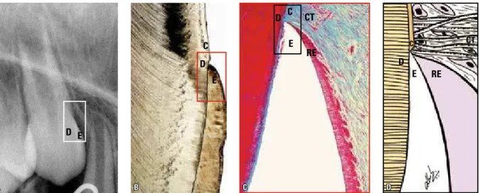
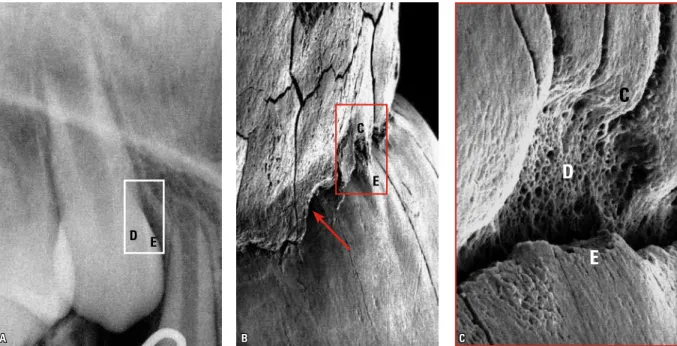
FIGURE 1 - The cervical region is a sensitive tooth structure due to the fragile junction between enamel and cementum (rectangles). In all human permanent and primary teeth, the circle formed by the cementoenamel junction line comprises exposed gaps or windows of dentin (D), which can only be observed mi-croscopically. In the dental follicle, the reduced epithelium (RE) of the enamel organ adheres to the enamel (E), while its connective tissue (CT) attaches itself to the root cementum (C) via collagen fibers. (B = section obtained by grinding and preserving the enamel; C = section obtained by demineralization, involving loss of the crystallized enamel structure and maintenance of its space).
it seamlessly with the surrounding bone (Fig 1). By surgically removing the follicle and detach-ing it from the surrounddetach-ing bone a tissue fragment is obtained which is organized in the form of a thin film and is therefore known as pericoronal membrane. This tissue fragment represented by the dental follicle, when observed in isolation, has the appearance of a sack, which contained the dental crown, and is thus also called pericoronal pouch. During removal of unerupted teeth, the follicle often remains adhered to the surrounding bone tissue or to the overlying soft tissue of the surgical flap. In surgical procedures, the follicle ad-heres to the enamel surface only occasionally.
After removing the dental follicle of un-erupted teeth, it becomes apparent that the fol-licle terminates in or attaches itself firmly to the cervical region of the tooth. The reduced epi-thelium of the enamel remains adhered to the cervical border of the enamel, while its connec-tive portion attaches itself to the cervical root cementum (Figs 1 and 2).
The cementoenamel junction lies between enamel and cementum. It is therefore reasonable to assert that the dental follicle in the cervical region overlies the line formed by the adjacent
relationship between enamel and cementum1,4,6,8
(Figs 1 and 2).
The cementoenamel junction exhibits dentin windows or gaps along the cervical circumference of all human teeth, from which dentinal tubules emerge5,6,8 (Fig 2), exposing inorganic and organic
dentinal components, particularly their proteins.
Surgical exposure and manipulation of the cementoenamel junction may induce Exter-nal Cervical Resorption
Some dentin proteins are deposited by odon-toblasts during tooth formation and during in-trauterine life, without ever having been directly exposed to immune system components. In other words, the immune system cannot recognize some dentin proteins as normal or as belonging to the body because during immunological memory
D E
E
E
E RE
CT
CT
RE D
D
D C
C
A B C D
C
C
D
E
E
E
development these proteins were not exhibited, contacted or exposed.
By depositing the dentin the odontoblasts cover it internally thus preventing any contact with other cells and body components. Dentinal proteins are therefore not recognized or cataloged during intrauterine life,2 unlike what typically
oc-curs with almost all other proteins in the body. If proteins not cataloged or not contacted by the body during intrauterine life later enter into close contact with the tissues they will be seen as foreign and approached as antigens. Once rec-ognized and located, antigens, or foreign proteins must be eliminated from the body and to this end cells perform phagocytosis and extracellular digestion, and make use of enzymes, toxins, re-sorption, etc. This occurs with bacteria and some transplants, for example.
In some tissues and organs of the body, as is the case with dentin, some proteins are isolat-ed, not cataloged or recognized by the immune
system, becoming known as sequestered anti-gens. Other examples are the proteins of the thyroid and sperm. If some time during their life these proteins or sequestered antigens are exposed to connective tissues due to external or internal agents, the cells and other compo-nents of the immune system will consider them foreign, or as antigens, and will tend to elimi-nate them. In the case of dentin, elimination will take place by resorption of the mineralized portion by isolating the foreign protein and dis-solving it. In this case, tooth resorption occurs.
A1
C1
B D
E G
H F I
C2 C3
A2 A3 A4 A5
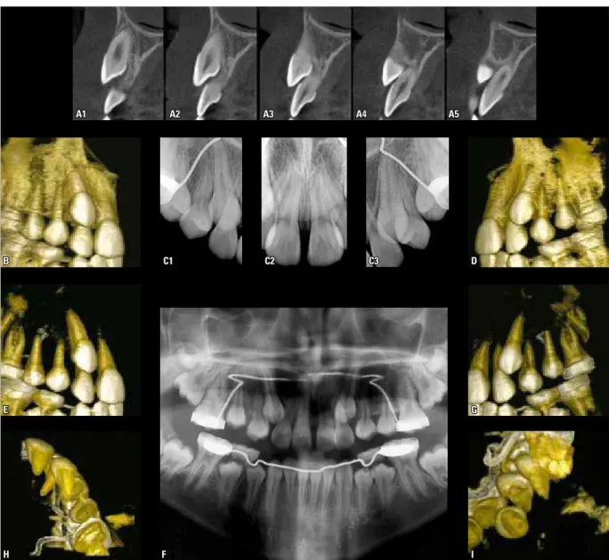
FIGURE 3 - Imaging aspects of unerupted maxillary canines, their position and relationship with adjacent teeth, as well as their spatial individualization providing a view of the cervical region from various observation angles.
During dental trauma as well as after inter-nal tooth bleaching,4 this type of resorption can
also occur because these situations also promote exposure of dentinal gaps to the gingival connec-tive tissue.
Procedure for traction of unerupted canines and External Cervical Resorption
If inadequately performed, surgical proce-dures for placing an orthodontic traction device in
unerupted maxillary canines may involve:
unerupted maxillary canines consisted in binding the dental cervix with wire. A twisted wire was used and a loop was placed around the tooth in the cer-vical region of the upper canine with which orth-odontic traction was accomplished. The force and displacement of the orthodontic wire in the neck of the tooth exposed the dentin gaps in the cementoe-namel junction, adding to the constant inflamma-tion that resulted from the continuous trauma.
Installing traction device on the crown and recovering of the surgical cavity: What now? The follicular tissues regenerate and repair themselves!
Epithelial cells undergo a constant process of proliferation and synthesis, and are therefore ap-propriately called labile cells.2 Given this
char-acteristic, the epithelial tissue features great re-generative capacity. When wounds and mucous membranes appear immediately after trauma or surgery, marginal epithelial cells expose all their surface receptors to large amounts of mediators released by the cells themselves, especially EGF (Epidermal or Epithelial Growth Factor), which induces them to proliferate and organize them-selves in layers that cover the altered surface.2
Typically, the closure of a wound by epithe-lial proliferation arising out of the surgical mar-gins appears in the shape of an iris or diaphragm, and gradually—within a few hours—decreases the diameter of the area of exposed underlying tissue.2 Below the epithelium, connective tissue
adjacent to the injured area produce granulation tissue which evolves within a few days, giving rise to newly-formed connective tissue that repopu-lates the region. At a distance, bone can once again form from that same granulation tissue.
When a window is opened into the tissues of the dental follicle in order to set up an orthodon-tic traction device, by analogy, one can envisage a wound with injured epithelium and exposed connective tissue turned towards the enamel. The reduced epithelium of the enamel organ tends to weeks or months. This can happen during
orth-odontic traction or after the tooth has reached the occlusal plane.
In many cases, detection tends to occur belat-edly. External Cervical Resorption is character-ized as a slow, painless, insidious process that does not compromise pulp tissues. In more advanced cases, it can lead to gingival inflammation and pulpitides secondary to bacterial contamination.
One way to prevent this traction effect of un-erupted maxillary canines is to allow at least 2 mm of soft tissue from the dental follicle to re-main adhered to the cervical region. It is essential to refrain from manipulating the cementoenamel junction, and to do so only if strictly necessary.
2. Applying excessively or extensively acids and other products to facilitate the bonding of devices necessary for attaching the traction wires. Excessive administration of these products may cause them to seep through to the cervical region where the dental follicle attaches itself to the ce-mentoenamel junction, affecting the cells and tis-sues chemically and thereby exposing, and even increasing the number of, dentin gaps and freeing the sequestered antigens into the adjacent con-nective tissue after closing the surgical wound. This situation may explain some cases of external resorption in maxillary canines subjected to orth-odontic traction.
3. Anchoring or fixing surgical instruments in the cervical region of unerupted maxillary ca-nines. This anchoring generally aims to achieve luxation or subluxation of the unerupted max-illary canine, as indicated in some procedures where alveolodental ankylosis is suspected. Sub-sequently, orthodontic traction is applied. The le-vers, chisels and tips of surgical instruments such as forceps can mechanically damage the follicle and periodontal tissues in the cervical region, and expose, or even increase the exposure of dentin in the cementoenamel junction, from where Exter-nal Cervical Resorption originates.
A
F G
I J
H B
C D1
E1 E2 E3 E4 E5
D2
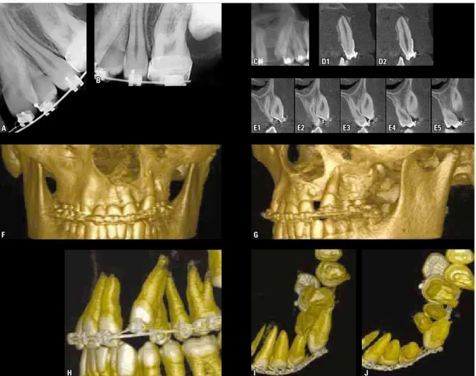
FIGURE 4 - Imaging aspects of unerupted canines undergoing orthodontic traction in cleft patients. It is worthy of note how one can view their position and relationship with adjacent teeth, as well as their spatial individualization from various observation angles.
proliferate rapidly, covering the enamel and trac-tion devices over a period of hours or days. The underlying connective tissue starts forming again from the granulation tissue that grows temporar-ily in the area. Thus, the enamel is not exposed to the connective tissue until the tooth reaches the oral environment.
Aren't the follicular tissues torn during orth-odontic traction?
During the extrusive tooth movement in-duced by traction of unerupted maxillary canines there should be no rupture of periodontal or den-tal follicle fibers, nor any tearing of their vessels
and nerves. If this happens, such dental trauma is named surgical or orthodontically induced avul-sion—often mistakenly called rapid traction or ex-trusion. Biologically, this can be defined as dental injury, which may result in conditions such as al-veolodental ankylosis and replacement resorption. Induced tooth movement consists of forces that are slowly applied and dissipated, consistent with normal biological tissue. Connective and ep-ithelial tissues are constantly remodeling, which gives them remarkable ability to adapt to new functional demands.
bone tissues remodel and adapt naturally to the presence of the crown and traction devices with-out rupturing or offering any physical resistance. No tissue laceration occurs due to the displace-ment of a traction device along with a tooth. Ves-sels and nerves do not rupture and the tissues are not "torn". Right angles, walls and corners of metal traction devices will not cause any trauma to adja-cent follicle tissues. Should tissue laceration occur, extrusion is not being caused by an orthodontic tooth movement per se, but rather by rapid tooth displacement, of a surgical or traumatic nature.
The junctional epithelium also forms during orthodontic traction
Given the proximity between follicle and oral mucosa, the reduced epithelium of the enamel or-gan will fuse together with the oral mucosa. In the central region of this extensive area of epithelial fusion necrosis will occur due to lack of nourish-ment because the source of such nourishnourish-ment, the connective tissue, is now distant. The incisal tip of the canine will appear at this site. The two epithelia now fused around the crown will give rise to the primary junctional epithelium to prevent the inter-nal environment—represented by the connective tissue—from being exposed to a highly contami-nated oral environment. This process also occurs in teeth that erupt in the oral environment with the aid of orthodontic treatment.
CT and 3D images as resources for diagnosing and assessing External Cervical Resorption
Compared with CT images reconstructed in 3D, radiographs provide a visual perception of im-ages at a more advanced stage in the process of loss of mineral components in bone tissue and teeth (Figs 3 and 4). For example, in an acute dentoal-veolar abscess, radiographic images have virtually lost their key features since it is generally accepted that in order to generate images, bone resorption in a particular location should be at least 10 days old.
In assessing the damage caused by root
re-sorption in maxillary lateral incisors due to the proximity of unerupted canines, it seems ap-propriate to highlight some of the evidence. Associated root resorption was found in the periapical radiographs of 3,000 patients be-tween 10 and 15 years of age. Moreover, 12.5% of their lateral incisors were located near ca-nines that had remained unerupted for longer than normal.7 The same cases were evaluated
using tomographic slices and reconstructions, and disclosed 25% impairment. Computed To-mography (CT) is the best method to accurate-ly assess the damage caused by canine traction to the roots of upper lateral incisors.
By extrapolation, CT and 3D images can promote a much earlier diagnosis of External Cervical Resorption in teeth subjected to orth-odontic traction. In practice, before starting the procedures and 6 months to 1 year after a given tooth has been allocated in the dental arch, CT and 3D images can reveal early cases of cervical resorption.
Early diagnosis of external cervical resorption determines what sort of treatment should be ad-ministered: By raising a gingival flap one can have access to areas of resorption and fill them with functional, biological and aesthetically pleasing materials, with excellent prognosis. The use of CT scans and 3D images before starting orthodontic traction might help in planning such traction, in addition to averting the pre-existence of processes like external cervical resorption, alveolodental an-kylosis and replacement resorption of the teeth subjected to traction (Figs 3 and 4).
1. Consolaro A. Caracterização microscópica de folículos pericoronários de dentes não irrompidos e parcialmente irrompidos. Sua relação com a idade. [dissertação]. Bauru (SP): Universidade de São Paulo; 1987.
2. Consolaro A. O tracionamento ortodôntico representa um movimento dentário induzido! Os 4 pontos cardeais da prevenção de problemas durante o tracionamento ortodôntico. Rev Clín Ortod Dental Press. 2010 ago-set; 9(4):105-10.
3. Consolaro A. Inlamação e reparo. Maringá: Dental Press;
2009.
4. Esberard R, Esberard RR, Esberard RM, Consolaro A, Pameijer CH. Effect of bleaching on the cemento-enamel junction. Am
J Dent. 2007 Aug;20(4):245-9.
REFERENCES
Contact address
Alberto Consolaro
E-mail: [email protected]
of CT scans and 3D images may allow a diagnosis of alveolodental ankylosis to be reached at a much earlier stage, when the root surface is still relatively preserved.
Final considerations
One of the possible consequences of maxillary unerupted canine traction is external cervical re-sorption. In planning and implementing the orth-odontic traction of unerupted maxillary canines, one is advised to:
a) Consider the fragile structure of the cemen-toenamel junction with its dentin "gaps" present in all teeth, including deciduous.
b) Avoid unnecessary surgical instrumental manipulation of the cervical region.
c) Do not spill or leak chemicals such as ac-ids, for example, used for bonding orthodontic traction devices.
When performing orthodontic traction of unerupted maxillary canines, a few hours and days after surgery, the epithelial, fibrous con-nective and bone tissues regenerate and repair themselves, in that order. Normal relationship is thus restored with epithelial covering of the enamel and metal devices, reconstruction of fibrous connective tissue and new peripheral bone formation. As the tooth moves in the oc-clusal direction, pericoronal tissues are not lac-erated or torn. Normal tissue remodeling fulfills functional demands and gradually adapts to this dental extrusion movement.
5. Francischone LA, Consolaro A. Clareação dentária externa:
importância e tipos de proteção da junção amelocementária.
Rev Clín Ortod Dental Press. 2005 out-nov;4(5):88-98.
6. Francischone LA, Consolaro A. Morphology of the
cementoenamel junction of primary teeth. J Dent Child. 2008
Sep-Dec;75(3):252-9.
7. Otto RL. Early and unusual incisor resorption due to impacted maxillary canines. Am J Orthod Dentofacial Orthop. 2003 Oct;124(4):446-9.
8. Neuvald L, Consolaro A. Cementoenamel junction: