CLINICAL SCIENCE
I Glaucoma Department, Zhongshan Ophthalmic Centre, State Key
Labora-tory of Ophthalmology, Sun Yat-sen University – Guangzhou, China.
II Sun Yat-sen University Tungwah Hospital – Dongguan, China.
Email: [email protected] Tel: 86-20-87331374
Received for publication on January 19, 2009 Accepted for publication on March 23, 2009
LONG-TERM SURGICAL OUTCOMES OF PRIMARY
CONGENITAL GLAUCOMA IN CHINA
Xiulan Zhang,I Shaolin Du,I,II Qian Fan,I Shouxiong Peng,I Minbin Yu,I Jian GeI
doi: 10.1590/S1807-59322009000600009
Zhang X, Du S, Fan Q, Peng S, Yu M, Ge J. Long-term surgical outcomes of primary congenital glaucoma in China. Clinics. 2009;64(6):543-51.
OBJECTIVE: To evaluate the long-term outcomes of three surgical procedures for the treatment of primary congenital glaucoma (PCG).
INTRODUCTION: PCG is one of the main causes of blindness in children. There is a paucity of contemporary data on PCG in China.
METHODS: A retrospective study of 48 patients (81 eyes) with PCG who underwent primary trabeculectomy, trabeculotomy, or combined trabeculotomy and trabeculectomy (CTT).
RESULTS: All patients were less than 4 years (yrs) of age, with a mean age of 2.08 ± 1.23 yrs. The mean duration of follow-up was 5.49 ± 3.09 yrs. The difference in success rates among the three surgical procedures at 1, 3, 6 and 9 yrs was not statistically significant (p = 0.492). However, in patients with over 4 yrs of follow-up, Kaplan-Meier survival analysis revealed that the success rates of trabeculectomy and CTT declined more slowly than that of trabeculotomy. Among the patients, 66.22% acquired good vision (VA ≥ 0.4), 17.57% acquired fair vision (VA = 0.1 - 0.3), and 16.22% acquired poor vision (VA < 0.1). The patients with good vision were mostly in the successful surgery group. Myopia was more prevalent postoperatively (p = 0.009). Reductions in the cup-disc ratio and corneal diameter were only seen in the successful surgery group (p = 0.000). In addition, the successful surgery group contained more patients that complied with a regular follow-up routine (p = 0.002).
DISCUSSION: Our cases were all primary surgeries. Primary trabeculectomy was performed in many cases because no treatment was sought until an advanced stage of disease had been reached.
CONCLUSIONS: In contrast to most reports, in the present study, trabeculectomy and CTT achieved higher long-term success rates than trabeculotomy. The patients with successful surgical results had better vision. Compliance with a routine of regular follow-up may increase the chances of a successful surgical outcome.
KEYWORDS: Primary congenital glaucoma (PCG); Trabeculectomy; Trabeculotomy; Combined trabeculotomy and trabeculec-tomy (CTT); China.
INTRODUCTION
Congenital glaucoma is one of main causes of blindness in children. In 1982, Mao et al.1 reported that congenital
glaucoma represented 1.3% of all congenital ocular disorders in China. In recent yrs, this number has increased to
5.1%.2 In a study from Brazil that evaluated 3,210 visually
impaired children, congenital glaucoma was responsible for 10.8% of the visual impairments.3 The incidence of
congenital glaucoma varied between counties, ranging from 1/10,000-1/30,200.4,5 Since at least 50% of eyes presenting
with primary congenital glaucoma (PCG) at birth will become legally blind, patients with PCG require not only prompt treatment but also follow-up examinations and care throughout their lives.
the predominant option for the initial treatment of PCG. Trabeculectomy, trabeculotomy, and combined trabeculotomy and trabeculectomy (CTT) are the three major surgical options for congenital glaucoma; goniotomy,deep sclerectomy, and viscotrabeculotomy, as well as glaucoma drainage tube shunts have also been applied in suitable cases.9-12 Trabeculotomy and goniotomy are considered to be
the procedures of first choice, and if they are not adequately controlled by IOP, trabeculectomy is usually the next course of action. Many studies have assessed the short and/or long-term visual outcomes associated with the various surgical options, as well as indications such as the type and severity of glaucoma.5-35 Surgical treatments can usually achieve safe
and effective results with congenital glaucoma if performed promptly and appropriately.
However, there is a paucity of contemporary clinical data regarding this condition in China.16, 22 Problems in
the management of congenital glaucoma in China include delayed acquisition of treatment, low surgical success rates, and poor follow-up. Many patients do not seek treatment until their condition is severe, and they thus face reduced surgical options. Additionally, long-term follow-up of childhood glaucoma is a long-lasting and difficult task. This study retrospectively evaluated the long-term effects of three surgical treatments exclusively in PCG patients who received management and follow-up at the Glaucoma Department, Zhongshan Ophthalmic Center, Guangzhou, China over an 11-year period.
METHODS
Subjects
The case files of 48 patients (81 eyes) who underwent primary surgical management during the 11 yrs from September 1996 to September 2007 in the Glaucoma Department, Zhongshan Ophthalmic Center of Sun Yat-sen University were retrospectively investigated. Thirty-two patients (56 eyes) were male, and 16 (25 eyes) were female. All patients were less than 4 yrs of age, with a mean age of 2.08 ± 1.23 yrs, when accepted for primary surgery, while the mean age at the last visit was 7.72 ± 3.38 yrs. The mean follow-up duration was 5.76 ± 3.19 yrs (range: 1-11 yrs), with 38 patients (79.17%) having had a minimum follow-up of 3 yrs, 31 (64.58%) with four 4 yrs, and 7 (14.58%) with 11 yrs.
In the present study, we only compared those patients who underwent one of the primary surgeries. There were 33 eyes that underwent trabeculectomy, 23 that underwent trabeculotomy, and 25 that underwent CTT. Eyes with previously failed glaucoma surgery, other congenital
ocular abnormalities, or less than 1 year of follow-up were not included in the study. The study was approved by the Institution Ethics Committee.
All surgeries were performed by glaucoma professionals using standard techniques: A) Trabeculectomy. A limbus-based conjunctival flap was dissected; a 4mm×3mm scleral flap was made; Mitomycin C (MMC) was applied with copious irrigation using a balanced salt solution (BBS, 150 - 200ml). MMC was applied at a concentration of 0.25 mg/ml - 0.4 mg/ml, and sustained for 2-5 min. An anterior paracentesis was made before the deep scleral flap was dissected, and then a 1.5 x 2mm deep scleral flap was cut, followed by an iridectomy. The scleral flap was sutured with #10-nylon. Two releasable sutures were made if necessary. The anterior chamber was reformed with BBS through the anterior paracentesis incision B) Trabeculotomy. A limbus-based or fornix-limbus-based conjunctival flap was dissected; a 3/4 thickness scleral flap 4mm × 3mm in size was made; a 2mm incision was cut along the bottom of the scleral flap and slowly deepened until the outer wall of Schlemm’s canal was opened and a seeping aqueous humor or pink liquid was seen; the outer wall incision was then extended by 1mm; the dissecting device was then rotated into the anterior chamber, and the inner wall of Schlemm’s canal and the trabecular meshwork were dissected 120° in both directions; the scleral flap and conjunctival flaps were then sutured. C) CTT: Trabeculotomy was first performed, followed by trabeculectomy. Mitomycin C was applied as above.
Surgical options are selected according to practice guidelines that have been used in China for decades: Trabeculotomy is the first choice for most PCG cases. When patient age exceeds 3 yrs or the corneal diameter exceeds 14 mm or other ocular abnormalities exist, then trabeculectomy or CTT is recommended. In these cases, if the cornea is clear and edema is mild, then CTT is preferred; if the cornea is cloudy or the edema is severe, then trabeculectomy alone is performed.
Evaluation items
evaluated and averaged using the LogMAR-value.34 In
patients who were able to cooperate with a VA test, VA was first assessed with the Tumbling E test (recorded as a decimal number, e.g., 0.1, 0.2… 1.5) and then transformed into LogMAR VA. Postoperative vision was assessed by the method used by Mendicino et al.25 A VA ≥ 0.4 was defined
as good, between 0.1 and 0.3 fair, and < 0.1 poor. Refractive error was measured by retinoscopy or retinoscopy combined with autorefractometry in cycloplegic situations.
Patient compliance was evaluated using the following definitions: (a) regular follow-up, the patient visited regularly and followed the physician’s advice; (b) irregular follow-up, the patient visited only when symptoms occurred and/or did not follow the physician’s advice.
Definition of surgical success
Surgical success was defined as a postoperative IOP ≤ 21 mmHg with or without medication, with no requirement for additional glaucoma surgery and no evidence of progression or enlargement of the C/D ratio or corneal diameter.10,16,30
These criteria were used to divide patients into two groups: the successful surgery group and the failed surgery group.
Statistical analyses
Data were analyzed with SPSS 13.0 statistical software. Pre- and postoperative VA, corneal diameter, IOP, C/D ratio, and refractive error were assessed with paired t-tests. The cumulative success rate was evaluated by Kaplan-Meier survival analysis and compared using the Log-Rank test. The difference between pre- and post-operative vision distribution was analyzed by the Chi-square test. Pre- and postoperative refractive status was also compared by the Chi-square test. The differences in refractive errors within individual groups were evaluated by analysis of variance (ANOVA). Correlations between surgical outcome, vision, refractive status, and compliance were assessed by correlation analysis. A P-value < 0.05 was considered as statistically significant.
RESULTS
Success rates
Kaplan-Meier survival analysis showed that success rates for all eyes at 1, 3, 6 and 9 yrs after surgery were 92.59%, 77.77%, 58.54%, and 53.29%, respectively.
With regard to individual treatment groups, the success rates at 1, 3, 6 and 9 yrs after trabeculectomy were 93.94%, 66.68%, 53.88%, and 53.88%; after trabeculotomy, they were 91.30%, 86.96%, 60.73%, and 37.70%; and after
CTT, they were 92.00%, 78.00%, 62.40%, and 62.40%, respectively. The Log-Rank test revealed no statistically significant difference among the three treatment groups (χ2
= 1.420, p= 0.492).
Success rates in those patients with over 4 yrs of follow-up were analyzed separately. Fifty eyes of 31 patients were followed-up for over 4 yrs, among which 18 eyes underwent trabeculectomy, 18 eyes underwent trabeculotomy, and 14 eyes underwent CTT. Evaluation by the Log-Rank test revealed that the difference between success rates among the three groups was statistically significant (χ2= 7.651,
p= 0.022). As evident from the curve, the success rates of CTT patients declined most slowly, followed by the trabeculectomy and trabeculotomy groups, which displayed the fastest decrease. Thus, trabeculectomy and CTT achieved higher success rates than did trabeculotomy (Figure 1).
IOP, corneal diameter, and C/D ratio
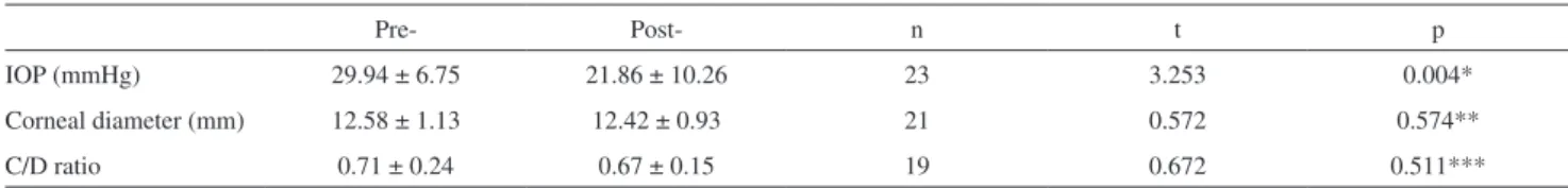
The mean IOP in all patients declined dramatically after the procedure (31.70 ± 9.16 mmHg preoperatively and 19.22 ± 8.67 mmHg postoperatively at the final visit; n = 81, t = 9.588, p = 0.000), but the corneal diameter (n = 78, t = 1.138, p = 0.259) and C/D ratio (n = 74, t = 0.341, p = 0.734) did not change significantly postoperatively. No effect on corneal diameter or C/D ratio was observed within the individual treatment groups, although IOP did decrease significantly in each of them (Tables 1, 2 and 3).
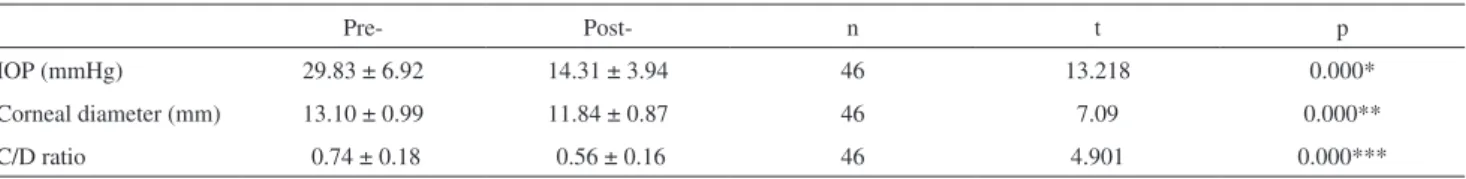
However, when the successful surgery group (46 eyes) was considered in isolation, a significant effect of the procedure on corneal diameter and C/D ratio was observed (Table 4). In this group, 25 eyes presented a reduction in corneal diameter, and 29 eyes presented a C/D ratio reversal. The mean corneal diameter of the 25 eyes was 13.10 ± 0.99 mm preoperatively and 11.84 ± 0.87 mm postoperatively (t = 7.09, p = 0.000). The mean age of the patients with a reduction in corneal diameter was 23 months (range: 12 - 36 months, median: 23 months). The mean C/D ratio of the 29 eyes was 0.74 ± 0.18 preoperatively and 0.56 ± 0.16 postoperatively (t = 4.901, p = 0.000). The mean age of the patients with a reversal in the C/D ratio was 12 months (range: 3 - 24 months, median: 12 months).
Evaluation of visual function
1. VA
Figure 1 - Kaplan–Meier survival curve (for eyes with over 4 yrs of follow-up). The cumulative success rates of three surgical techniques for the eyes of PCG patients with over 4 yrs of follow-up were evaluated by Kaplan-Meier survival analysis. The success rates declined over time, with that of CTT declining the most slowly, followed by trabeculectomy, and trabeculotomy, which demonstrated the fastest decrease. The differences between them were statistically significant according to the Log-Rank test (χ2
= 7.651, p = 0.022). This indicates that long-term follow-up (≥ 4 yrs) was a superior metric for surgical outcome. Compared with trabeculotomy, trabeculectomy and CTT achieved better long-term surgical outcomes. PCG, primary congenital glaucoma; CTT, combined trabeculotomy and trabeculectomy.
Table 1 - Pre- and postoperative IOP, corneal diameter, and C/D ratio in the trabeculectomy group (–x ±s)
Pre- Post- n t p
IOP (mmHg) 32.08 ± 10.82 19.25 ± 7.59 33 5.783 0.000*
Corneal diameter (mm) 12.64 ± 1.10 12.62 ± 1.29 32 0.087 0.931**
C/D ratio 0.62 ± 0.19 0.65 ± 0.23 31 -0.814 0.422***
* statistically significant difference (p< 0.05); **,*** no statistically significant difference (p> 0.05); IOP, intraocular pressure; C/D ratio, cup-disc ratio.
Table 2 - Pre- and postoperative IOP, corneal diameter, and C/D ratio in the trabeculotomy group (–x ±s)
Pre- Post- n t p
IOP (mmHg) 29.94 ± 6.75 21.86 ± 10.26 23 3.253 0.004*
Corneal diameter (mm) 12.58 ± 1.13 12.42 ± 0.93 21 0.572 0.574**
C/D ratio 0.71 ± 0.24 0.67 ± 0.15 19 0.672 0.511***
* statistically significant difference (p< 0.05); **,*** no statistically significant difference (p> 0.05); IOP, intraocular pressure; C/D ratio, cup-disc ratio.
Table 3 - Pre- and postoperative IOP, corneal diameter, and C/D ratio in the CTT group (–x ±s)
Pre- Post- n t p
IOP(mmHg) 32.74 ± 8.71 16.75 ± 8.00 25 8.836 0.000*
Corneal diameter (mm) 12.66 ± 1.08 12.40 ± 1.00 25 1.520 0.142**
C/D ratio 0.66 ± 0.22 0.62 ± 0.22 24 0.890 0.383***
74 eyes that were tested at their last visit was 0.29 ± 0.57. There was no statistically significant difference between these groups when tested by the paired t test (t = 1.849, p = 0.071).
To compare the VA of patients pre- and postoperatively, postoperative changes in the distribution of LogMAR VA scores were evaluated using the method described by Mendicino et al.25 (Table 5). This analysis revealed a
statistically significant effect of surgery on VA (χ2= 11.340,
p= 0.010), with 66.22% postoperative patients presenting good vision, 17.57% fair, and 16.22% poor.
The correlation between surgery outcome and postoperative LogMAR VA was analyzed by the Chi-square test, revealing a significant difference between the two outcome groups (Table 6, χ2= 7.219, p= 0.027). In the
successful surgical group, 67.3% of the eyes presented good vision postoperatively, while 75.0% of the eyes in the failed surgical group presented poor vision postoperatively.
2. Refractive Error
Refractive error was measured before surgery in 53 eyes, and the mean value was 0.39 ± 3.96 D. According to the classification of Alsheikheh et al.,26 53 eyes were found
to be emmetropic (0 ± 1 D, 17 eyes, 32.08%), myopic (< -1 D, 11 eyes, 20.75%), or hyperopic (> +1 D, 25 eyes, 47.17%). During the follow-up, 76 eyes were measured, and the mean refractive error was -3.05 ± 4.67 D, including 20
emmetropic (26.32%), 52 myopic (68.42%), and 4 hyperopic eyes (5.26%). The difference between pre- and postoperative refractive status was statistically significant (t = 4.380, p = 0.000). This analysis indicated that hyperopic eyes were predominant before the surgery while myopic ones were more prevalent after the surgery (χ2= 9.201, p= 0.009).
Pre- and postoperative differences in refractive error within individual treatment groups were also evaluated by analysis of variance (ANOVA). This analysis revealed a significant effect of surgery only in the trabeculotomy and CTT groups (t= 2.402, p= 0.032), and also that postoperative myopia was more prevalent in the CTT group.
COMPLICATIONS
The complications of trabeculectomy and CTT were similar. A shallow anterior chamber was observed in 2 eyes (6.06%) in the trabeculectomy group and in 1 (4.00%) in the CTT group; hypotony (< 5 mmHg) was seen in 4 eyes (12.12%) in the trabeculectomy group and in 2 (8.00%) in the CTT group; thin-wall blebs were seen in 2 eyes (6.06%) in the trabeculectomy group and in 1 (4.00%) in the CTT group; scarring filtering blebs occurred in 9 eyes (27.27%) in the trabeculectomy group and in 3 (12.00%) in the CTT group. No malignant glaucoma was detected in either group. Hyphema occurred in 2 eyes (8.69%) in the trabeculotomy group. There was no endophthalmitis in any of the eyes assessed.
Table 4 - Pre- and postoperative IOP, corneal diameter, and C/D ratio in the successful surgical group (–x ±s)
Pre- Post- n t p
IOP (mmHg) 29.83 ± 6.92 14.31 ± 3.94 46 13.218 0.000*
Corneal diameter (mm) 13.10 ± 0.99 11.84 ± 0.87 46 7.09 0.000**
C/D ratio 0.74 ± 0.18 0.56 ± 0.16 46 4.901 0.000***
*,**,*** statistically significant difference (p< 0.05); IOP, intraocular pressure; C/D ratio, cup-disc ratio.
Table 5 - Pre- and postoperative VA distributions
Good (≥ 0.4) Fair (0.1- 0.3) Poor (< 0.1) n χ2 p
Preoperative VA 9 (42.86%) 6 (28.57%) 6 (28.57%) 21 11.340 0.010*
Postoperative VA 49 (66.22%) 13 (17.57%) 12 (16.22%) 74
* statistically significant difference (p< 0.05); VA, visual acuity.
Table 6 - Correlation between surgical outcome and the postoperative VA distribution
Postoperative vision distribution Successful surgery group Failure surgery group χ2 p
Good 33 (67.3%) 16 (32.7%) 7.219 0.027*
Fair 7 (53.8%) 6 (46.2%)
Poor 3 (25.0%) 9 (75.0%)
Correlation between patient compliance and postopera-tive surgical outcomes
The correlation between patient compliance and surgical outcome is presented in Table 7. Patients in the successful surgery group were more likely to follow-up, while patients in the failed surgery group were less likely to follow-up. The Chi-square test demonstrated that this difference was statistically significant (χ2 = 9.311, p = 0.002).
DISCUSSION
The surgical treatment of PCG in China is consistent with that of most other countries. Trabeculotomy and goniotomy are the first choices for the majority of PCG cases.8-10,15,16,24,25 Trabeculectomy with or without
anti-metabolites is usually chosen for those with a previous failed surgery or those lacking a surgical history but with a corneal diameter >14 mm or more than 3 yrs of age.5, 19, 21, 35
In this study, 33 patients underwent primary trabeculectomy. The reason so many patients underwent this procedure is that most PCG cases in China are discovered late, when the condition is severe. These patients lose the opportunity to undergo the first-choice procedures. CTT offers a viable surgical option for PCG, and it has been widely performed in recent yrs, with results that are sufficiently safe, effective, and predictable for it to be considered as the first choice for corneal edema and for advanced cases.6,12,14,17,18,24,28,35
Trabeculotomy, trabeculectomy, and CTT are the most frequent surgical treatments for PCG, and their surgical outcomes have been intensively studied. A German study (2000) reported trabeculotomy success rates of 92.6% after 1 year of follow-up, 83.3% after 2 yrs, 66.7% after 3 yrs, and 50% after 4 yrs.15 In Turkey in 2007, Yalvac et al.8
reported that the success rates of trabeculotomy after 12, 24, and 36 months were 92%, 82%, and 74%, respectively. In 2008, a study in Brazil reported trabeculectomy success rates at 24, 36, 48, and 60 months of 90.2%, 78.7%, 60.7%, and 50.8%, respectively.5 In the U.S., Mendicino et al.25
compared 360° trabeculotomy and goniotomy and concluded that 360° trabeculotomy is a highly effective procedure that results in excellent pressure control and better vision than is reported in the literature for standard angle procedures.25, 31 In addition, Catrinu et al.23 compared trabeculotomy and
trabeculectomy for the treatment of early PCG and found that both procedures produced similar effects.
For trabeculectomy, Sidoti et al.19 from the U.S. reported
that the 12-, 24-, and 36-month life-table success rates for IOP control were 82%, 59%, and 59%, respectively. Agbeja-Baiyeroju et al.20 from Nigeria reported attaining an
IOP of 21 mmHg or less in 91.1% of cases, with or without additional medical therapy. Koraszewska-Matuszewska et al.21 from Poland reported that trabeculectomy normalized
IOP in 68.9% of the treated eyes of infants from 7 days to 12 months of age, whereas in older children (3 to 7 yrs of age), IOP was normalized in 50% of treated eyes.
Mandal et al.6 from Indiareported that the success rates
of CTT were 89.4% after 12 months of follow-up, 83.6% after 24 months, and 71.7% after 36 months, respectively. Kaplan-Meier survival analysis revealed the 3-, 6-, 9-, 12-, 24-, 48-, and 72-month success rates to be 86.5%, 80.5%, 78.1%, 75.5%, 71.1%, and 60.5%, respectively.18 Al-Hazmi
et al.12 from Saudi Arabia compared the effects of three
different surgical procedures on PCG cases of varying severity. Goniotomy, trabeculotomy, and CTT with MMC yielded 13%, 40%, and 80% success rates, respectively, for eyes classified as moderate glaucoma. The success rates for severe PCG were 10% and 70% for trabeculotomy and combined surgery, respectively. These results demonstrate that CTT with MMC provides the best results in moderate and severe cases of PCG.
Although China has many PCG patients and the diagnosis and treatment of this condition are commonplace, very few Chinese PCG studies have been reported in English-language journals. In 2004, Cai et al.16 from Beijing
presented a retrospective study in which the trabeculotomy success rates were 97.0%, 93.2%, and 74.5% after 1, 3 and 5 yrs of follow-up, respectively. Qiao et al.22 from Shandong
in 1999 reported that the success rates of trabeculectomy after a single surgery were 70.2% at 6 months, 63.8% at 12 months, 57.5% at 18 months, and 55.3% at 24 months. Our retrospective study of treatment at the Glaucoma Department of the Zhongshan Ophthalmic Center in Guangzhou compared the outcomes achieved by the three primary surgical procedures used to treat PCG in China and, to the best of our knowledge, followed patients for a longer period of time (up to 11 yrs) than previous Chinese studies. Postoperative success rates after 1, 3, 6 and 9 yrs of
follow-Table 7 - Compliance and postoperative surgical outcomes
Compliance Successful surgery group Failed surgery group χ2 P
a 31(70.45%) 11 (29.55%) 9.311 0.002*
b 15(41.66%) 24 (58.34%)
up in the trabeculectomy group were 93.94%, 66.68%, 53.88%, and 53.88%, respectively; in the trabeculotomy group, they were 91.30%, 86.96%, 60.73%, and 37.70%; and in the CTT group, they were 92.00%, 78.00%, 62.40%, and 62.40%. Although there were no significant differences between the treatment groups, the postoperative reductions in IOP were comparable to those described in the literature.6, 8, 12, 15, 16, 18, 23, 25 Most importantly, a focus on the success rates
of patients followed-up for 4 or more yrs revealed significant differences between the treatment groups. This suggests that short-term follow-up (less than 4 yrs) does not convincingly reveal the differences, and that long-term follow-up is a superior metric for assessing the surgical options. As seen in Figure 1, the success rates of all three surgical groups declined with time, but the trabeculectomy and CTT groups declined relatively slowly while the trabeculotomy group dropped more rapidly. This indicates that trabeculectomy and CTT achieve better long-term surgical outcomes, which is consistent with some previous studies,5, 12, 14, 24 but is in
contrast to most of the published literature.
With regard to IOP, even though Goldmann aplanation tonometry is more accurate than Shiotz tonometry, we chose the Shiotz tonometer for all patients and all stages pre- and post-operation because all of our patients were less than 4 yrs of age and most would not cooperate well with Goldmann aplanation tonometry. This approach eliminated any possibility of bias due to the use of different tonometry techniques in different patients.
Endophthalmitis is one of the major complications associated with trabeculectomy.5, 20 In the present study,
no bleb-related endophthalmitis was recorded. Mandal et al. also reported a study in which no cases of bleb-related endophthalmitis occurred.17 In our study, this was probably
due to the relatively low concentration of MMC (0.25 - 0.40 mg/ml for 2-5 min) used.
In addition to the type of surgery employed, the type and severity of glaucoma are also important factors that influence surgical outcomes. Al-Hamzi et al.12 showed
that patients with mild glaucoma had a high chance of surgical success, regardless of the procedure chosen. CTT demonstrated better performance on infantile glaucoma than on juvenile glaucoma.13 In the present study, we found
that patient compliance correlated with the success rate. Specifically, more patients who followed-up regularly were classified as successful surgeries. Although it is true that, in a retrospective study, it is difficult to distinguish between cause and effect (i.e., whether patients followed-up more regularly because they had had more successful surgeries, or whether patients had more successful surgical outcomes because they followed-up more regularly), we believe that regular postoperative follow-up is important for maintaining
optimum surgical outcomes in younger patients with PCG. Many studies have demonstrated significant declines in IOP following trabeculectomy, trabeculotomy, and CTT,8, 15, 16, 18, 22, 23, 26-29 but significant changes in corneal diameter and
the C/D ratio have been reported less frequently.8, 10,16,18,26-30 Some reports showed a significant reduction in corneal
diameter after trabeculotomy,10,16,30 but others did not achieve
significant changes.8, 29 Alsheikheh et al.26 showed that the
C/D ratio remains constant in about 40% of patients, while in 31.2% it decreased and in 29.5% it increases.Wu et al.30
found that 61.1% of patients experience a reversal in optic disc cupping. The mean time to stabilization of the cupping reversal was 4.8 (SD 2.8) months (range 2 - 12 months). The mean age of patients with reductions in cupping was 6.9 months (range 3 - 15 months). Optic disc cupping can be reversed at an early stage of PCG following successful reduction of IOP. Young age at the time of surgery correlated with reversal of cupping.30 Our study found that reduced
corneal diameters and reversal of the C/D ratio occurred only in the successful surgery group. We found that the mean age of patients exhibiting these changes was higher than in the previous report: the mean age of the 29 eyes with C/D ratio reversal was 12 months (range 3 – 24 months), while the mean age of the 25 eyes with corneal diameter reduction was 23 months (range 12 - 36 months). Since optic disc cupping was assessed under direct ophthalmoscopy, there was possibility of bias.
Relatively few studies have addressed long-term VA in treated PCG patients. One reason for this is that most PCG patients received surgical treatment in their infancy, and it is difficult to follow the progression of their visual function. In fact, VA in patients who have received successful operations did not always reach the same level of improvement. As such, VA status was not included in the indices used to evaluate the success rate of childhood glaucoma.30 The majority
of surgical PCG treatments improve vision.15,16,18,25-27,31,32
Cai et al.16 reported that among patients with successful
trabeculotomy, 41.7% displayed a corrected VA ≥ 0.4, 29.1% between 0.1 and 0.3, and 29.2% < 0.1. Mendicino et al.25 reported that 79% of trabeculotomized eyes had vision
of 20/50 or better. Mandal et al.18 showed that about
one-third of patients achieved a VA of 20/60 or better after CTT. Alsheikheh et al.26 also showed that 53% of patients received
a corrected VA of 0.32 or better at their final visit. However, they also found that the best corrected VA at the last visit was 0.25 +/- 4.6 lines; considering that the normal range of VA at this age was from 0.4 +/- 4.0 lines to 0.8 +/- 3.0 lines, the VA remained quite low in most cases, even though good long-term IOP control was usually achieved. Using the method of Mendicino et al.,25 we found that 66.22% of postoperative
had poor vision. Moreover, in the successful surgery group, 67.35% of eyes had good vision; in the failed surgery group, 75% of eyes had poor vision. This indicated that the patients with successful surgical outcomes had better vision.
Previous studies have shown characteristic changes in refractive error before and after surgical treatment in patients with PCG.6,18, 25,26,29,31,32 Mandal et al.6 found that
approximately half of the eyes (53.8%) were myopic after CTT. Alsheikheh et al.26 showed that myopia was present
in 57.4% of the eyes after surgery, with a mean spherical equivalent of -6.1 +/-3.9 D. MacKinnon et al.31 from Australia
reported that myopia was frequently found in patients who had been treated for primary infantile glaucoma. The mean spherical equivalent was –3.25 D, with a maximum myopic refractive error of –19.00 D. It is thought that increased IOP causes myopia through axial elongation.27,31 The elasticity
of the infant eye also allows an increase in corneal diameter as a result of excessive IOP. By the age of 2 (or at the most 3 ), the elasticity of the eye has reduced, and scleral rigidity prevents development of buphthalmos.31 Our study showed
that hyperopia (47.17%) was the most prevalent refractive error in preoperative patients, while myopia (68.42%) was predominant after surgery, corresponding well with other reports.6, 26, 32 Mendicino et al.25 reported that a higher level
of myopia was seen in the goniotomy group compared to the trabeculotomy group. Mandal et al.18 reported that the
majority (80.5%) of the patients were myopes after CTT, with a mean spherical equivalent of -5.8+/-4.5 D (range, 0.75 - 22.0 D). Our study found that myopia was mostly seen in the CTT group. Some reports have also demonstrated a prevalence of hyperopia after surgery.6, 15, 26, 32 In contrast,
one study found a small influence of normalized IOP after
trabeculectomy on both eyeball size and refraction in children with PCG.29 There was no correlation between IOP
and refraction changes in the long-term follow-up.29 Since
younger patients with PCG are too young to have their refractive error determined and we could only obtain results from those who were available for examination, this result must be confirmed by further study.
In conclusion, our study assessed long-term surgical outcomes of Chinese PCG patients treated with one of the three primary surgical treatments for this disease. In contrast to most of the published literature, primary trabeculectomy and CTT achieved better long-term surgical outcomes than trabeculotomy. More importantly, long-term follow-up (≥ 4 yrs) was a superior metric for assessing the effectiveness of the various surgical options. Our study also found that postoperative corneal diameter reduction and C/D ratio reversal were only observed in patients in the successful surgery group. Better vision was also achieved in the successful surgery group. Myopia was the predominant postoperative refractive error, while hyperopia was common preoperatively, corresponding well with previous studies. Our results also indicate that patient compliance with a regular follow-up routine might lead to better surgical results. Although it is difficult to convincingly assess the comparability of three management techniques with a retrospective study, we believe that our analysis has revealed some useful information about the long-term control of PCG.
ACKNOWLEDGEMENTS
This study was supported by the Science and Technology Projest of Guangdong Province 2008 (2008B060600028).
REFERENCES
1. Mao WS, Pang GX, Qin M. A primary analysis of the hereditary ocular diseases [in Chinese]. Zhonghua Ya Ke Za Zhi. 1982;18:170-2. 2. Liu B, Huang W, He M, Zheng Y. An investigation on the causes of
blindness and low vision of students in blind school in Guangzhou [in Chinese]. Yan Ke Xue Bao. 2007;23:117-20.
3. Haddad MA, Sei M, Sampaio MW, Kara-José N. Causes of visual impairment in children: a study of 3,210 cases.J Pediatr Ophthalmol Strabismus. 2007;44:232-40.
4. Papadopoulos M, Cable N, Rahi J, Khaw PT; BIG Eye Study Investigators. The British Infantile and Childhood Glaucoma (BIG) Eye Study. Invest Ophthalmol Vis Sci. 2007;48:4100-6.
5. Giampani J Jr, Borges-Giampani AS, Carani JC, Oltrogge EW, Susanna R Jr. Efficacy and safety of trabeculectomy with mitomycin C for childhood glaucoma: a study of results with long-term follow-up. Clinics. 2008;63:421-6
6. Mandal AK, Gothwal VK, Bagga H, Nutheti R, Mansoori T. Outcome of surgery on infants younger than 1 month with congenital glaucoma. Ophthalmology. 2003;110:1909-15.
7. Denis D, Pommier S, Coste R, Fogliarini C, Benso C, Cornand E. Deep sclerectomy in congenital glaucoma: results of a study lasting more than 3 yrs. J Fr Ophtalmol. 2008;31:173-9.
8. Yalvac IS, Satana B, Suveren A, Eksioglu U, Duman S. Success of trabeculotomy in patients with congenital glaucoma operated on within 3 months of birth. Eye. 2007;21:459-64.
9. Tamcelik N, Ozkiris A. Long-term results of viscotrabeculotomy in congenital glaucoma: comparison to classical trabeculotomy. Br J Ophthalmol. 2008;92:36-9.
11. O’Malley Schotthoefer E, Yanovitch TL, Freedman SF. Aqueous drainage device surgery in refractory pediatric glaucomas: I. Long-term outcomes. J AAPOS. 2008;12:33-9.
12. Hazmi A, Awad A, Zwaan J, Mesfer SA, Jadaan I, Al-Mohammed A. Correlation between surgical success rate and severity of congenital glaucoma. Br J Ophthalmol. 2005;89: 449-53.
13. Mandal AK, Naduvilath TJ, Jayagandan A. Surgical results of combined trabeculotomy and trabeculectomy for developmental glaucoma. Ophthalmology. 1998;105: 974-82.
14. Mullaney PB, Selleck C, Al-Awad A, Al-Mesfer S, Zwaan J. Combined trabeculotomy and trabeculectomy as an initial procedure in uncomplicated congenital glaucoma. Arch Ophthalmol.1999;117:457-60.
15. , Schwenn O, Pfeiffer N, Grehn F. Trabeculotomy in congenital glaucoma. Graefes Arch Clin Exp Ophthalmol. 2000;238:207-13. 16. Cai Y, Li MY, Shen YY, Liu LN. Long-term effect of trabeculotomy on
primary congenital glaucoma [in Chinese]. Zhonghua Yan Ke Za Zhi. 2004;40:733-36.
17. Mandal AK, Bhatia PG, Bhaskar A, Nutheti R. Long-term surgical and visual outcomes in Indian children with developmental glaucoma operated on within 6 months of birth.Ophthalmology. 2004;111:283-90.
18. Mandal AK, Matalia JH, Nutheti R, Krishnaiah S. Combined trabeculotomy and trabeculectomy in advanced primary developmental glaucoma with corneal diameter of 14 mm or more. Eye. 2006;20:135-43.
19. Sidoti PA, Belmonte SJ, Liebmann JM, Ritch R. Trabeculectomy with mitomycin-C in the treatment of pediatric glaucomas. Ophthalmology. 2000;107:422-9.
20. Agbeja-Baiyeroju AM, Owoaje ET, Omoruyi M. Trabeculectomy in young Nigerian patients. Afr J Med Med Sci. 2002;31:33-5.
21. Koraszewska-Matuszewska B, Samochowiec-Donocik E, Pieczara E, Lange E. Trabeculectomy as the initial procedure in primary congenital glaucoma. Klin Oczna. 2000;102: 331-4.
22. Qiao Z, Li W, Li S, Hu P. Long-term effect of extra-trabeculectomy in treatment of congenital glaucoma [in Chinese]. Zhonghua Yan Ke Za Zhi. 1999;35:369-70.
23. Catrinu L, Vancea PP. Research about surgical treatment in primary congenital glaucoma. Oftalmologia. 2005;49:59-65.
24. Khan AO. Trabeculotomy versus trabeculotomy-trabeculectomy for congenital glaucoma. Br J Ophthalmol. 2006;90:125.
25. Mendicino ME, Lynch MG, Drack A, Beck AD, Harbin T, Pollard Z et al. Long-term surgical and visual outcomes in primary congenital glaucoma: 360 degrees trabeculotomy versus goniotomy. J AAPOS. 2000;4:205-10.
26. Alsheikheh A, Klink J, Klink T, Steffen H, Grehn F. Long-term results of surgery in childhood glaucoma. Graefe’s Arch Clin Exp Ophthalmol. 2007;245:195–203.
27. Filous A, Brunová B. Results of the modified trabeculotomy in the treatment of primary congenital glaucoma. J AAPOS. 2002;6:182-6. 28. Mandal AK, Bhatia PG, Gothwal VK, Reddy VM, Sriramulu P, Prasad
MS et al. Safety and efficacy of simultaneous bilateral primary combined trabeculotomy-trabeculectomy for developmental glaucoma. Indian J Ophthalmol. 2002;50:13-9.
29. Koraszewska-Matuszewska B, Smochowiec-Donocik E. Eye growth in children with primary congenital glaucoma after trabeculectomy. Klin Oczna. 2002;104:211-3.
30. Wu SC, Huang SC, Kuo CL, Lin KK, Lin SM. Reversal of optic disc cupping after trabeculotomy in primary congenital glaucoma. Can J Ophthalmol. 2002;37:337-41.
31. MacKinnon JR, Giubilato A, Elder JE, Craig JE, Mackey DA. Primary infantile glaucoma in an Australian population. Clin Experiment Ophthalmol. 2004;32:14-8.
32. Dannheim R, Haas H. Visual acuity and intraocular pressure after surgery in congenital glaucoma. Klin Monatsbl Augenheikd. 1980;177:296-303.
33. Onwasigwe EN, Ezegwui IR, Onwasigwe CN, Aghaji AE. Management of primary congenital glaucoma by trabeculectomy in Nigeria. Ann Trop Paediatr. 2008;28:49-52
34. Bourne RR, Rosser DA, Sukudom P, Dineen B, Laidlaw DA, Johnson GJ et al. Evaluating a new logMAR chart designed to improve visual acuity assessment in population-based surveys. Eye. 2003;17:754-8. 35. Tanimoto SA, Brandt JD. Options in pediatric glaucoma after angle