DOI: 10.1590/0004-282X20150003
ARTICLE
Survival analysis in patients with metastatic
spinal disease: the influence of surgery,
histology, clinical and neurologic status
Análise de sobrevida em pacientes com doença metastática espinhal: influência da
cirurgia, histologia, status clínico e neurológico
Matheus Fernandes de Oliveira, Jose Marcus Rotta, Ricardo Vieira Botelho
The spine is the most common site for skeletal metas-tasis in patients with malignancy. Approximately half of all patients with an advanced malignancy have evidence of spinal involvement. Surgical treatment remains a source of debate, and prognostic factors need to be well estab-lished to allow an appropriate decision regarding treat-ment modalities1,2,3,4,5.
Vertebral involvement in such patients is associated with worse morbidity and mortality rates, impairing prognosis.
Quantiication of vertebral involvement, vertebral canal in -vasion, neurological status, general health status, and ma-lignancy prognosis determined by primary tumor histology
are paramount factors for surgical planning and deinition of
therapeutic targets6,7,8,9,10.
We conducted a survival analysis in a sample of patients with metastatic spinal disease to determine factors associat-ed with survival.
METHOD
he study sample consisted of patients with vertebral me -tastases consecutively admitted from July 2010 to January
Hospital do Servidor Público Estadual de São Paulo, Departamento de Neurocirurgia, Sao Paulo SP, Brazil.
Correspondence: Matheus Fernandes de Oliveira; Rua Estado de Israel, 70 / ap. 51, Vila Clementino; 04022-002 São Paulo SP, Brasil; E-mail: [email protected] Conflict of interest: There is no conflict of interest to declare.
Received 29 September 2014; Received in final form 21 November 2014; Accepted 11 December 2014.
ABSTRACT
Spine is the most common site for skeletal metastasis in patients with malignancy. Vertebral involvement quantification, neurological status, general health status and primary tumor histology are factors to set surgical planning and therapeutic targets. We evaluated the impact of general clinical and neurological status, histologic type and surgery in survival. Method: The study sample consisted of consecutive patients admitted from July 2010 to January 2013 for treatment. Results: Sixty eight patients were evaluated. 23 were female and 45 were male. Main primary neoplasic sites were: breast, prostate, lung/pleura and linfoproliferative. Thirty three out of 68 received surgical treatment, 2 received percutaneous biopsy and 33 had nonsurgical treatment. Survival: Log Rank curves revealed no statistical significant difference according to histological type, surgical approach and Frankel Score. Karnofsky Score was statistically different. Conclusion: Histological type and clinical status were statistically associated with life expectancy in vertebral metastatic disease.
Keywords: neoplasm metastasis, spinal diseases, prognosis.
RESUMO
A coluna vertebral é o sítio mais comum de metastases ósseas. A quantificação do acometimento vertebral, o status neurológico, status clínico e histologia do tumor primário são fatores importantes para planejamento cirúrgico e metas terapêuticas. Nós avaliamos o impacto do status clinico geral e neurológico, tipo histológico e cirurgia na sobrevida de pacientes com metástases espinhais. Método: A amostra consistiu de pacientes consecutivamente admitidos de Julho de 2010 a Janeiro de 2013. Resultados: Sessenta e oito pacientes foram avali-ados. 23 eram mulheres e 45 eram homens. Os principais sítios primários foram mama, próstata, pulmão e linfoproliferativos. Trinta e três realizaram tratamento cirúrgico, 2 realizaram biópsia percutânea e 33 tiveram tratamento conservador e radioterapia. Conclusão: As curvas Log Rank não revelaram significância quanto à cirurgia e escore de Frankel, mas revelaram associação com Karnofsky e tipo histológico.
2013 (30 months) at Hospital do Servidor Público Estadual de São Paulo (HSPE).
he project was approved by the HSPE Research Ethics
Committee.
he factors of interest potentially related to survival were
histological cancer type, neurological status (represented by the Frankel grade), performance status (as determined by the
Karnofsky scale), and treatment strategy. he duration of fol -low-up ranged from 1 to 64 months, with a mean fol-low-up of 13.8 months.
Statistics
Parametric numerical data were expressed as mean ± standard deviation, and nonparametric data, as median and percentages.
Data distribution was evaluated by the Kolmogorov-Smirnov test. Student’s t-test was used for the paired and unpaired groups as appropriate. Kaplan-Meier estimates were used for wrank tests were used to compare
Kaplan-Meier curves. he signiicance level was established
as p < 0.05. All analyses were performed in the SPSS 20.0 soft-ware package.
RESULTS
Sample data
Sixty-eight consecutively enrolled patients with spinal
metastasis, 23 female and 45 male, were evaluated. he mean
age was 60.52 ± 11.69 years for women and 63.12 ± 10.28 years
for men. here was no statistically signiicant diference in
age between the two groups (p > 0.05).
Of the 68 patients, only two were asymptomatic at initial presentation, both of whom were referred from the oncology department after an active search for metastasis. Twenty-one patients presented with spinal pain, 23 with neurological
def-icits, and 22 with both pain and neurological deicit.
Neurologically, six patients presented with complete def-icit (Frankel grade A), three with Frankel grade B, 25 with Frankel grade C, 15 with Frankel grade D, and 19 were neuro-logically intact (Frankel grade E).
he Karnofsky score (KS) ranged from 30 to 100. hree
patients (5%) presented with a KS of 30, seven with KS = 40, 13 patients with KS = 50, 23 patients with KS = 60, eight pa-tients with KS = 70, seven papa-tients with KS = 80, six papa-tients with KS = 90, and one patient with a score of 100.
Spinal metastases were located in the thoracic spine in 79% of cases, in the lumbar spine in 42%, in the cervical spine in 16%, and in the sacral spine in 10% of cases.
he main primary sites of neoplasia were: breast (n = 18); prostate (n = 16); lung/pleura (n = 11); non-Hodgkin lym
-phoma (n = 8); multiple myeloma (n = 5); colon (n = 3); and
kidney (n = 2). Five other patients had other primary cancers (thyroid, larynx, biliary, gallbladder, and testicular cancer).
hirty-three of the 68 patients (48.5%) underwent sur -gical intervention (23 posterior approaches, one combined anterior and posterior, and one anterior alone), and two
un-derwent percutaneous biopsy. he reasons for surgery were
spinal cord decompression, vertebral instability, and biopsy.
Patients without cord compression, neurological deicit, or
vertebral instability and with known primary cancer (n = 33)
were treated in accordance with their speciic tumor type and with radiotherapy (RT).
Surgical approach was planned according to spinal cord
compression pattern and spinal instability. he majority of pa -tients were submitted only to laminectomy and transpedicular decompression, without instrumentation, due to ongoing spinal cord compression syndrome and risk for evolution. In those
pa-tients with mainly anterior compression and/or higher instabil -ity, an anterior or combined approach was performed.
Survival
At the end of evaluation (January 2013 - 30 months), 57.3% of patients had died (39 patients). Among those who died, the median survival was 20 months from diagnosis of primary dis-ease and 7 months from diagnosis of spinal disdis-ease.
Breast cancer
Of the 18 patients with breast cancer, 12 (66%) died be-fore last follow-up. The median survival among patients who died was 28 months from diagnosis of primary dis-ease and 10.5 months from diagnosis of spinal metastasis.
Prostate cancer
Of the 16 patients with prostate cancer, 12 (75%) had died
by the end of evaluation. he median survival among patients
who died was 27 months from diagnosis of primary disease and 5.5 months from diagnosis of spinal metastasis.
Lung/pleural cancer
Of the 11 patients with lung/pleural cancer, seven (63%) were dead by the end of evaluation. he median survival
among patients who died was 12.5 months from diagnosis of primary disease and 4 months from diagnosis of spinal
me-tastasis. he median survival among patients still alive at i -nal evaluation was 69 months from diagnosis of primary dis-ease and 5 months from diagnosis of spinal metastasis.
Lymphoproliferative neoplasms Lymphomas
Of the eight patients with non-Hodgkin lymphoma, two (25%) had died by the end of evaluation, and six (75%) were
alive. he median survival among patients who died was 58.5
Myeloma
Of the ive patients with multiple myeloma, one (20%)
was dead by the end of evaluation, and four (80%) were alive.
he median survival in the patient who died was 1 month
from diagnosis of primary disease and 1 month from
diag-nosis of spinal metastasis. he median survival among
still-living patients was 46 months from diagnosis of primary dis-ease and 2 months from diagnosis of spinal metastasis.
Other primary sites
Other histological cancer types were scarce in this sample,
and so could not be pooled for analysis. hree patients had
colon cancer, with a mortality rate of 33% during evaluation. Median survival in the patient who died was 115 months from diagnosis of primary disease and 1 month from diagnosis of
spinal metastasis. he median survival among still-living pa -tients was 48 months from diagnosis of primary disease and 18 months from diagnosis of spinal metastasis. Two patients had kidney cancer, with 100% mortality and a median survival of 15 months from diagnosis of primary disease and 11 months from diagnosis of spinal metastasis. One patient had biliary cancer
and was still alive at inal evaluation, with a survival time of
82 months from diagnosis of primary disease and 24 months from diagnosis of spinal metastasis. One patient had
gallblad-der cancer and was dead by inal evaluation, with a survival
time of 22 months from diagnosis of primary disease and 7 months from diagnosis of spinal metastasis. One patient had
laryngeal cancer and also died before inal evaluation, with a
survival time of 10 months from diagnosis of primary disease and 7 months from diagnosis of spinal metastasis. One patient
had Hürthle cell carcinoma of the thyroid and was alive at inal
evaluation, with a survival time of 2 months from diagnosis of primary disease and 2 months from diagnosis of spinal metas-tasis. Finally, one patient has seminomatous and
non-semi-nomatous testicular cancer and had died by inal evaluation,
with a survival time of 8 months from diagnosis of primary dis-ease and 8 months from diagnosis of spinal metastasis.
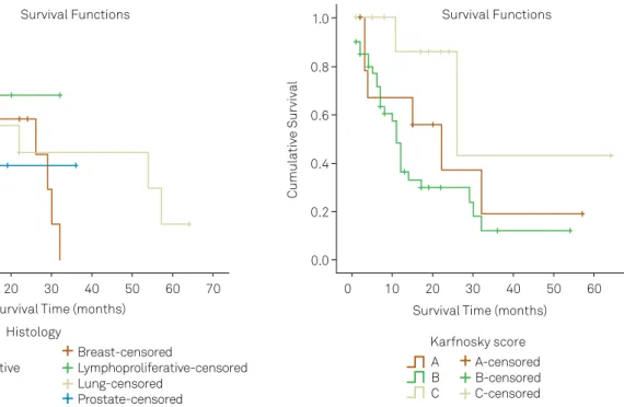
Survival comparison (Figure 1)
he log-rank curves of diferent histological cancer types
are shown below. Survival was longer for lymphoproliferative cancers than for other types (p < 0.001).
Karnofsky score (Figure 2)
We divided patients’’ Karnofsky scores into three groups for
analysis: 10-40 (Group A), 40-70 (Group B), and > 70 (Group C). he median survival after spinal metastasis diagnosis was 4.5 months in Group A, 8.2 months in Group B, and 11.7 months in Group C. he diference was statistically signiicant (p = 0.016).
Frankel grade (Figure 3)
When evaluating Frankel scale grades, median
surviv-al was 7.6 months among Frankel A patients; 7.9 months among Frankel B patients; 6.4 months among Frankel C pa
-tients; 8.4 months among Frankel D pa-tients; and 8.8 months among Frankel E patients. he diference was not statistically signiicant (p = 0.769).
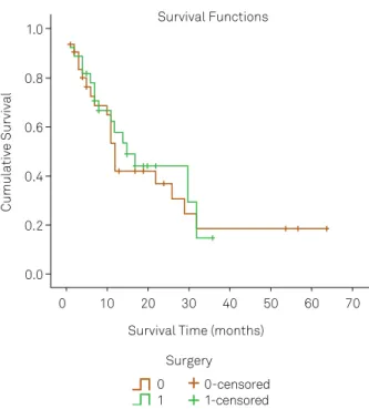
Survival difference between surgically and conservatively treated patients (Figure 4)
By the end of follow-up, 18 of 33 patients who under-went surgical treatment had died, as had 21 of 35 who were
Survival Time (months) Survival Functions
Histology Breast
Lymphoproliferative Lung
Prostate
Cumulative Survival
0 0.0 0.2 0.4 0.6 0.8 1.0
10 20 30 40 50 60 70
Breast-censored
Lymphoproliferative-censored Lung-censored
Prostate-censored
Figure 1. Survival by primary cancer site.
Survival Time (months) Survival Functions
Karfnosky score A
B C
Cumulative Survival
0 0.0 0.2 0.4 0.6 0.8 1.0
10 20 30 40 50 60 70
A-censored B-censored C-censored
treated conservatively. here was no diference in surviv -al between the operative and conservative management groups (p = 0.713).
DISCUSSION
A variety of surgical methods are available for the treat-ment of spinal metastases. Dorsal spinal decompression and stabilization are the most frequent surgical techniques used to treat metastatic disease of the thoracic and lumbar spine1,2,3,4,5,6,7,8,9,10. For patients with a solitary spinal metastasis
without vertebral canal invasion and who are in good general health with a long life expectancy, tumor resection through
en bloc spondylectomy/total vertebrectomy with primary
stabilizing instrumentation has been suggested9,10,11,12,13. A
re-cent paper reported that candidates for en bloc
spondylec-tomy are not frequently encountered14,15,16.
In all scenarios, RT is a valuable tool to treat systemic dis
-ease and control emboligenic foci. Radiosurgery is also efec
-tive as either primary or adjunc-tive treatment of metastatic
tumors of the spine. Preoperative embolization techniques can also be employed with the intention of controlling intra-operative bleeding5,6,17,18,19,20.
Prognosis can be predicted in to determine the best therapeutic approach. For this purpose, many systems have been proposed to guide decision-making, such as the Sioutos, Tomita, Van der Linden, Bauer, and Tokuhashi scores. In all cases, the main parameters used to estimate prognosis are preoperative ambulatory capacity, Karnofsky
performance status, RT, primary cancer type, presence of
extraspinal metastasis, and number of spinal segments with metastasis. Additionally, pain on presentation and body
mass index 25-30 kg/m2 have been identiied as predictors
of poor outcome21,22,23,24,25.
Our results conirm that the histologic diagnosis of spinal
metastasis is an independent prognostic factor in the course of malignancy.
Although major advances in cancer treatment have been
achieved in recent years, patients with vertebral metastasis
still exhibit short survival. his suggests that vertebral in -volvement is indicative of aggressive cancer behavior.
We demonstrated an association between histologi-cal cancer type and survival, with lymphoproliferative neo-plasms having the best survival rate. Additionally, Karnofsky score at presentation was also related to survival.
RT alone is usually advocated in patients with intact neu -rological status and patients with stable spines, who may be in good or poor clinical status. Surgery is generally indi-cated when neurological status and stability issues must be promptly addressed. In both situations, there is wide varia-tion in prognostic factors. We found similar survival in
pa-tients treated with surgery plus RT and in those treated with RT alone5,6,26.
Current evidence shows that surgery plays a decisive
role in quality of life and survival rates, and inal outcomes in patients receiving both surgery and RT are better than RT alone. hese studies suggest that surgery can provide pain
relief, neurologic improvement (in aspects such as ambula-tion), and aid recovery of general condition. Other reports
Survival Time (months) Survival Functions
Neurological seficit
Frankel A & B Frankel C Frankel D Frankel E
Frankel A & B-censored Frankel C-censored Frankel D-censored Frankel E-censored
Cumulative Survival
0 0.0 0.2 0.4 0.6 0.8 1.0
10 20 30 40 50 60 70
Figure 3. Survival according to Frankel Score.
Survival Time (months) Survival Functions
Surgery 0 1
0-censored 1-censored
Cumulative Survival
0 0.0 0.2 0.4 0.6 0.8 1.0
10 20 30 40 50 60 70
Figure 4. Survival according to treatment modality
have discussed the importance of patient age and revealed
that, in patients over 60 years old, beneits may not be sig
-niicant, especially due to the high risk of complications. In our sample, there was no diference in outcome between RT alone or RT + surgery5,6.
Surgery was not beneficial in terms of improving sur-vival. Cancer type, aging, and advanced staging have greater influence on life expectancy than treatment type. Surgical complications, although not addressed in the present study, should not be neglected as a possible factor in lowering life expectancy.
It also bears stressing that preservation of ambulation, pain control, and functional independence are achieved more quickly after surgery than with conservative treatment.
his may help patients face their disease in a more positive
manner, which could entail better psychological status and superior treatment adherence.
Several limitations of this study must be addressed. First, our sample was recruited from a single tertiary referral cen-ter. Second, although sample size was robust, the number of
patients in each subgroup should be greater. hen, there is
an important question concerning linfoproliferative
diseas-es. here is usually a discussion if vertebral lesions presented
in these diseases are primary or metastatic. Once these dis-eases origin from hematopoietic elements and are present
usually with difuse vertebral and systemic involvement, we
considered them as secondary lesions, similarly to other
pre-vious reports from literature3,13,16,21,22,23,24. Indeed, if
linfopro-liferative neoplasms were considered primary lesions in the
present study, we would not reach statistical signiicant dif -ference among survival rates of remaining histological types. Finally, we did not apply in our sample any quality of life (QOL) assessment tool. We strongly believe QOL is very im-portant in the context of such patients. However, our aim was to determine how much time they lived after
metasta-sis diagnometasta-sis. As we conirmed, their survival is limited and
surely is associated with poor QOL.
Current scientiic evidence shows that advanced cancer
staging is directly associated with poor prognosis. Patients with metastatic cancer are challenging to treat, because me-tastasis represents an advanced stage of the disease and, hence, is indicative of poor prognosis1,2,3,9,18,19.
Knowledge of individual patient survival proiles has the potential to interfere with therapeutic targets and thus inlu -ence surgical and clinical treatment planning.
References
1. Akbar M, Ayache A, Eichler M, Klotz M, Wiedenhöfer B, Lehner B. [Management of spinal metastases, strategies and surgical indications]. Orthopade. 2012;41(8):632-9. German. http://dx.doi.org/10.1007/s00132-012-1910-2
2. Bilsky MH, Lis E, Raizer J, Lee H, Boland P. The diagnosis and treatment of metastatic spinal tumor. J Neurosurg. 1992;177(1):90-5.
3. Botelho RV, Oliveira MF, Rotta JM. Quantification of vertebral involvement in metastatic spinal disease. Open Orthop J.
2013;7(7):286-91. http://dx.doi.org/10.2174/1874325001307010286
4. Chataigner H, Onimus M. Surgery in spinal metastasis without spinal cord compression: indications and strategy related to the risk of recurrence. Eur Spine J. 2000;9(6):523-7. http://dx.doi.org/10.1007/s005860000163
5. Chi JH, Gokaslan Z, McCormick P, Tibbs PA, Kryscio RJ, Patchell RA. Selecting treatment for patients with malignant epidural spinal cord compression-does age matter?: results from a randomized clinical trial. Spine. (Phila Pa 1976). 2009;34(5):431-5. http://dx.doi.org/10.1097/BRS.0b013e318193a25b
6. Delank KS, Wendtner C, Eich HT, Eysel P The treatment of spinal metastases. Dtsch Arztebl Int. 2011;108(5):71-80. http://dx.doi.org/10.3238/arztebl.2011.0071
7. Demura S, Kawahara N, Murakami H, Abdel-Wanis ME, Kato S, Yoshioka K et al. Total en bloc spondylectomy for spinal metastases in thyroid carcinoma. J Neurosurg Spine. 2011;14(2):172-6. http://dx.doi.org/10.3171/2010.9.SPINE09878
8. Fehlings MG, David KS, Vialle L, Vialle E, Setzer M, Vrionis FD Decision making in the surgical treatment of cervical spine metastases. Spine (Phila Pa 1976). 2009;34(22 Suppl):S108-17. http://dx.doi.org/10.1097/BRS.0b013e3181bae1d2
9. Hessler C, Burkhardt T, Raimund F, Regelsberger J, Vettorazzi E, Madert J et al. Dynamics of neurological deficit after surgical decompression of symptomatic vertebral metastases. Spine. (Phila Pa 1976). 2009;34(6):566-71. http://dx.doi.org/10.1097/BRS.0b013e31819a825d
10. Itshayek E, Yamada J, Bilsky M, Schmidt M, Shaffrey C, Gerszten P et al. Timing of surgery and radiotherapy in the management of metastatic spine disease: a systematic review. Int J Oncol. 2010;36(3):533-44. http://dx.doi.org/10.3892/ijo_00000527
11. Kilbride L, Cox M, Kennedy CM, Lee SH, Grant R. Metastatic spinal cord compression: a review of practice and care. J Clin Nurs. 2010;19(13-14):1767-83. http://dx.doi.org/10.1111/j.1365-2702.2010.03236.x
12. Lau D, Leach MR, La Marca F, Park P. Independent predictors of survival and the impact of repeat surgery in patients undergoing surgical treatment of spinal metastasis. J Neurosurg Spine. 2012;17(6):565-76. http://dx.doi.org/10.3171/2012.8.SPINE12449
13. Leithner A, Radl R, Gruber G, Hochegger M, Leithner K, Welkerling H et al. Predictive value of seven preoperative prognostic scoring systems for spinal metastases. Eur Spine J. 2008;17(11):1488-95. http://dx.doi.org/10.1007/s00586-008-0763-1
14. Liang T, Wan Y, Zou X, Peng X, Liu S. Is surgery for spine metastasis reasonable in patients older than 60 years? Clin Orthop Relat Res. 2013;471(2):628-39. http://dx.doi.org/10.1007/s11999-012-2699-3
15. Murakami H, Kawahara N, Demura S, Kato S, Yoshioka K, Tomita K. Total en bloc spondylectomy for lung cancer metastasis to the spine. J Neurosurg Spine. 2010;13(4):414-7. http://dx.doi.org/10.3171/2010.4.SPINE09365
17. Papastefanou S, Alpantaki K, Akra G, Katonis P. Predictive value of Tokuhashi and Tomita scores in patients with metastatic spine disease. Acta Orthop Traumatol Turc. 2012;46(1):50-6. http://dx.doi.org/10.3944/AOTT.2012.2645
18. Sciubba DM, Petteys RJ, Dekutoski MB, Fisher CG, Fehlings MG, Ondra SL et al. Diagnosis and management of metastatic spine disease: a review. J Neurosurg Spine. 2010;13(1):94-108. http://dx.doi.org/10.3171/2010.3.SPINE09202
19. Sheehan JP, Shaffrey CI, Schlesinger D, Williams BJ, Arlet V, Larner J. Radiosurgery in the treatment of spinal metastases: tumor control, survival, and quality of life after helical tomotherapy. Neurosurgery. 2009;65(6):1052-62. http://dx.doi.org/10.1227/01.NEU.0000359315.20268.73
20. Thomas KC, Nosyk B, Fisher CG, Dvorak M, Patchell RA, Regine WF et al. Cost-effectiveness of surgery plus radiotherapy versus radiotherapy alone for metastatic epidural spinal cord compression. Int J Radiat Oncol Biol Phys. 2006;66(4):1212-8. http://dx.doi.org/10.1016/j.ijrobp.2006.06.021
21. Tokuhashi Y, Matsuzaki H, Oda H, Oshima M, Ryu J. A revised scoring system for preoperative evaluation of metastatic spine tumor prognosis. Spine. 2001;30(19):2186-91. http://dx.doi.org/10.1097/01.brs.0000180401.06919.a5
22. Tokuhashi Y, Matsuzaki H, Toriyama S, Kawano H, Ohsaka S. Scoring system for the preoperative evaluation of metastatic spine tumor prognosis. Spine. 1990;15(11):1110-3. http://dx.doi.org/10.1097/00007632-199011010-00005
23. Tomita K, Kawahara N, Baba H, Tsuchiya H, Fujita T, Toribatake Y. Total en bloc spondylectomy. A new surgical technique for primary malignant vertebral tumors. Spine. 1997;22(3):324-33. http://dx.doi.org/10.1097/00007632-199702010-00018
24. Tomita K, Kawahara N, Kobayashi T, Yoshida A, Murakami H, Akamaru T. Surgical strategy for spinal metastases. Spine. 2001;26:298-306. http://dx.doi.org/10.1097/00007632-200102010-00016
25. Ulmar B, Huch K, Naumann U, Catalkaya S, Cakir B, Gerstner S et al. Evaluation of the Tokuhashi prognosis score and its modifications in 217 patients with vertebral metastases. Eur J Surg Oncol. 2007;33(7):914-9. http://dx.doi.org/10.1016/j.ejso.2006.11.018