GASTROINTESTINAL STROMAL TUMOR: CLINICAL, RADIOLOGIC
AND PATHOLOGIC FEATURES*
Leonardo Lopes de Macedo1
, Lucas Rios Torres2
,Rafael Artigas Faucz3
, Olger de Souza Tornin4
, Fábio Mota Gonzalez5
, Igor Motta de Aquino3
, Carlos Alberto Marcovechio Fonseca6
,
Alexandre Pescioto7
, Ricardo Pires de Souza8
OBJECTIVE: To investigate and describe clinical, radiologic and pathologic findings of gastrointestinal stro-mal tumors. MATERIALS AND METHODS: In the period between December 2000 and March 2006, 16 patients were submitted to surgery for gastrointestinal stromal tumors in our institution. The following variables were taken into consideration: sex and age, signs and symptoms at presentation, tumor site and size, radiological and pathological features, and presence of metastasis. RESULTS: The study population was constituted by nine men and seven women. The primary tumor sites of origin were: stomach (n = 5), rectum (n = 4), small bowel (n = 3), mesentery (n = 3), and colon (n = 1). Mean primary tumor size was 9 cm. Computed to-mography was the main radiological method utilized. Circumscribed, lobulated and heterogeneously con-trast-enhanced mass was the main image finding. Metastasis was found in nine patients (56% of cases) at presentation or tumor recurrence was observed during the follow-up period (mean = 32 months). CONCLU-SION: Gastrointestinal stromal tumor occurs in middle-age adults and the elderly, and must be taken into consideration as differential diagnosis for abdominal masses. Early diagnosis, adequate therapy, and rigor-ous follow-up are essential, considering the high probability of malignancy of these neoplasms as demon-strated by the present study.
Keywords: Gastrointestinal stromal tumor; Gastrointestinal neoplasms; Gastrointestinal diseases; Sarcoma.
Tumor do estroma gastrintestinal: achados clínicos, radiológicos e patológicos.
OBJETIVO: Investigar e descrever os achados clínicos, radiológicos e anatomopatológicos dos tumores do estroma gastrintestinal. MATERIAIS E MÉTODOS: De dezembro de 2000 a março de 2006, 16 pacientes foram operados por tumores do estroma gastrintestinal em nossa instituição. As variáveis analisadas foram sexo e idade dos pacientes, sinais e sintomas na consulta inicial, localização e tamanho do tumor, achados radiológicos, características anatomopatológicas e a ocorrência de metástases. RESULTADOS: A população em estudo constou de nove homens e sete mulheres. Os locais de origem dos tumores primários foram o estômago (n = 5), o reto (n = 4), o intestino delgado (n = 3), o mesentério (n = 3) e o cólon sigmóide (n = 1). Tomografia computadorizada foi o principal método radiológico empregado. Massa circunscrita, de contornos lobulados e que sofre realce heterogêneo pelo meio de contraste foi o principal achado por ima-gem. Em nosso estudo, nove pacientes (56% dos casos) apresentaram metástases ao diagnóstico ou recor-rência do tumor num período médio de dois anos e oito meses. CONCLUSÃO: O tumor do estroma gastrin-testinal acomete adultos de meia-idade e idosos e deve ser lembrado no diagnóstico diferencial das massas abdominais. Diagnóstico precoce, tratamento correto e acompanhamento rigoroso são fundamentais, pois, como demonstrado em nosso trabalho, essas neoplasias apresentam alta tendência à malignidade. Unitermos: Tumor do estroma gastrintestinal; Neoplasias gastrintestinais; Doenças gastrintestinais; Sarcoma.
Abstract
Resumo
* Study developed in the Service of Radiology and Diagnostic Imaging, Hospital Heliópolis, São Paulo, SP, Brazil.
1. MD, Resident in Radiology at the Service of Radiology and Diagnostic Imaging, Fellow Master degree in Sciences of Health, Post-graduation at Hospital Heliópolis, São Paulo, SP, Brazil.
2. MD, Resident in Radiology at the Service of Radiology and Diagnostic Imaging, Hospital Heliópolis, São Paulo, SP, Brazil.
3. MD, Radiologist, formerly Resident for the Service of Radi-ology and Diagnostic Imaging, Hospital Heliópolis, São Paulo, SP, Brazil.
4. MD, Radiologist, Master in Sciences of Health, Post-gra-duation at Hospital Heliópolis, São Paulo, SP, Brazil.
5. MD, Radiologist, Fellow Master degree in Sciences of Health, Post-graduation at Hospital Heliópolis, São Paulo, SP, Brazil.
6. PhD, Universidade Federal de São Paulo/Escola Paulista de Medicina (Unifesp/EPM), Physician Assistant for the Service of General Surgery , Hospital Heliópolis, São Paulo, SP, Brazil.
7. Master in Sciences of Health, Post-graduation at Hospital Heliópolis, Physician Assistant for the Service of General Surgery, Hospital Heliópolis, São Paulo, SP, Brazil.
INTRODUCTION
Gastrointestinal stromal tumors (GIST) are the most frequent mesenchymal neo-plasms of the gastrointestinal tract, charac-terized by the expression of the C-KIT pro-tein (CD117), a membrane receptor with a
tyrosine kynase component(1–3). Although they may occur in any site of the gas-trointestinal tract, they correspond to only 1% of tumors in these organs(2).These tu-mors affect subjects above 50 years of age, and rarely are found before the age of 40 years(4).
Symptoms are non-specific, and com-puted tomography (CT) is the method of choice for the diagnosis of this lesion(5).
Previously, GISTs were classified into a group of smooth muscle tumors includ-ing leiomyomas, leiomyosarcomas, and leiomyoblastomas(2). With the introduction 8. PhD in Radiology, Universidade de São Paulo (USP),
Coor-dinator for Medical Residency, Service of Radiology and Diagnos-tic Imaging, Hospital Heliópolis, São Paulo, SP, Brazil.
Mailing address: Dr. Leonardo Lopes de Macedo. Rua Marti-niano de Carvalho, 1049, ap. 253C, Bela Vista. São Paulo, SP, Brazil, 01321-001. E-mail: [email protected]
of immuno-histochemical staining tech-niques and the breakthrough of markers such as the C-KIT, currently these tumors are recognized as a distinct, new class of tumors, which is extremely important, con-sidering the differences in their prognosis and treatment(6).
GISTs presentations may range from small, asymptomatic, incidentally detected lesions to masses large enough to cause symptoms, including multiple metasta-ses(2). Metastases, usually, affect the liver and the peritoneum, but rarely lymph nodes(1,5,7).
In case of localized tumors, surgical re-section is the therapy of choice(5). In pa-tients with inoperable or metastatic dis-ease, immediate imatinib therapy (STI571 — a tyrosine kynase inhibitor) is indi-cated(5,7,8).
Considering that this is a recently de-scribed disease, we have tried to demon-strate the relevance of imaging studies in the detection of these tumors, besides evaluating the role of these methods for aiding in the differential anatomopatholo-gical diagnosis.
MATERIALS AND METHODS
The present study was retrospectively performed, utilizing the non-experimental (observational) model. Data from 16 pa-tients operated for GIST in our institution
in the period between December/2000 and March/2006 were evaluated. Only lesions with histopathological and immuno-his-tochemical (C-KIT-positive) patterns com-patible with GIST were included. All the C-KIT-negative patients were excluded. The variables analyzed were the following: patients’ sex and age, signs and symptoms at the initial presentation, primary site and size of the tumor, radiological findings, anatomopathological features of the lesion, presence of metastasis at diagnosis, and incidence of metastasis or tumor recurrence in the follow-up of the patients.
Imaging studies (12 CT and two eso-phagogastroduodenal – EGD series) of 12 patients in the sample of 16 were evaluated by two radiologists (specialist title by Colégio Brasileiro de Radiologia e Diag-nóstico por Imagem) of our institution. From the other four patients whose CT studies were not available, we could only to recover the CT reports. The radiological signs evaluated were: site and size of the lesion, contrast-enhancement, margins, contours, central hypodensity, calcification and presence of metastases.
RESULTS
The study population included nine men and seven women. Mean age among men was 49 years (ranging between 25 and 66 years), and among women was 69 years
(ranging between 63 and 75 years). The group mean age was 58 years. Initially, the main complaints of patients were: body weight loss (n = 8), followed by
abdomi-nal pain (n = 7), nausea (n = 5), emesis (n
= 3), upper digestive hemorrhage (n = 1),
hematochezia (n = 1) and melena (n = 1).
Two patients were admitted into the hospi-tal with acute obstructive abdomen, and one with intestinal subocclusion.
In the present study, primary tumors sites of origin were: stomach (n = 5),
rec-tum (n = 4), small bowel (n = 3),
mesen-tery (n = 3) and sigmoid (n = 1). The tumors
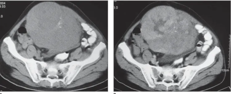
size ranged between 2 cm and 20 cm (mean = 9 cm), with stomach tumors presenting mean 3 cm and the mesenteric ones (Fig-ure 1), mean 15 cm.
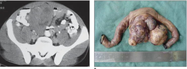
At CT all of the tumors presented het-erogeneous contrast-enhancement. The gastric neoplasms presented circumscribed margins and slightly lobulated contour, with only one tumor (the largest, with 6 cm) with a central hypodense area. The mesen-teric tumors, as well as the small bowel tumors (Figure 2), produced a mass effect causing compression of adjacent structures. They were larger, with lobulated margins, and only one of them did not present a cen-tral hypodense area. A mural mass causing the mucosa to bulge, with mildly lobulated margins was the main presentation of rec-tal tumors (Figure 3). Calcification was found in two mesenteric tumors.
Figure 1. Mesenteric GIST. A: Well-defined mass with lobulated margins and some calcifications. B: The mass presents heterogeneous contrast-enhance-ment.
In both cases evaluated by EGD series, a circumscribed, typically submucosal le-sion without signs of ulceration was found (Figure 4).
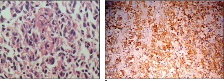
Histopathologically, 13 patients pre-sented with spindle cells tumors (Figure 5A), and other three with epithelioid cells (one in the small bowel, and two in the rec-tum). The immuno-histochemical analysis was essential for diagnosis confirmation, and in all of the cases presented C-KIT positivity (Figure 5B).
Four patients present with liver metasta-sis at the moment of the diagnometasta-sis and other five patients presented tumor recurrence, four of them in the peritoneum, and one in the liver, within two months to eight years and eight months (mean period = two years
and eight months). Liver metastases were hypoattenuating as compared with the well-defined, adjacent normal parenchyma.
DISCUSSION
GISTs are the most frequent mesenchy-mal neoplasms occurring at any site of the gastrointestinal tract(1,3). Approximately 40%–70% of GISTs affect the stomach, ac-counting for 2.5% of gastric tumors, 20%– 40% affect the small bowel, and the re-mainders occur in other sites such as esophagus, colon, rectum, mesentery and omentum(9,10).
These tumors affect subjects above 50 years of age, and rarely are found before the age of 40 years, with a slightly higher male
prevalence(4,7). In the present casuistic, the mean age at the moment of the diagnosis amongst men was markedly lower than amongst women, raising the hypothesis that GISTs affect men at an earlier age. This data is not reported in the literature.
Clinical symptoms are non-specific and are basically associated with the site and size of the lesion. Abdominal pain, disten-sion, gastrointestinal bleeding, anemia, body weight loss and palpable mass are some of possible signs of the disease(4,7). These tumors may achieve large dimen-sions, with size usually ranging between 3 cm and 10 cm(1), and because of a predomi-nantly extraluminal growth, they rarely cause obstructive symptoms(1,4,11). In the present study, stomach tumors presented
Figure 2. Small bowel GIST. A: Mass presenting soft tissues density, well-defined limits, lobulated margins, and some central, hypoattenuating areas in close contact with intestinal loops. B: Mass attached to the small bowel wall with predominant extraluminal component.
Figure 3. Rectal GIST. Heterogeneously contrast-enhanced, well-defined mass with soft tissues density. The mass is predominantly extraluminal and, despite its size (8 cm), it does not cause significant rectal stenosis.
B A
considerably smaller at diagnosis, as com-pared with the mesenteric tumors, which corroborates the literature(12,13).
Histologically, GISTs are classified ac-cording to the predominant cellular type, as follows: spindle cells (70%), epithelioid cells (20%), and mixed (10%)(6).
Immuno-histochemical evaluation may detect the C-KIT (CD117), a tyrosine kynase receptor, the most important GIST marker(6,14). The majority of lesions also present CD34 positivity. Other possible markers include vimentine, actin, S-100 protein and (rarely) desmin(1,4,6,9). These markers are extremely useful in the dif-ferentiation of these tumors from others of similar origin, such as leiomyomas, leio-myoblastomas, leiomyosarcomas, and schwannomas(3). Some tumors such as lei-omyosarcomas may show radiological and histological presentations very similar to GISTs, however C-KIT is GIST-specific(15). GIST may be benign or malignant, and major negative prognostic factors include distal intestinal location, tumor size, high mitotic activity, and presence of metasta-sis(6,10). There is no correlation between degree of necrosis, hemorrhage or pattern of contrast-enhancement on CT indicating a higher or lower malignant potential(4). Notwithstanding some studies demonstrate that less than 50% of primary, localized tumors do not recur in a five-year-pe-riod(10), it is known that in cases of tumor recurrence in the liver or peritoneum (the two most frequent sites of metastasis) the
prognosis is poor(6). In the present study, nine patients (56% of cases) presented with metastasis at diagnosis, or tumor recurrence in a mean period of two years and eight months, which demonstrates a high pro-pensity to malignancy. Considering that this is a recently described disease, studies reporting a long lasting follow-up of a con-siderable number of patients are still to be published. Currently, these tumors are con-sidered as potentially malignant and, there-fore, all the patients affected by this disease should be carefully treated and followed-up(5,6,10).
Amongst the currently available imag-ing methods, CT remains as the method of choice for evaluation of abdominal masses or biopsy-confirmed GISTs, especially if the wide availability of the method is con-sidered(5).
Generally, these tumors present as a well circumscribed mass, frequently origi-nating from the stomach or small bowel, with heterogeneous contrast-enhance-ment(7,11). Small foci of calcifications, fre-quently related to malignant lesions may be observed(12). Areas of central attenuation may correspond to cystic degeneration, hemorrhage or tumor necrosis(4,9,13), which includes this neoplasm in the differential diagnosis of cystic or necrotic lesions re-lated to the stomach or adjacent struc-tures(11). Mucosal alteration may be found in up to 50% of gastric tumors(1) and aneurismatic dilatation of small bowel loops, previously related to lymphoma,
may be found in up to 33% of enteric GISTs(11). Most of times, mesenteric GISTs present well-defined margins, lobulated contour, large dimensions (10 cm to 27 cm) and areas of low central attenuation(13).
In their most aggressive feature, these tumors may generate metastases, the liver and peritoneum being the most affected sites. More rarely, the tumor may spread to lymph nodes, bones and lungs. At CT, liver metastases present contrast-enhancement, because of their usually hypervascular na-ture(1,7). It is important to note that, during the CT portal phase, hepatic metastases may become imperceptible, which makes the performance of the arterial phase ex-tremely important(5). The cystic pattern appearance after na adequate chemo-therapy is typical and has already been de-scribed in the literature, and should not be erroneously interpreted as a disease pro-gression or as new lesions(1,5,16).
GISTs can be cured only by surgery(5). Considering the absence of a true capsule, the tumor must be block-resected with a free 2-3 cm margin as possible. Lymphade-nectomy is unnecessary since these tumors rarely produce lymph nodes metastasis(2, 3,5). The follow-up of these patients must
include CT every six months, considering the potentially malignant nature of the dis-ease(5,10).
In cases of inoperable or metastatic tu-mors, the therapy of choice is with imatinib (STI571), a tyrosine kynase inhibitor, and there is no indication for radiotherapy or
Figure 5.GIST histological and immuno-histochemical findings. A: Photomicrography shows fusiform neoplastic cell of mesenchymal origin (hematoxilin-eosin, 20× increase). B: Immuno-histochemical analysis showing cytoplasm cells stained in brown, indicating C-KIT (main GIST marker) positivity.
chemotherapy. The drug administration should be initiated upon the diagnosis of metastatic or advanced disease, and main-tained until the patient develops intolerance or progressive disease(3,5). Recent studies have demonstrated that more than 50% of patients with advanced disease are respon-sive to the medicamentous treatment(8,17,18). CT remains as the method of choice for evaluation of the patients´ response to the therapy, although positron emission tomog-raphy (PET) has shown high sensitivity for demonstrating na early response of the tu-mor(19). Progressive hypoattenuation of the mass, decrease in nodular and vasculariza-tion enhancement are parameters indicative of a good response of the tumor to the therapy(8). However, it should be high-lighted that some tumors increase in size during the first six months of therapy, de-spite the significant clinical improvement and regression visualized by PET(5,19).
CONCLUSION
GISTs, although relatively rare, are the most frequent mesenchymal neoplasms in the gastrointestinal tract. These tumors af-fect middle-aged adults and elders, and notwithstanding the patients present with non-specific symptoms, they should be considered in the differential diagnosis of solid of solid/cystic masses in the abdomi-nal cavity.
In the present study, the most frequent site of GISTs was the stomach. Gastric tu-mors presented reduced dimensions as compared with small bowel and mesenteric tumors. Central hypodensity was observed in 50% of cases and in larger tumors. Cal-cification was not a common finding. Oc-currence of metastasis or tumor reOc-currence was observed in the majority of cases.
The main finding at CT was heteroge-neously contrast-enhanced circumscribed mass, with lobulated contour. The EGD
series identified circumscribed and typi-cally submucosal mass. These findings corroborate the literature(1,4,7).
Two patients presented with acute ob-structive abdomen, and one with intestinal subocclusion at the initial presentation. These are uncommon findings at the first presentation, however they should be con-sidered in the differential diagnosis of ab-dominal lesions.
Spindle cells pattern was the main his-tological tumor type, followed by the epi-thelioid cell type, also corroborated in the literature(4,6).
As demonstrated in the present study, GISTs present a high tendency to malig-nancy. Therefore, early diagnosis, an appro-priate therapy and careful follow-up are essential for the management of the dis-ease.
Finally, amongst the differential diag-noses, the radiologist’s suspicion is essen-tial to reduce the morbidity or even the mortality of patients with GIST.
REFERENCES
1. Sandrasegaran K, Rajesh A, Rydberg J, Rushing DA, Akisik FM, Henley JD. Gastrointestinal stro-mal tumors: clinical, radiologic, and pathologic features. AJR Am J Roentgenol 2005;184:803– 811.
2. Sugár I, Forgács B, István G, Bognár G, Sápy Z, Ondrejka P. Gastrointestinal stromal tumors (GIST). Hepatogastroenterology 2005;52:409– 413.
3. Shinomura Y, Kinoshita K, Tsutsui S, Hirota S. Pathophysiology, diagnosis, and treatment of gas-trointestinal stromal tumors. J Gastroenterol 2005;40:775–780.
4. Levy AD, Remotti HE, Thompson WM, Sobin LH, Miettinen M. Gastrointestinal stromal tu-mors: radiologic features with pathologic corre-lation. RadioGraphics 2003;23:283–304. 5. Blay JY, Bonvalot S, Casali P, et al. Consensus
meeting for the management of gastrointestinal stromal tumors. Report of the GIST Consensus Conference of 20-21 March 2004, under the aus-pices of ESMO. Ann Oncol 2005;16:566–578. 6. Fletcher CDM, Berman JJ, Corless C, et al.
Di-agnosis of gastrointestinal stromal tumors: a con-sensus approach. Hum Pathol 2002;33:459–465. 7. Burkill GJC, Badran M, Al-Muderis O, et al.
Ma-lignant gastrointestinal stromal tumor: distribu-tion, imaging features, and pattern of metastatic spread. Radiology 2003;226:527–532. 8. Dagher R, Cohen M, Williams G, et al. Approval
summary: imatinib mesylate in the treatment of metastatic and/or unresectable malignant gas-trointestinal stromal tumors. Clin Cancer Res 2002;8:3034–3038.
9. Sharp RM, Ansel HF, Keel SB. Best cases of the AFIP: gastrointestinal stromal tumor. Armed Forces Institute of Pathology. RadioGraphics 2001;21:1557–1560.
10. DeMatteo RP, Lewis JJ, Leung D, Mudan SS, Woodruff JM, Brennan MF. Two hundred gas-trointestinal stromal tumors: recurrence patterns and prognostic factors for survival. Ann Surg 2000;231:51–58.
11. Sandrasegaran K, Rajesh A, Rushing DA, Rydberg J, Akisik FM, Henley JD. Gastrointesti-nal stromal tumors: CT and MRI findings. Eur Radiol 2005;15:1407–1414.
12. Kim HC, Lee JM, Kim KW, et al. Gastrointesti-nal stromal tumors of the stomach: CT findings and prediction of malignancy. AJR Am J Roent-genol 2004;183:893–898.
13. Kim HC, Lee JM, Kim SH, et al. Primary gas-trointestinal stromal tumors in the omentum and mesentery: CT findings and pathologic correla-tions. AJR Am J Roentgenol 2004;182:1463– 1467.
14. Medeiros F, Corless CL, Duensing A, et al. KIT-negative gastrointestinal stromal tumors: proof of concept and therapeutic implications. Am J Surg Pathol 2004;28:889–894.
15. Miettinen M, Sarlomo-Rikala M, Sobin LH, Lasota J. Gastrointestinal stromal tumors and lei-omyosarcomas in the colon: a clinicopathologic, immunohistochemical, and molecular genetic study of 44 cases. Am J Surg Pathol 2000;24: 1339–1352.
16. Chen MY, Bechtold RE, Savage PD. Cystic changes in hepatic metastases from gastrointes-tinal stromal tumors (GISTs) treated with Gleevec (imatinib mesylate). AJR Am J Roentgenol 2002; 179:1059–1062.
17. Demetri GD, von Mehren M, Blanke CD, et al. Efficacy and safety of imatinib mesylate in ad-vanced gastrointestinal stromal tumors. N Engl J Med 2002;347:472–480.
18. van Oosterom AT, Judson I, Verweij J, et al. Safety and efficacy of imatinib (STI571) in metastatic gastrointestinal stromal tumours: a phase I study. Lancet 2001;358:1421–1423.