Original Article
3 4 8 Arq Bras Oftalmol. 2015;78(6):348-51 http://dx.doi.org/10.5935/0004-2749.20150092
INTRODUCTION
Pseudoexfoliation syndrome (PEX) is an age-related disease cha-racterized by the deposit of anomalous ibrillar extracellular material on intraocular structures in the anterior segment of the eye(1). Pseu-doexfoliation material (PEM) is detected by electron microscopy and indirect immunoluorescence in the stromal connective tissue of the conjunctiva, ciliary body, iris, lens capsule, and cornea. The presence of PEM on the anterior lens surface is the most consistent and impor-tant clinical diagnostic feature of PEX(2).
In vivo corneal confocal microscopy enables real-time, high re so-lution microscopic imaging of the cornea and has been increa singly used to evaluate the corneal changes in PEX(3,4). Previously, the only method of delineating the structural features of this disease was pa-thological examination after corneal biopsy. In vivo corneal confocal microscopy allows the examination of PEX-related corneal altera-tions. Using in vivo confocal microscopy, Zheng et al. showed that eyes with PEX, as well as their contralateral eyes, have signiicantly lower cell densities in the basal epithelium, anterior and posterior
stroma, and endothelium(3). They also demonstrated the deposition of hyperrelective material on the endothelium, which they presumed to be PEM, on eyes with PEX and the contralateral eyes(3).
To our knowledge, no study has compared the density of cells of corneal layers and the presence of hyperrelective endothelial de-posits in patients with PEX. The aim of this study was to measure the density of cells in diferent layers of the cornea and determine whether there are morphologic changes of the sub-basal corneal nerve plexus in eyes with PEX.
METHODS
One eye each of 37 patients diagnosed with PEX without glauco-ma and of 20 healthy control subjects was studied. In the patients with PEX, the eyes with more PEM as determined by biomicroscopic examination were used in the present study The study protocols were approved by the local ethics committee and follow the tenets of the Declaration of Helsinki. All patients underwent a complete
Characteristics of the cornea in patients with pseudoexfoliation syndrome
Características da córnea em pacientes com síndrome de pseudoexfoliação
Refik Oltulu¹, Gunhal SatiRtav¹, emine tinkiR kayitmazbatiR¹, Gulfidan bitiRGen¹, ahmet OzkaGnici¹, adnan kaRaibRahimOGlu²
Submitted for publication: May 27, 2015 Accepted for publication: August 23, 2015
1 Ophthalmology Department, Necmettin Erbakan University Faculty of Medicine, Konya, Turkey. 2 Biostatistics Unit, Medical Education and Informatics Department, Necmettin Erbakan University
Faculty of Medicine, Konya, Turkey.
Funding: No specific financial support was available for this study.
Disclosure of potential conflicts of interest: None of the authors have any potential conflict of interest to disclose.
Corresponding author: Refik Oltulu. Ophthalmology Department. Necmettin Erbakan University Meram Faculty of Medicine. Meram, Konya 42080 - Turkey - E-mail: [email protected]
Approved by the following research ethics committee: Necmettin Erbakan University Meram Faculty of Medicine. Project number: 2013/96.
ABSTRACT
Purpose: To quantify the morphological alterations in corneal nerve fibers and
cells in patients with pseudoexfoliation syndrome (PEX) and their relationship with the presence of hyperreflective endothelial deposits observed using in vivo confocal microscopy.
Methods: One eye each of 37 patients with PEX and 20 age-matched healthy
control subjects was evaluated by in vivo corneal confocal microscopy. Patients with PEX were further classified into two groups: those with and without hyper-reflective endothelial deposits. We evaluated the densities of basal epithelial cells, anterior and posterior stromal keratocytes, and endothelial cells and structure of sub-basal nerve fibers.
Results: The mean anterior and posterior stromal keratocyte and endothelial cell
densities and corneal sub-basal nerve plexus variables were significantly lower in patients with PEX compared with those in healthy control subjects. The mean basal epithelial cell density did not significantly differ.
Conclusion: Eyes with PEX presented decreased corneal sub-basal nerve plexus
variables and cell densities in all corneas, except for the mean basal epithelial cell density. Further, a trend of lower corneal sub-basal nerve plexus measurements in patients with hyperreflective endothelial deposits compared with those without endothelial deposits was observed.
Keywords: Cornea/physiopathology; Cornea/innervation; Exfoliation syndrome/
diagnosis; Confocal microscopy; Nerve fibers/pathology
RESUMO
Objetivo: Quantificar as alterações morfológicas nas fibras nervosas e nas células da córnea em pacientes com síndrome de pseudoexfoliação (PEX) e sua relação com a presença de depósitos endoteliais hiper-refletivos, observados por meio da micros-copia confocal in vivo.
Métodos: Trinta e sete olhos de 37 pacientes portadores de PEX e 20 olhos de 20 indivíduos saudáveis, pareados por idade, foram avaliados por meio da microsco -pia confocal de córnea. Os pacientes com PEX foram classificados em dois grupos: pacientes sem depósitos endoteliais hiper-refletivos e pacientes com depósitos en-doteliais hiper-refletivos. Células basais epiteliais, ceratócitos do estroma anterior e posterior, e densidades celulares endoteliais assim como a estrutura das fibras nervosas sub-basais foram avaliadas.
Resultados: A média de ceratócitos do estroma anterior e posterior, as densidades celulares endoteliais, bem como as variáveis de plexo nervo sub-basal foram significa-tivamente menores nos pacientes com PEX em comparação com indivíduos saudáveis. A densidade celular epitelial basal não diferiu significativamente.
Conclusões: Os olhos com PEX apresentaram diminuição das variáveis do plexo do nervo sub-basal e das densidades celulares em toda a córnea, exceto na camada basal das células epiteliais. Além disso, foi notada uma tendência para valores mais baixos nas variáveis do plexo do nervoso sub-basal em pacientes com depósitos endoteliais hiper-refletivos em comparação àqueles sem depósitos endoteliais.
Oltulu R, et al.
3 4 9 Arq Bras Oftalmol. 2015;78(6):348-51 ophthalmic examination, and in vivo corneal confocal microscopy
was performed. The diagnosis of PEX was made by visualizing PEM on the anterior lens surface and/or pupillary margin on slit-lamp biomicroscopy following pupillary dilation. On biomicroscopic exa-mination, pigment granules were not observed on the endothelium in any patient. Based on the in vivo confocal microscopy images, one independent observer assessed the patients for hyperrelective endothelial deposits and classiied the patients into two groups: patients without hyperrelective endothelial deposits (group 1, n=20) and patients with hyperrelective endothelial deposits (group 2, n=17). The subjects in the control group had no history of intraocular or refractive surgery and glaucoma, no previous or active ocular disea-se, no corneal pathology, no prior contact lens udisea-se, and no systemic disease that may have afected the cornea. Laser scanning in vivo
corneal confocal microscopy was performed on all subjects using the Rostock Corneal Module/Heidelberg Retina Tomograph III (RCM/ HRT III; Heidelberg Engineering GmBH, Dossenheim, Germany). The RCM uses an entirely digital image capture system. During the in vivo
corneal confocal microscopy examination, proparacaine was instil-led, and a contact lens was placed on the cornea while the patient was asked to ixate on a distant target aligned to enable examination of the central cornea. Two frames per location that contained the clearest images were selected from each of the following layers: ba-sal epithelium, anterior stroma, posterior stroma, and endothelium. A standard central counting frame size of 200 × 200 µm was used for all epithelial and endothelial images, and a frame size of 300 × 300 µm was used for the stromal images. Two frames were analyzed for each corneal layer, and an average was taken. The number of cells/mm2 was calculated by the proprietary software within the RCM/ HRT III. Three-to-ive high-quality images of the sub-basal nerve plexus from the center of the cornea were assessed from each sub-ject. For all sub-basal nerve plexus images, the full 400 × 400-µm fra-me was used. Automatic CCMetrics software, version 1.0 (University of Manchester, Manchester, UK) was used for the quantitative analysis of the nerve ibers. Three parameters were quantiied: the corneal nerve iber density (NFD), i.e., the total number of major nerves per square millimeter; the total length of the nerve ibers (TLNF) and branches (millimeters per square millimeter); and the nerve branch density (NBD), i.e., the number of branches emanating from the major nerve trunks per square millimeter(5). All images were analyzed by one observer (GB), who was unaware of whether the images belonged to a patient with PEX or a control subject.
Statistical analyses were performed using SPSS software version 16.0 (SPSS Inc., Chicago, IL, USA). Descriptive analyses were presented
using means and standard deviations for basal epithelial cell, anterior stromal keratocyte, posterior stromal keratocyte, and endothelial cell densities, as well as the NFD, NBD, and TLNF, and using frequencies for PEX and control groups. Statistical signiicance was determined by the non-parametric Kruskal-Wallis test and Mann-Whitney U test.
P-values <0.05 were considered statistically signiicant.
RESULTS
The mean age of the subjects was 68.7 ± 4.4 years (range, 61-76 years) in group 1, 70 ± 5.4 years (range, 59-85 years) in group 2, and 67.8 ± 2.6 years (range, 64-74 years) in the control group. None of the enrolled eyes had remarkable pathology of the cornea on slit-lamp biomicroscopy. There was no statistically signiicant diference within the three groups in terms of age (p=0.196) and sex (p=0.52). Table 1 shows a summary of the quantitative in vivo corneal confocal micros-copy indings of the cell density measurements and nerve iber varia-bles in patients with PEX and healthy control subjects. Figures 1 and 2 illustrate in vivo corneal confocal microscopy images of the sub-basal corneal nerve plexus and endothelial layer, respectively, in a healthy subject, a patient without hyperrelective endothelial deposits, and a patient with hyperrelective endothelial deposits.
The mean anterior stromal keratocyte, posterior stromal keratocyte, endothelial cell densities, and the corneal sub-basal nerve plex us variables (NFD, NBD, and TLNF) were signiicantly lower in patients with PEX compared with those in healthy control subjects (p<0.001); conversely, the mean basal epithelial cell density did not difer signi-icantly between the two groups (p=0.173).
When groups 1 and 2 were compared, no statistically signiicant diferences for the corneal cell density and corneal sub-basal nerve plexus variables (p>0.907 and p>0.32, respectively), were observed. Group 2 presented signiicantly lower TLNF (p=0.024).
When comparing the group without hyperrelective endothelial deposits (group 1) with the control group, the mean anterior stromal keratocyte, posterior stromal keratocyte, and endothelial cell densi-ties as well as the corneal sub-basal nerve plexus variables except for the NFD (p=0.310) were signiicantly lower in group 1 subjects. There was no statistically signiicant diference between the two groups for the mean basal epithelial cell density (p=0.53).
When comparing the group with hyperrelective endothelial deposits (group 2) with the control group, there were statistically sig-niicant diferences for the corneal sub-basal nerve plexus variables and all of the corneal cell density variables except for the mean basal epithelial cell density (p=0.447).
Table 1. Corneal confocal microscopic variables in patients with PEX and healthy control subjects
Basal epithelium (cells/mm2)
Anterior stroma (cells/mm2)
Posterior stroma (cells/mm2)
Endothelium (cells/mm2)
Nerve iber density
(ibers/mm2)
Nerve branch density
(branches/mm2)
Total length of the nerve ibers
(mm/mm2)
Control subjects (n=20) 5913 ± 480 764 ± 139 280 ± 49 2570 ± 369 34.6 ± 08.4 73.4 ± 32.1 25.0 ± 2.7
PEX (n=37) 5722 ± 531 434 ± 131 227 ± 45 2124 ± 346 28.6 ± 09.6 37.6 ± 21.4 17.3 ± 4.2
Group 1 (n=20) 5736 ± 464 433 ± 131 229 ± 48 2067 ± 289 30.6 ± 10.4 41.3 ± 19.9 19.0 ± 4.4 Group 2 (n=17) 5705 ± 616 436 ± 134 225 ± 42 2190 ± 403 26.2 ± 08.2 33.3 ± 22.9 15.3 ± 3.0
P values
Control subjects vs. PEX 0.173 <0.001 <0.001 <0.001 <0.001 <0.001 <0.001 Control subjects vs.
group 1 0.530 <0.001 <0.010 <0.001 <0.310 <0.004 <0.001
Control subjects vs.
group 2 0.447 <0.001 <0.004 <0.027 <0.004 <0.001 <0.001
Group 1 vs. group 2 0.982 1 1 <0.907 <0.278 <0.759 <0.024
Characteristics of the cornea in patients with pseudoexfoliation syndrome
350 Arq Bras Oftalmol. 2015;78(6):348-51 DISCUSSION
In vivo confocal microscopy ofers direct, non-invasive visual ac-cess to the cornea at the cellular level and also permits evaluation of the sub-basal nerve plexus(6). A few studies in the literature have used in vivo corneal confocal microscopy to study the corneas of patients with PEX. Martone et al. detected hyperrelective deposits and den dritic cells iniltrating the basal epithelial cell layer in one case using in vivo corneal confocal microscopy(7). They also found ibrillar subepithelial structures, and the endothelial layer showed cellular anomalies. In a prospective observational case series, Sbeity et al. used noncontact in vivo corneal confocal microscopy to detect PEM on the lens surfaces and corneal endothelium of PEX eyes and their fellow eyes(8). Moreover, Zheng et al. demonstrated a decrease in corneal sensitivity as well asall of the corneal cell density and corneal sub-basal nerve plexus variables in patients with PEX(3).
In our study, we studied the cell density in diferent layers of the cornea and aimed to determine whether there are morphologic dife-rences in the sub-basal corneal nerve plexus between eyes with and without PEX. We found no signiicant diference in the mean basal epithelial cell density between groups 1 and 2 and between patients with PEX and healthy control subjects. This inding contradicts those of previous studies, which showed signiicant diferences in the mean basal epithelial cell density between patients with PEX and healthy control subjects(3,4).
We further observed signiicantly lower anterior and posterior stro-mal keratocyte densities in patients with PEX compared with heal thy control subjects. However, these variables did not signiicantly difer between groups 1 and 2. Zheng et al. observed PEM deposits in the anterior stroma of PEX eyes and suggested that the PEM may so-mehow cause PEX, perhaps by inducing apoptosis of the keratocytes as well as other pathogenic factors, such as altered levels of cytokines or chemokines in the cornea(3).
Oxidative stress is generally induced through the formation of multiple reactive oxygen species, including superoxide, hydrogen pe roxide, and hydroxyl radicals, which can initiate and propagate free radicals. The net oxidative burden between the prooxidant and antio-xidant systems represents the oxidative stress that damages cellular and tissue macromolecules, such as lipids, proteins, and nucleic acids, and results in cellular and tissue dysfunction and cellular death(9). The numbers of keratocytes and other cells of the cornea may decline with age in response to cumulative oxidative insults or declining an -tio xidant protec-tion(10). Demirdögen et al. reported that the total antioxidant status (TAS) levels were signiicantly lower in patients with PEX and pseudoexfoliation glaucoma (PG) compared with that in controls, and that lack of antioxidants may play an important role in the development of PEX/PG(11). This reduced level of antioxidants may be a contributing factor in the lower keratocyte density in patients with PEX compared with that in control subjects found in
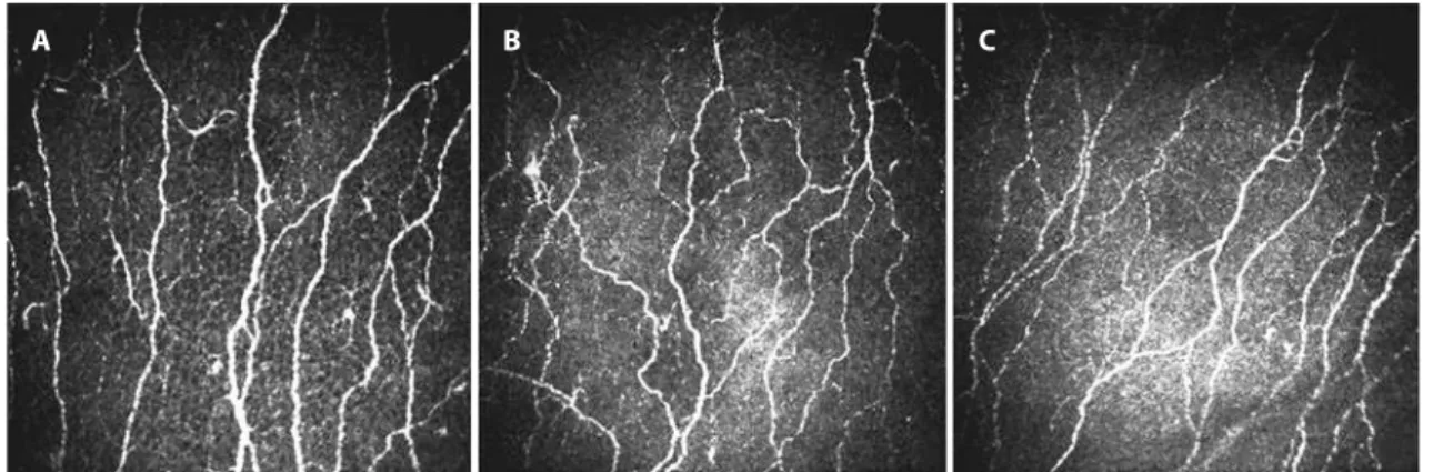
Figure 1.In vivo confocal microscopic images of the sub-basal nerve plexus. (A) Representative image from a healthy control subject with normal corneal nerve ibers. (B) Representative image from a patient without hyperrelective endothelial deposits showing lower nerve branch density (branches/mm2) and the total length of the nerve ibers (mm/mm2) compared with the control. (C) Representative image from a patient with
hyper-relective endothelial deposits showing lower nerve iber density (ibers/mm2), nerve branch density (branches/mm2), and the total length of the
nerve ibers (mm/mm2) compared with the control.
A B C
A B C
Oltulu R, et al.
351
Arq Bras Oftalmol. 2015;78(6):348-51 our study, although stromal deposits were not found in any of the
patients. Additionally, the lack of any diference between groups 1 and 2 concerning the keratocyte density, together with the previou-sly reported decreased TAS levels in patients with PEX, supports the altered cytokine and chemokine level hypothesis suggested by Zheng et al.(3). Furthermore, groups 1 and 2 did not difer with regard to the cell numbers in the corneal layers, and therefore, the presence or ab-sence of PEM on the endothelium does not afect the number of cells; instead, a secondary mechanism is thought to afect the cell numbers. Various studies have described the changes in the histopatholo-gical endothelial characteristics(3,7,12), although few used in vivo cor neal confocal microscopy to analyze the possible diferences between patients with and without PEX. Zheng et al. observed not only lower endothelial cell density but also increased degrees of pleomorphism and polymegathism and a higher coeicient of variation in the cell sizes in the patients with PEX compared with those in the healthy control subjects(3). We found that the mean endothelial cell density was lower in patients with PEX compared with that in healthy control subjects. Although we observed qualitative variation in the size and shape of the endothelial cells in eyes with PEX, quantitative analysis of pleomorphism and polymegathism was not performed in the current study, which limits detailed analysis. Deposition of PEM has been described as irregular clumps deposited on the corneal endo-thelium and has been associated with a decrease in the endothelial cell count(13). However, there was no statistically signiicant diference in the mean endothelial cell density when comparing group 1 with group 2. This suggests that factors other than the presence of PEM may be associated with the reduction of the endothelial cell density. Abnormalities of the sub-basal nerve plexus were another im-portant inding of the present study. We found that NFD, NBD, and TLNF were signiicantly lower in patients with PEX compared with those in healthy controls; additionally, we further showed that TLNF was signiicantly lower in group 2 compared with that in group 1. Additionally, neither NFD nor NBD difered signiicantly among the study subgroups, although both of them tended to be lower in group 2 compared with those in group 1. The reason for this may be that the two groups were at diferent stages of the PEX process; the eyes in group 1 may have been at an early stage of PEX.
There were some limitations in this study. First, the quantitative analysis of the in vivo corneal confocal microscopy images was per-formed by an observer who was unmasked regarding whether the images belonged to a patient with PEX or a control subject. Secondly, the study groups had small sample sizes. However, the sample sizes of our report are comparable with those of similar studies in the lite-rature. Another factor that could be considered as a limitation in this study is the possible efect of the presence of hyperrelective endo-thelial spots on the accuracy of measurements of the sub-basal nerve plexus variables. Further studies are needed to address whether the
etiology of the diferences between the measurements of the groups was due to an actual efect of the presence of hyperrelective spots on the nerve variables or an artifact efect of these deposits that limited the accuracy of the measurements.
In conclusion, in vivo corneal confocal microscopy permits in vivo
imaging of microstructural changes in the corneas of patients with PEX. Our study demonstrated that eyes with PEX have lower cell den-sities in all layers of the cornea except for the basal epithelium, as well as the corneal sub-basal nerve plexus variables, compared with those in control subjects. Further, we found trends toward lower corneal sub-basal nerve plexus measurements in patients with hyperrelec-tive endothelial deposits compared with those in subjects without hyperrelective endothelial deposits. Further studies are needed to elucidate the relationships between hyperrelective endothelial de-posits and in vivo corneal confocal microscopy indings.
REFERENCES
1. Schlötzer-Schrehardt U, Naumann GO. Ocular and systemic pseudoexfoliation syn-drome. Am J Ophthalmol. 2006;141(5):921-37.
2. Schlötzer-Schrehardt U, von der Mark K, Sakai LY, Naumann GO. Increased extracellu-lar deposition of ibrillin-containing ibrils in pseudoexfoliation syndrome. Invest Oph thalmol Vis Sci. 1997;38(5):970-84.
3. Zheng X, Shiraishi A, Okuma S, Mizoue S, Goto T, Kawasaki S, et al. In vivo confocal microscopic evidence of keratopathy in patients with pseudoexfoliation syndrome. Invest Ophthalmol Vis Sci. 2011;52(3):1755-61.
4. Zheng X. New indings for an old disease: morphological studies on pseudoexfolia-tion syndrome-related keratopathy and binocular asymmetry. Cornea. 2013;32(Suppl 1): S84-90.
5. Dabbah MA, Graham J, Petropoulos IN, Tavakoli M, Malik RA. Automatic analysis of diabetic peripheral neuropathy using multi-scale quantitative morphology of nerve ibres in corneal confocal microscopy imaging. Med Image Anal. 2011;15(5):738-47. 6. Guthof RF, Zhivov A, Stachs O. In vivo confocal microscopy, an inner vision of the
cornea-a major review. Clin Exp Ophthalmol. 2009;37(1):100-17.
7. Martone G, Casprini F, Traaversi C, Lepri F, Pichierri P, Caporossi A. Pseudoexfoliation syn-drome: in vivo confocal microscopy analysis. Clin Exp Ophthalmol. 2007;35(6):582-5. 8. Sbeity Z, Palmiero PM, Tello C, Liebmann JM, Ritch R. Non-contact in vivo confocal
scanning laser microscopy in exfoliation syndrome, exfoliation syndrome suspect and normal eyes. Acta Ophthalmol. 2011;89(3):241-7.
9. Tanito M, Kaidzu S, Takai Y, Ohira A. Status of systemic oxidative stresses in patients with primary open-angle glaucoma and pseudoexfoliation syndrome. PLoS One. 2012; 7(11):e49680.
10. Patel S, McLaren J, Hodge D, Bourne W. Normal human keratocyte density and corneal thickness measurement by using confocal microscopy in vivo. Invest Ophthalmol Vis Sci. 2001;42(2):333-9.
11. Demirdögen BC, Ceylan OM, Işikoğlu S, Mumcuoğlu T, Erel O. Evaluation of oxidative stress and paraoxonase phenotypes in pseudoexfoliation syndrome and pseudoexfo-liation glaucoma. Clin Lab. 2014;60(1):79-86.
12. Quiroga L, Lansingh VC, Samudio M, Peña FY, Carter MJ. Characteristics of the corneal endothelium and pseudoexfoliation syndrome in patients with senile cataract. Clin Experiment Ophthalmol. 2010;38(5):449-55.