Case Report
3 8 2 Arq Bras Oftalmol. 2015;78(6):382-4 http://dx.doi.org/10.5935/0004-2749.20150101
INTRODUCTION
Acute lymphoblastic leukemia (ALL) is a malignant hematopoie-tic neoplasia, which is rare in adults(1,2). In this condition, lymphoid
precursors proliferate and replace normal marrow elements. Lym-phoblasts also proliferate in other organs, most commonly in the liver, spleen, and lymph nodes(1). Patients with ALL frequently present with
fever and other symptoms related to either the lack of normal marrow cells (anemia, neutropenia, and thrombocytopenia) or symptoms related to the direct infiltration of the marrow or other organs by leukemic cells(1). In contrast to pediatric ALL, and despite intensive
treatment, only 20%-46% of adults with ALL are cured(1,3).
Almost any ocular structure can be affected by leukemia, either through direct infiltration, hemorrhage, or ischemia (due to ane mia, thrombocytopenia, or leukostasis) or as a consequence of oppor tu nistic infections(4). The fundus of patients with ALL often exhibit in traretinal
and/or white-centered hemorrhage, tortuous dilated veins, cotton wool spots, vascular sheathing, and leukemic infiltrates(4,5);
mean-while, serous retinal detachment is a rare complication(4,5). Although
fundus alterations are present in up to 90% of ALL patients over the course of the disease(5) and up to 50% at the time of diagnosis(6),
these are usually asymptomatic and are rarely presenting sign of the disease(5).
The purpose of our study was to describe a case of ALL presenting with bilateral serous macular detachment in an adult.
CASE REPORT
We describe the case of a 63-year-old female who was admitted to the emergency unit with bilateral, painless, and progressive loss of visual acuity developing over two weeks. The complete clinical his-tory was recorded, and physical examination revealed fever (38.3°C) and cervical lymphadenopathy.
Best-corrected visual acuity (BCVA) was 2/10 in the right eye and 3/10 in the left eye. Fundus examination (Figure 1 A, B) showed bi-lateral macular serous detachment, which was confirmed by optical coherence tomography (Figure 1 C, D). Central macular thickness on the right and left eye was 638 µm and 423 µm, respectively. Fluores-cein angiography (Figure 1 E, F) revealed hyperfluorescent pinpoints in the posterior poles. The limits of the macular detachment were revealed in the late phase of the angiogram.
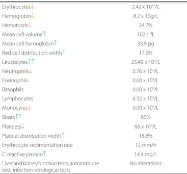
Blood tests (Table 1) were performed and revealed anemia, neu-tropenia with 80% blast cells in the white cell series, thrombocyto-penia, and marginally increased C-reactive protein (CRP) levels. Based on these findings, the patient was referred to the Hematology De-partment, where bone marrow examination was performed (Figure 2), revealing increased cellularity with 97% blast cells. The patient was diagnosed with ALL type B (CD10+) using immunophenotyping.
Intensive systemic chemotherapy (rituximab-hyperCVAD (fractio-nated cyclophosphamide, vincristine, doxorubicin, and dexame tha-sone) regimen plus intrathecal therapy (methotrexate/cytara bine) was
Acute lymphoblastic leukemia presenting with bilateral serous macular detachment
Leucemia linfoblástica aguda com descolamento macular seroso bilateral
Luisa Vieira1, NuNo aguiar siLVa1, Marco Dutra MeDeiros1,2, rita FLores1,2, Vitor MaDuro1,2
Submitted for publication: September 19, 2014 Accepted for publication: February 3, 2015
1 Central Lisbon Hospital Center, Lisbon, Portugal. 2 NOVA Medical School, Lisbon, Portugal.
Funding: No specific financial support was available for this study.
Disclosure of potential conflicts of interest: None of the authors have any potential conflict of interest to disclose.
Corresponding author: Luísa Vieira. José António Serrano St. 1150-199 Lisbon, Portugal E-mail: [email protected]
ABSTRACT
Acute lymphoblastic leukemia is a malignant hematopoietic neoplasia, which is rare in adults. Although ocular fundus alterations may be commonly observed in the course of the disease, such alterations are rarely the presenting signs of the disease. Here we describe the case of a patient with painless and progressive loss of visual acuity (right eye, 2/10; left eye, 3/10) developing over two weeks, accompanied by fever and cervical lymphadenopathy. Fundus examination showed bilateral macular serous detachment, which was confirmed by optical coherence tomography. Fluorescein angiography revealed hyperfluorescent pinpoints in the posterior poles. The limits of the macular detachment were revealed in the late phase of the angiogram. The results of blood count analysis triggered a thorough, systematic patient examination. The diagnosis of acute lymphoblastic leukemia B (CD10+) was established, and intensive systemic chemotherapy was immediately initiated. One year after the diagnosis, the patient remains in complete remission without any ophthalmologic alterations.
Keywords: Acute lymphoblastic leukemia; Serous macular detachment; Visual acuity; Optical coherence tomography; Fluorescein angiography
RESUMO
A leucemia linfoblástica aguda é uma neoplasia maligna das células hematopoié-ticas, incomum em adultos. Apesar da maioria dos casos apresentar alterações no fundo ocular no decurso da doença, estas são raramente forma de apresentação da mesma. Descreve-se o caso de uma doente com diminuição progressiva e indolor da acuidade visual (OD 2/10 e OE 3/10), que apresentava concomitantemente febre e adenopatias cervicais, com duas semanas de evolução. À oftalmoscopia apresentava descolamento seroso macular bilateral, confirmado por tomografia de coerência ótica. A angiografia fluoresceínica revelou pequenas lesões hiperfluorescentes tipo pinpoints no polo posterior. Nos tempos médios e tardios do exame adivinham-se os limites da bolsa do descolamento do neuroepitélio. As alterações encontradas no hemograma suscitaram um estudo sistêmico extenso. O diagnóstico de leucemia linfoblástica aguda B (CD10+) foi efetuado, iniciando-se, de imediato, quimioterapia sistêmica intensiva. Um ano após o diagnóstico a doente continua em remissão e sem alterações oftalmológicas de novo.
Vieira L, et al.
3 8 3 Arq Bras Oftalmol. 2015;78(6):382-4
Figure 2. Giemsa-stained smear of bone marrow aspirate. A lymphoblast is indi-cated by the arrow.
Figure 3. Findings after the second cycle of chemotherapy: A) and B) Fundus photographs of the right and left eye, respectively, with no signiicant alterations. C) and D) Optical coherence tomography (horizontal sections) of the right and left eye, respectively, at the level of the fovea, showing reattached maculas.
A
C
B
D A
C
E
B
D
F
Figure 1. Findings at presentation: A) and B) Fundus photographs of the right and left eye, respectively, showing shallow serous macular detachment. C) and D) Optical coherence tomography (horizontal sections) of the right and left eye, respectively, at the level of the fovea, showing neurosensory detachment, with central macular thickness of 638 µm and 423 µm in the right and left eye, respectively. E) and F) Fluo-rescein angiogram of the right and left eye, respectively, showing hyperluorescent pinpoints in the posterior poles. The limits of the macular detachment were revealed in the late phase of the angiogram.
Table 1. Blood tests performed upon admission
Erythrocytes↓ 2.42 x 10¹²/L
Hemoglobin↓ 8.2 x 10g/L
Hematocrit↓ 24.7%
Mean cell volume↑ 102.1 fL
Mean cell hemoglobin↑ 33.9 pg
Red cell distribution width↑ 17.5%
Leucocytes↑↑ 25.40 x 10⁹/L
Neutrophils↓ 0.76 x 10⁹/L
Eosinophils 0.00 x 10⁹/L
Basophils 0.00 x 10⁹/L
Lymphocytes 4.32 x 10⁹/L
Monocytes↓ 0.00 x 10⁹/L
Blasts↑↑ 80%
Platelets↓ 66 x 109/L
Platelet distribution width↑ 18.8%
Erythrocyte sedimentation rate 12 mm/h
C-reactive protein↑ 14.4 mg/L
Liver and kidney function tests, autoimmune test, infection serological tests
No alterations
scheduled and initiated immediately. After the first che mo therapy cycle, complete remission was achieved, and a BCVA of 10/10 was obtained in both eyes with reattachment of the macula (Figure 3) after the second cycle. One year after the diagnosis, the patient remains in complete remission without any ophthalmologic alterations.
DISCUSSION
Ophthalmological involvement in leukemia, which is more common in acute than in chronic disease(4,6), is not unusual but is rarely a
pre-senting sign of the disease(5). Although leukemic retinopathy
(intra-retinal hemorrhages, white-centered hemorrhages and cotton-wool spots) is the most frequent fundus finding and retinal infiltrates are easily seen, choroidal infiltration is rarely observed clinically (5). Ho-wever, the choroid is affected in 30% to 93% of the cases as demons-trated by histopathological studies(2,4,7), and decreased thickness after
chemotherapy has been reported by Bajenova et al.(7).
When the choroidal infiltration is severe, it leads to serous retinal detachment, which is reported to be shallow in the posterior pole, more common in adults, and bilateral(5).
lite-Acute lymphoblastic leukemia presenting with bilateral serous macular detachment
384 Arq Bras Oftalmol. 2015;78(6):382-4
rature(5). Moreover, the resolution of the macular detachment with
systemic chemotherapy was in favor of our theory that serous macular detach ment was due to leukemic choroidal infi ltration.
The physiopathological mechanism postulated(5) is that choroidal
infi ltration leads to a decreased blood fl ow in the choriocapillaris, re-sulting in ischemia and the disruption of the intercellular tight junctions or the necrosis of the retinal pigment epithelium, as evidenced by the multifocal hyperfl uorescent pinpoints in the early phases of fl uo-res cein angiography. This decompensated posterior blood-retinal barrier leads to exudation as evidenced by the diff use subretinal accumulation of fl uorescein in the late phase of the angiogram.
This rare case illustrates the importance of a systematic examina-tion in managing a case of bilateral macular serous detachment, with no other signs suggestive of local injury. This is particularly important when early treatment is imperative and is critical to patient survival.
ACKNOWLEDGEMENTS
We would like to thank the Department of Hematology for precious contribution in the follow-up of this patient, and the Department of
Orthotics for valuable assistance in the acquisition of images for this article.
REFERENCES
1. Seiter K. Acute lymphoblastic leukemia. Available at http://emedicine.medscape.com/ article/207631-overview
2. Berthou C, Roncin S, Colin J, Abgrall JF. Ocular sites of acute leukemias. J Fr Ophtalmol. 1996;19(6-7):470-8.
3. Kim DY, Moon JH, Joo YD. Current status and future directions of clinical research and practice in adult acute lymphoblastic leukemia patients in Korea. Blood Res. 2014 Jun; 49(2):80-2.
4. Gordon KB, Rugo HS, Duncan JL, Irvine AR, Howes EL, Jr, O’Brien JM, Carter SR. Ocular manifestations of leukemia: leukemic infi ltration versus infectious process. Ophthal-mology. 2001;108(12):2293-300.
5. Riss JM, Kaplanski G, Righini-Chossegros M, Harle JR, Escoffi er P, Saracco JB. Bilateral serous detachment of neuroepithelium of the posterior pole disclosing acute leuke-mia. J Fr Ophtalmol. 1990;13(11-12):563-8.
6. Reddy SC, Jackson N. Retinopathy in acute leukaemia at initial diagnosis: correlation of fundus lesions and haematological parameters. Acta Ophthalmol Scand. 2004 Feb; 82(1):81-5.
7. Bajenova NV, Vanderbeek BL, Johnson MW. Change in choroidal thickness after che-mo therapy in leukemic choroidopathy. Retina. 2012 Jan;32(1):203-5.