Arq Neuropsiquiatr 2011;69(2-A):188-191
188
Article
Radicular dysfunction preponderance
at early phase clinical evaluation in
myelitis by Schistosoma mansoni
Claudio Henrique Fernandes Vidal1,2, Joacil Carlos da Silva1,2, Jeferson Jane Oliveira Souza2, Sara Pimentel Belleza Bernardino3, Maria Lucia Brito Ferreira4ABSTRACT
In neuroschistosomiasis, the spinal cord is the most common place of the disease. In high prevalent areas for schistosomiasis mansoni, the clinical alertness is important for an early diagnostic, in order to decrease the final neurological damage. This study provides some useful neurologic information about a series of patients with schistosomal myelitis. Method: The sample consisted of 13 schistosomiasis mansoni carriers examined at the moment of the diagnosis of myelitis. Results: The classical triad (lumbago, weakness at the lower limbs and urinary dysfunctions) was documented in 11 (86.61%) patients. The distribution of the clinical forms was: myeloradicular in six patients (46.15%), radicular in four (30.76%) and myelitic in three (23.07%). Conclusion: The radicular dysfunction and their clinical associated forms were the most prominent pattern during the early phase of this disease.
Key words: schistosomiasis mansoni, neuroschistosomiasis, myelitis, neurologic exami-nation, medical history taking.
Preponderância de disfunções radiculares na avaliação clínica precoce da mielite pelo
Schistosoma mansoni
RESUMO
Na neuroesquistossomose, a medula espinhal é o sítio de predileção da doença. Em áreas de alta prevalência para esquistossomose mansoni, o conhecimento clínico dessa condição é importante para o seu diagnóstico precoce e consequente redução da lesão neurológica definitiva. Este estudo provê informações neurológicas relevantes pertinentes a uma série clínica de pacientes com mielite esquistossomótica. Método: A amostra consistiu de 13 pacientes portadores de esquistossomose mansoni examinados no momento do diagnóstico de sua forma mielítica. Resultados: A tríade clássica (lombalgia, fraqueza nos membros inferiores e disfunções urinárias) foi documentada em 11 (86,61%) pacientes. Quanto à distribuição das formas clínicas, se observou a ocorrência da mieloradicular em seis pacientes (46,15%), da radicular em quatro (30,76%) pacientes e da mielítica em três (23,07%) pacientes. Conclusão: A disfunção radicular e suas formas clínicas associadas foram o padrão mais frequente durante a avaliação na fase precoce da doença.
Palavras-chave: esquistossomose mansoni, neuroesquistossomose, mielite, exame neurológico, anamnese.
Correspondence Claudio Henrique F. Vidal Rua Francisco da Cunha 206 51020-041 Recife PE - Brasil E-mail: [email protected]
Received 6 July 2010
Received in final form 3 October 2010 Accepted 14 October 2010
1Departamento de Pós-graduação em Neuropsiquiatria e Ciências Comportamentais do Centro de Ciências da Saúde da
Universidade Federal de Pernambuco (UFPE), Recife PE, Brazil; 2Neurosurgeon; 3Resident of Neurology; 4Head of Neurology,
Hospital da Restauração, Recife PE, Brazil.
Schistosomiasis is one of the older and more diffused parasitic diseases of the world, and nowadays represents a
Arq Neuropsiquiatr 2011;69(2-A)
189
S. mansoni myelitis: radicular dysfunction Vidal et al.
hypertension among the young population. Around the world, it is believed that there are 200 million people in-fected by the specimens mansoni, haematobium, japon-icum, intercalatum or mekong1,2.
In neuroschistosomiasis, the spinal cord is the most common place of the disease3. he exact frequency of myelopathy in these patients has never been properly studied, in part because of the wide variations in clinical expression4. Nowadays, neuroschistosomiasis is consid-ered the third most common cause of myelopathy, after traumatic and tumor involvements5,6.
Originally from the African continent, Schistosoma mansoni (SM) was brought to Brazil during the slavery period, and found favorable weather conditions and a large number of hosts for its proliferation. Currently, bad sanitary conditions in Brazil perpetuate the endemic status of this severe parasitic disease2.
The first reference to myelitis by SM was made in 1930 when a German citizen that lived in Brazil devel-oped spinal cord afection at the thoracic and lumbar levels. he necropsy study showed eggs of SM. In 1945, Gama registered the irst case of schistosomal myelitis in the Brazilian medical literature, and the diagnosis was made by the histopathological study of surgical tissue. In 1956, Martinez recommended a high level of suspi-cion in cases of transverse myelitis that came from en-demic zones for SM. In 1964, Oliveira classiied the dis-ease in three diferent syndromes: pseudotumoral spinal cord compression, myeloradiculopathy and myelobulbar, apud Santos7.
In high prevalent areas for schistosomiasis, clinical alertness and the use of pertinent exams for the diag-nosis must be performed as soon as possible, for all pa-tients with complaints of: acute or subacute lumbago, paresthesic sensation at the lower limbs and urinary dys-function3,4,8. he physical exam can reveal motor deicit in lower limbs and altered sensation on dermatomes of lumbosacral roots.
he recent advances in neurological radiology and in immunology have increased the knowledge of the phys-iopathology and clinical aspects of this intriguing con-dition. herefore, this diagnosis has become more fre-quent8. his study adds information about the clinical exam of patients with the spinal form of schistosomiasis at the moment of the development of their symptoms.
METHOD
he study protocol was approved by the institutional review board of the participating center (Hospital da Restauração, CAAE- 0033.0.102.172-05) and patients gave written consent for data analysis.
his study was a sectional and descriptive evaluation (in a series of cases), from a hospital base, where the
sample consisted of schistosomiasis mansoni carriers at the moment of the diagnosis of myelitis. he patients were hospitalized in the neurological inirmary between July 2005 and June 2006.
he authors used as inclusion criteria, the recom-mendations for diagnosis from the Centers for Disease Control (CDC)9: dysfunction of thoracic or lumbosa-cral spinal cord; parasitological conirmation of the in-fection by SM and exclusion of other causes for trans-versal myelitis.
he neurological form of the disease was deined by some clinical indings during examination. For the spinal cord form, the features considered were: symmetry of the deicits, hyperactive deeper tendon relex, and deinition of a sensitive or motor spinal level. he radicular forms were considered when the motor deficits were asym-metric, deeper tendon relex was decreased or absent, and with radicular (in distribution) sensitive deicits.
RESULTS
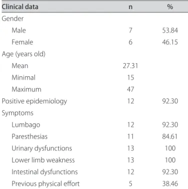
A total of 13 patients were evaluated during the pe-riod of study. he anamnesis data are shown in Table 1. he classical triad (lumbago, weakness at the lower limbs and urinary dysfunctions) was documented in 11 (86.61%) patients.
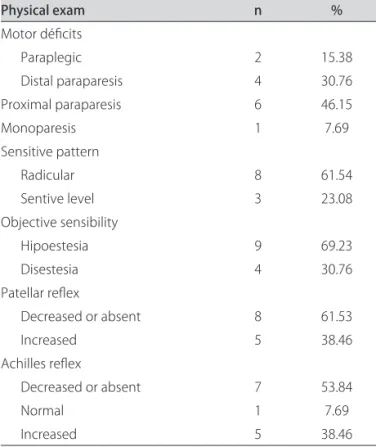
he results of the physical examination of the patients are shown in Table 2. Whatever the motor deicits, in six cases (46.15%) they were considered symmetric, and asymmetric in the others (seven cases - 53.84%). Sellar sensitive alterations occurred in nine (69.23%) patients.
Concerning the clinical forms, they are described in Table 3.
Table 1. Anamnesis data of the patients.
Clinical data n %
Gender
Male 7 53.84
Female 6 46.15
Age (years old)
Mean 27.31
Minimal 15
Maximum 47
Positive epidemiology 12 92.30
Symptoms
Lumbago 12 92.30
Paresthesias 11 84.61
Urinary dysfunctions 13 100
Lower limb weakness 13 100
Intestinal dysfunctions 12 92.30
Arq Neuropsiquiatr 2011;69(2-A)
190
S. mansoni myelitis: radicular dysfunction Vidal et al.
DISCUSSION
he distribution, whatever the gender, was very sim-ilar (53.84% of males and 46.15% of females), and di-verges from the literature, that refers to men a greater risk of acquiring this condition. his occurs because of social and economic factors as recreational activities and heavy work, and consequently a higher frequency of physical eforts, that cause higher levels of abdominal inner pressure4,8,10.
he mean age was 27.31 years (ranging from 15 to 47 years). hese results are in accordance with other re-ports3,7, and relect the most prevalent age for schistoso-miasis (between 10 and 40 years of age)11. hese young individuals usually are more exposed to infected waters, moreover they usually are involved in more physical ac-tivities, and therefore are more susceptible to higher ab-dominal inner pressure and eggs migration.
A positive epidemiological history for schistosomiasis was present in 12 (92.3%) patients, and this was deined by the contact with natural reservoirs. Eight (61.53%)
pa-tients had had direct contact with the snail (Biomphal-aria). he only case that didn’t fulill this criterion came from a high prevalent zone for this disease (Forest zone - Zona da Mata- of Pernambuco State), and had the clas-sical clinical picture with parasitic conirmation in stools and positive reactions for schistosomiasis in the cerebro-spinal luid. hese data are diicult to confront with the ones of other series, because they usually are not shown. In the anamnesis, all the patients had complaints of weakness of the lower limbs and urinary dysfunction. hese symptoms have been considered very typical of this disease4. Urinary retention was found in 12 (92.3%) of the patients and was attributed to damage at the conus medullaris (mictional center), cauda equine (aferent and efferent pathways) or just efferent pathways. The two former situations are called autonomic bladder and the last one, motor-paralytic bladder. One of the patients showed urinary incontinence (by reflex bladder), and this patient had a pure spinal cord form of the disease. here was a high occurrence of fecal retention (92.3% of the cases). Similar data was supported by Correia12 and Asano’s13 casuistic in which sphincters symptoms were the most prevalent.
Lumbago was described in 12 (92.3%) of the cases, and this was in accordance with the literature consulted, that described this symptom from 72.2% up to100% of the patients3,4,10,12,14,15. he classical triad of complaints (lumbago, urinary dysfunction and paresthesias in lower limbs), was identiied in 11 (84.61%) patients in the present study. Peregrino et al.3,8 emphasized the ne-cessity that health care professionals that work in en-demic zones for schistosomiasis be familiarized with these complaints. his surveillance would promote an early diagnosis, and probably would decrease the occur-rence of deinitive neurological damage.
he motor exam revealed that two (15.38%) patients were paraplegic, and four (30.77%) had predominant distal paraparesis whereas six (46.15%), had predominant prox-imal paraparesis. Crural monoparesis was observed in just one (7.69%) patient. hus, there were 10 paraparetic patients, of whom six (60%) had proximal deicit and four (40%) had distal deicit. In six cases (46.15%) the deicits were considered symmetric, and in the other seven cases (53.84%) asymmetric. An equivalent amount of parapa-retic patients was reported by Santos et al.4, but all those patients had a distal and symmetric distribution. hese patterns diverge from the present report. Maybe a prox-imal and asymmetric distribution, as it occurred in our patients, can be better associated with the multi-radicular compromising usually observed in schistosomal myelitis. Whatever the deep tendon reflex, patellar and Achilles relex were decreased or absent in 8 (61.53%) and in 7 (53.84%) of the patients, respectively. hey were
Table 2. Physical examination of the patients.
Physical exam n %
Motor déicits
Paraplegic 2 15.38
Distal paraparesis 4 30.76
Proximal paraparesis 6 46.15
Monoparesis 1 7.69
Sensitive pattern
Radicular 8 61.54
Sentive level 3 23.08
Objective sensibility
Hipoestesia 9 69.23
Disestesia 4 30.76
Patellar relex
Decreased or absent 8 61.53
Increased 5 38.46
Achilles relex
Decreased or absent 7 53.84
Normal 1 7.69
Increased 5 38.46
Table 3. Clinical forms of schistosomal myelitis.
Clinical forms n %
Radicular 4 30.76
Myeloradicular 6 46.15
Myelitic 3 23.07
Arq Neuropsiquiatr 2011;69(2-A)
191
S. mansoni myelitis: radicular dysfunction Vidal et al.
increased in 5 (38.46%) patients for each relex. Santos et al.4 found similar results in their analyses. Instead of high spinal cord involvement detected in an MRI during this study (12 patients, 92.3%), the long pathways dysfunction was blunted during clinical evaluation, probably by con-comitant radicular involvement (46.15% of the myelora-dicular form and 30.76% of just the ramyelora-dicular form). An-other inding was the short period of time between the beginning of the symptoms and the neurological exam. his may explain the low expression of liberation pyra-midal signs. We believe that this brief evolution also jus-tiies the absence of pseudotumoral forms.
he sensitive disturbances were: deicitary syndrome (hypoesthesia) in 9 (69.23%) cases, and irritative syn-dromes like paresthesias in 4 (30.76%). A pure spinal cord sensitive level could just be deined in 3 (23.08%) cases, whereas in the remaining, some radicular patterns were observed which is in accordance with Galvão10 data. Sellar sensitive alterations occurred in 9 (69.23%) pa-tients, corroborating the high index of dysfunction at the conus medullaris and cauda equine.
he myelitic form of the disease was observed in just 3 (23.07%) of the patients, and the myeloradicular form was present in 6 (46.15%) cases. A pure radicular form was documented in 4 (30.76%) patients. he pseudotu-moral form wasn’t present in the current study. Peregrino et al.8 and Asano13 reported the myeloradicular form in 72.5% and 96.5% of their patients, respectively. Probably in these series, the clinical neurological evaluation was done in a moment that the signals of liberation of the superior motor neuron dysfunction, dominated the clinical setting.
In a survey of the literature available until 2001, Santos et al.4 catalogued the following distribution: my-eloradicular (71 cases/44.4%), myelitic (44 cases/27.5%) and pseudotumoral (39 cases/24.4%). hese results are closer to the indings of the present study. Valença, Barros and Ataíde16 after analyses of 60 cases, observed that the myelitic form was preponderant (62%), but the signals of motor neuron compromising (radicular) were also common, especially during the acute phase of the disease.
he nature of this study (sectional) forbids any in-ference about the course of the clinical manifestations over time. Probably in a coorte study, the sample might change their indings during the illness, with the radic-ular pattern being more common at an early phase of the neuroschistosomiasis and myelitic features might pre-ponderate at a late phase.
REFERENCES
1. Doumenge JP. Atlas de la répartition mondiale des schistosomiasis. Ge-nève: OMS/WHO; 1987.
2. Coutinho AD, Domingues ALC. Esquistossomose mansoni. In: Dani R, Castro LP (Eds). Gastroenterologia clínica. Rio de Janeiro: Guanabara Koogan; 1993:1697-1728.
3. Peregrino AJP, Oliveira SP, Porto CA, et al. Meningomielorradiculite por Shistosoma mansoni: protocolo de investigação e registro de 21 casos. Arq Neuropsiquiatr 1988;46:49-60.
4. Santos EC, Campos GB, Diniz AC, Leal JC, Rocha MOC. Peril clínico e cri-térios diagnósticos da mielorradiculopatia esquistossomótica. Arq Neu-ropsiquiatr 2001;59:772-777.
5. Spina-França A, Salum PNB, Limongi JCP, Berger A, Losso ER. Mielopatias: aspectos diagnósticos. Arq Neuropsiquiatr 1980;38:360-366.
6. Brito JCF, Nóbrega PV. Mielopatias: considerações clínicas e aspectos eti-ológicos. Arq Neuropsiquiatr 2003;61: 816-821.
7. Santos EC. Peril diagnóstico da mielorradiculopatia esquistossomótica em três hospitais de Belo Horizonte: estudo retrospectivo - 1972 a 1992. [Dissertação Mestrado]. Belo Horizonte: Faculdade de Medicina da Uni-versidade Federal de Minas Gerais; 1994.
8. Peregrino AJP, Puglia PMK, Nóbrega JPS, Livramento JA, Marques-Dias MJ, Scaf M. Esquistossomose medular: análise de 80 casos. Arq Neurop-siquiatr 2002;60:603-608.
9. Centers for Disease Control and Prevention: acute schistosomiasis with transverse myelitis in American students returning from Kenya [editorial note]. Morb Mortal Wkly Rep 1984;33:446-447.
10. Galvão ACR. Milelopatias esquistossomóticas; aspectos clínicos e labora-toriais. [Dissertação de Mestrado]. São Paulo: Faculdade de Medicina da Universidade de São Paulo; 1983.
11. Raso P. Esquistossomose Mansônica. In: Filho GB (Ed). Bogliolo Patologia. Rio de Janeiro: Editora Guanabara Kookan SA; 2000:1186-1207. 12. Correia CC. Aspectos clínicos, eletromiográicos e de evolução de
paci-entes com neuroesquistossomose. [Dissertação de Mestrado]. Recife: Fac-uldade de Medicina da Universidade de Pernambuco; 2004.
13. Asano NMJ. Neuroesquistossomose: Aspectos clínicos, laboratoriais e de imunodiagnóstico. [Dissertação de Mestrado]. Recife: Faculdade de Me-dicina da Universidade Federal de Pernambuco; 1992.
14. Ferrari TCA, Moreira PRR, Ferrari MLA, et al. Clinical and immunolog-ical study of schistomal myelorradiculopathy. Ann Trop Med Parasitol 1993;87:295-297.
15. Costa RO, Gameleira FT, Tenório RB, Bras LH, Costa VB, Jr JMP. Neuroesquis-tossomose em Alagoas. Rev Bras Neurol 1992;28:79-84.