Management of degenerative cervical myelopathy – An update
ANDREI F. JOAQUIM1*, ENRICO GHIZONI1, HELDER TEDESCHI1, WELLINGTON K. HSU2, ALPESH A. PATEL3
1MD, PhD. Department of Neurology, Neurosurgery Division, Universidade Estadual de Campinas (Unicamp), Campinas, SP, Brazil 2MD. Department of Orthopaedic Surgery, Northwestern University, Chicago, IL, USA
3MD, FACS. Department of Orthopaedic Surgery, Northwestern University, Chicago, IL, USA
SUMMARY
Study conducted at Universidade Estadual de Campinas (Unicamp),
Campinas, SP, Brazil
Article received: 8/12/2015
Accepted for publication: 1/10/2016
*Correspondence:
Departamento de Neurologia Address: Rua Tessália
Vieira de Camargo, 126, Cidade Universitária Zeferino Vaz
Campinas, SP – Brazil Postal code: 13083-887
http://dx.doi.org/10.1590/1806-9282.62.09.886
Introduction: Degenerative cervical myelopathy (DCM) is the most common cause of spinal cord dysfunction in adult patients. Patients generally present with a slow, progressive neurological decline or a stepwise deterioration pattern. In this paper, we discuss the most important factors involved in the management of DCM, including a discussion about the surgical approaches.
Method: The authors performed an extensive review of the peer-reviewed litera-ture addressing the aforementioned objectives.
Results: Although the diagnosis is clinical, magnetic resonance imaging (MRI) is the study of choice to conirm stenosis and also to exclude the differential diagnosis. The severity the clinical symptoms of DCM are evaluated by different scales, but the modiied Japanese Orthopedic Association (mJOA) and the Nürick scale are probably the most commonly used. Spontaneous clinical improvement is rare and surgery is the main treatment form in an attempt to prevent further neurological deterioration and, potentially, to provide some improvement in symptoms and function. Anterior, posterior or combined cervical approaches are used to decompress the spinal cord, with adjunctive fusion being com-monly performed. The choice of one approach over the other depends on patient characteristics (such as number of involved levels, site of compression, cervical alignment, previous surgeries, bone quality, presence of instability, among oth-ers) as well as surgeon preference and experience.
Conclusion: Spine surgeons must understand the advantages and disadvan-tages of all surgical techniques to choose the best procedure for their patients. Further comparative studies are necessary to establish the superiority of one approach over the other when multiple options are available.
Keywords: cervical myelopathy, spondylotic myelopathy, surgical approach, anterior approach, posterior approach.
I
NTRODUCTIONChronic cervical degeneration or spondylosis (CS) is a natural consequence of aging, resulting in arthritic chang-es in the intervertebral discs, facet joints, ligaments and vertebral bodies.1,2 These include abnormal bony spurs
on the vertebrae, disc and joints degeneration, ligament hypertrophy and ossiication. However, although the vast majority of the general population will have cervical spon-dylosis, only the minority of patients will have clinical symptoms.2
Degenerative cervical myelopathy (DCM) is the most common cause of spinal cord dysfunction in adult patients and a clinical presentation of spondylosis resulting in spinal cord compression.2-4 The degenerative changes, at
approach is generally individualized according to many factors involved in each case and surgeon’s experience.5
In this paper, we discuss the most important factors involved in the surgical management of DCM, including the approaches.
N
ATURAL HISTORYThe clinical history of patients is quite variable, similarly to its radiological presentation.2 Some patients may
pres-ent with a slow and progressive neurological decline, whereas others may have a stepwise pattern with periods of quiescent disease prior to deterioration.2,5,6 An acute
presentation is not common, with exception for cervical trauma or acute disc herniation.2
It is well known that patients with symptoms will be unlikely to improve without surgical treatment. Non operative management techniques, while utilized to ad-dress concurrent neck pain, do not improve the underly-ing disease state of compression, demyelination, macro and micro-vascular architecture changes, neuronal and oligodendrocyte apoptosis and destruction of the blood-spinal cord barrier. Under chronic pressure, necrosis of white and gray matter ensues in the cervical spinal cord.2,6
Finally, almost all patients untreated surgically will wors-en and, in some reports, over 50% will progress to severe clinical disability.7-10
C
LINICAL PRESENTATIONPhysical examination of a DCM patient may include at least one long-tract sign localized to the cervical spinal cord (Babinski, Oppenheim and Hoffman signs, clonus, hyperrelexia, crossed abductor sign, coordination impair-ment and gait dysfunction). Symptomatically, patients can complain of tingling and numbness in the arms and hands, muscle weakness, gait dificulty, neck and arm pain, loss of coordination, heavy feelings in the legs, and deterioration in ine motor skills (such as buttoning a
shirt).11 Neck pain and stiffness are also common, with
restriction to movements. Finally, some patients may uncommonly present with bladder dysfunction.
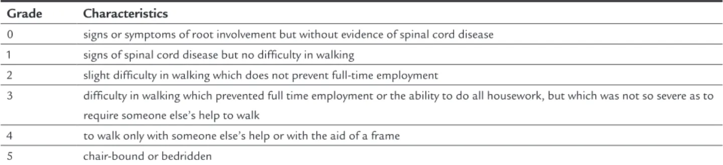
The two most common systems used to evaluate the severity of DCM are the Nürick grade system (Table 1) and the Japanese Orthopedic Association (JOA).12,13 Of
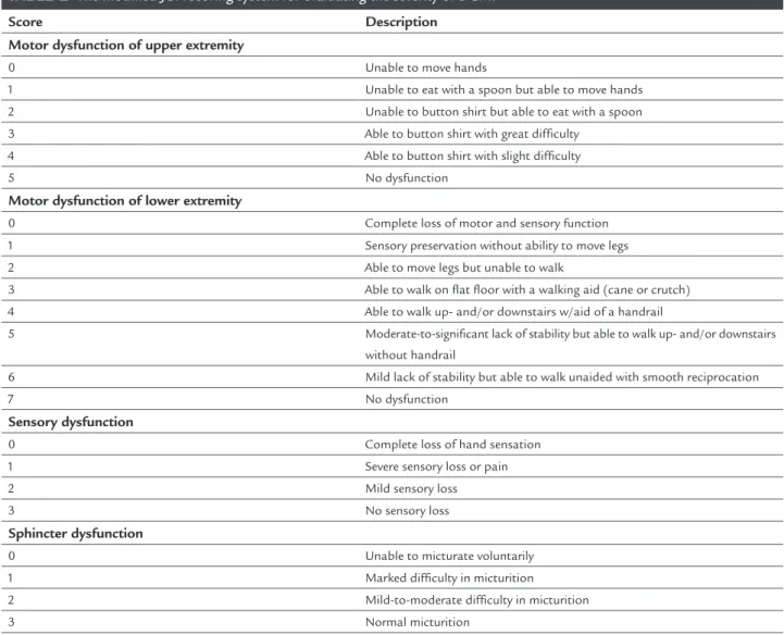
note, a modiied version of the JOA has been proposed and validated for western cultures (Table 2).14
The JOA scale is an objecting assessment scale of the severity of DCM.12 It is based on six domain scores: 1)
Motor dysfunction of the upper extremities; 2) Motor dysfunction of the lower extremities; 3) Sensory dysfunc-tion in the upper extremities; 4) Sensory funcdysfunc-tion in the trunk; 5) Sensory function in the lower extremities and 6) Bladder function.
The JOA score can range from 0 to a maximum of 17 points. Yonenobu et al. deined the myelopathy as mild if the JOA is larger than 12, moderate from 9 to 13 and severe when the JOA is less than 9 points.15
A modiied JOA score, which improves applicability towards Western cultures, replaces the use of chopsticks with a spoon to evaluate motor function in the upper extremities, assessing only motor dysfunction in the up-per and lower extremities, sensory function in the upup-per extremities and bladder function, excluding sensory func-tion in the trunk and lower extremities (four domain scores).14 Each one of the four scales varies, respectively,
from 0 to 5, 7, 3 and 3, ranging from 0 to 18 points. Feh-lings et al. proposed classifying the severity of the my-elopathy using the mJOA as mild (15 or more), moderate (12 to 14) and severe (less than 12).16 Of note, a score of
17 in the JOA scale or 18 in the mJOA scale relects a normal neurological function.
Finally, Kato et al. performed a prospective multicenter study and reported that the both JOA and the mJOA have a good correlation, but it was not ideal to use them inter-changeably.17
TABLE 1 The Nürick grade system – Six grades of severity based on “difficulty in walking”.
Grade Characteristics
0 signs or symptoms of root involvement but without evidence of spinal cord disease 1 signs of spinal cord disease but no difficulty in walking
2 slight difficulty in walking which does not prevent full-time employment
3 difficulty in walking which prevented full time employment or the ability to do all housework, but which was not so severe as to require someone else’s help to walk
4 to walk only with someone else’s help or with the aid of a frame
A
DDITIONAL WORK-
UPPlain radiographies
Initial investigation of DCM is based on simple plain cervical radiographs, including lexion-extension exams to detect occult instability.18 CS is characterized by
osteo-phyte formation, loss of cervical disc space, loss of normal cervical alignment, uncovertebral and facet joints hyper-trophy.
The Pavlov ratio is measured based on simple plain cervical radiographies to estimate congenital narrowing of the spinal canal, a major risk of DCM.19,20 Using the
lateral incidence, the spinal canal / vertebral body ratio is determined (a/b), where “a” is the distance from the posterior surface of the vertebral body to the nearest point of the spinal laminar line and “b” is deined as the mid-point between the anterior surface and the posterior
sur-face of the vertebral body. A normal Pavlov ratio is about 1.0, whereas < 0.8 suggests congenital cervical stenosis,
and spondylotic compression occurs when < 0.4. Although the ratio is useful for screening, further radiological in-vestigation is needed when the ratio is < 0.8, as large ver-tebral bodies may skew the inal ratio and yield false positive results.21
Also useful information obtained with plain cervical radiography is the mean value of the spinal canal in the antero-posterior (AP) diameter. In normal adult males, the mean value of the AP diameter of the spinal canal measured on lateral cervical radiograph is 17 to 18 mm at C3-5 and 12 to 14 mm at C6-7. Severe cervical stenosis is presumed when the diameter is less than 10 mm in the lower cervical spine and 10 to 13 in the upper cervical spine.22
TABLE 2 The modified JOA scoring system for evaluating the severity of DCM.
Score Description
Motor dysfunction of upper extremity
0 Unable to move hands
1 Unable to eat with a spoon but able to move hands 2 Unable to button shirt but able to eat with a spoon 3 Able to button shirt with great difficulty
4 Able to button shirt with slight difficulty
5 No dysfunction
Motor dysfunction of lower extremity
0 Complete loss of motor and sensory function 1 Sensory preservation without ability to move legs
2 Able to move legs but unable to walk
3 Able to walk on flat floor with a walking aid (cane or crutch) 4 Able to walk up- and/or downstairs w/aid of a handrail
5 Moderate-to-significant lack of stability but able to walk up- and/or downstairs without handrail
6 Mild lack of stability but able to walk unaided with smooth reciprocation
7 No dysfunction
Sensory dysfunction
0 Complete loss of hand sensation
1 Severe sensory loss or pain
2 Mild sensory loss
3 No sensory loss
Sphincter dysfunction
0 Unable to micturate voluntarily
1 Marked difficulty in micturition
2 Mild-to-moderate difficulty in micturition
Additionally, in cases where cervical deformity is pres-ent, measuring the C2-7 sagittal vertical axis (SVA) is important for outcome purposes. The horizontal distance from two plumb lines, one from the mid-vertebral body of C2 and the second from the posterior superior corner of the C7 vertebral body, greater than 4 cm postopera-tively is associated with poor neck disability scores.18
Finally, 36 inch long cassette X-rays may be useful for assessment of global sagittal balance in patients with severe cervical deformity and concordant thoracolumbar deformity and dynamic lexion-extension x-rays are also used for detecting segmental instability and also assess-ment of cervical lexibility.18
Magnetic resonance imaging
Magnetic resonance imaging (MRI) is the modality of choice to evaluate the neural elements, with multiplanar images and a high accuracy in detecting spinal cord compression secondary to degenerative changes. It is the gold standard modality for conirming the diagnosis of DCM. In addition to diagnosis, MRI can provide prognostic information: patients with high T2 signal intensity and concomitant a low T1 signal intensity in the spinal cord generally have unfavorable outcomes. Some new studies using new MRI techniques, such as diffusion tensor imaging (DTI) and iber tractography can demonstrate spinal cord changes and DCM earlier than conventional sequences.23
CT scan
Conventional CT scan can demonstrate a small diameter of the spinal canal, osteophytes and degenerative chang-es but has a poor visualization of the spinal cord. It pro-vides detailed information of the bone anatomy, with superiority when compared to X-rays and even MRI.24 CT
scan is important for surgical planning when spinal in-strumentation will be required for preoperative anatom-ical study including that of the upper cervanatom-ical spine.
In patients with contra-indications for an MRI study or those with previous surgeries and spinal instrumenta-tion that may affect image quality, CT myelography can demonstrate indirect cord and nerve root compression.24,25
CT myelography can also be used for calculating the com-pression ratio, a relationship between the smallest sagit-tal diameter of the spinal canal / transverse diameter x 100 (%). A compression rate of less than 0.4 correlates with clinical evidence of DCM and with a less favorable clinical outcome. By this reasons, some authors proposed that surgery should be indicated before this degree of deformation occurs into the spinal cord.26
Electromyography
Although limited in the assessment myelopathy,
electro-myography (EMG) may demonstrate radiculopathy.27 It
is, however, most useful in assessing differential diagno-ses, such as motor neuron diseases (such as amyotrophic lateral sclerosis) and peripheral neuropathy. Addition-ally, EMG may offer some prognostic information: patients without myelopathy (mJOA scale of 18 or normal) but with severe stenosis and abnormal indings on the EMG have been shown in one study to eventually develop symp-tomatic DCM.26
Somatosensory and motor evoked potential (SSEP and MEP)
In patients with cervical cord compression, the somato-sensory evoked potential (SSEP) are delayed or have low amplitude.26 Cortical motor evoked potential (MEP) are
more sensitive than SSEP in assessing early spinal cord dysfunction.28 Bednarik et al. demonstrated that patients
with cervical stenosis without clinical myelopathy who had abnormal SSEP had statistically more chances of developed DCM than those with normal SSEP, suggesting a predictable importance of this exam.26
N
ORMAL CERVICAL ALIGNMENTAssessment of cervical alignment is important for plan-ning surgical treatment in DCM. The normal cervical lordosis between C2 to C7 range from 20 to 35 degrees, but these measures depends on patient’s population, age, radiological modality, etc.29 Benzel et al. proposed that
cervical kyphosis can be diagnosed if any vertebra from C3 to C6 crosses a line drawn from the posteroinferior aspect of the body of C2 in the midsagittal plane to the posteroinferior aspect of the body of C7.30 Maintenance
of cervical alignment is important, once it is associated with patient’s outcome.
Lastly, in the cervical spine, about two thirds of the weight-bearing axis lies posterior to the vertebral bodies in the posterior column.30 This emphasizes the importance
of the integrity of the posterior ligamentous and bony structures in the maintenance of cervical alignment.
D
IFFERENTIAL DIAGNOSISDifferential diagnosis of DCM may include:31 primary
A thorough clinical examination, an adequate assess-ment of patient medical history, and the MRI is critical for differential diagnosis of DCM and other pathologies.
S
URGICAL TREATMENTIn cases with moderate or severe DCM, surgical treatment is clearly indicated as non-operative management will lead to continued, and possibly irreversible, neurological decline. Additionally, although recovery of lost function after surgery is generally uncertain, the best surgical results are generally associated with patients with mild DCM and early symptoms.26 Considering these points, some
authors have proposed surgery for all patients with diag-nosis of DCM. However, Kadanka et al. have demon-strated that patients with mild DCM were successfully treat (no signiicant deterioration in the mJOA score, recovery ratio, or timed 10 m walk within either group during the 2 years of follow-up) with close clinical obser-vation in one randomized controlled study comparing conservative versus surgery in spondylotic cervical my-elopathy.32 Of note, a limitation of this study is that,
al-though patients did not deteriorate, they did not have any clinical improvement with clinical treatment.
The main goals of surgical treatment in DCM are to decompress the spinal cord, maintain or restore cervical alignment and stabilize the involved segments when needed. To achieve these goals, many different surgical techniques were proposed, using anterior, posterior or combined approaches. The choice of each one is based on patient characteristics (age, clinical symptoms, num-ber of involved levels, site of compression, cervical align-ment, previous surgeries, patients functional status, bone quality, presence of instability, among others), surgeon’s
preference and analysis of risk and beneits of each ap-proach over the other considering the case.
S
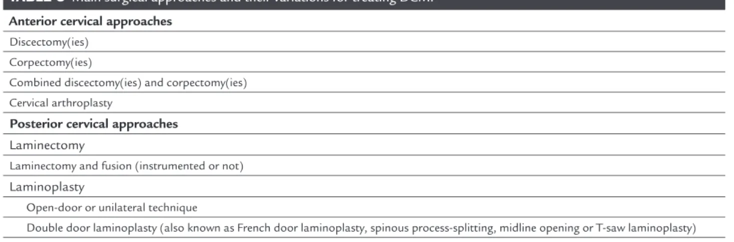
URGICAL APPROACHESConsidering all the surgical goals, we discussed the key components of the most common surgical approaches used to treat DCM, summarized in Table 3.
A
NTERIORCERVICAL APPROACHESAnterior cervical surgery is one of the most frequent spi-nal surgeries performed in the US, mostly commonly used to treat DCM in one, two, or three level disease. The anterior cervical approach was described by Smith and Robinson in 1955.33 It allows an excellent exposure of the
ventral aspect of the spinal cord without touching the neural elements. With the anterior cervical approach, it is also possible to perform disc space distraction that can enlarge the neural foramen and the spinal canal, with direct decompression and restoring cervical lordosis.34,35
The use of autografts or allografts into the disc space is important to achieve fusion.34,35 Fusion is important to
maintain the disc space height without collapse, main-taining cervical lordosis, and also for stabilization of the involved segments, avoiding further new compression.34,35
In cases where ventral cord compression involves a signiicant component of the vertebral body, beyond the disc boundaries, a cervical corpectomy is necessary to ad-equately decompress the spinal cord, followed by recon-struction and stabilization. An interbody device such as a titanium, PEEK, allograft or autograft (harvest from the iliac crest or ibula) is used for reconstruction and fusion.34,35
A combination of discectomies and corpectomies can be used according to patient need and sites of compression.35-37
TABLE 3 Main surgical approaches and their variations for treating DCM.
Anterior cervical approaches
Discectomy(ies) Corpectomy(ies)
Combined discectomy(ies) and corpectomy(ies)
Cervical arthroplasty
Posterior cervical approaches Laminectomy
Laminectomy and fusion (instrumented or not) Laminoplasty
Open-door or unilateral technique
Double door laminoplasty (also known as French door laminoplasty, spinous process-splitting, midline opening or T-saw laminoplasty)
Comparison of anterior discectomies versus corpectomy
Clinical outcomes [JOA, visual analog sclace (VAS) for arm and neck pain] for two levels discectomies were com-parable with one level corpectomy when both options are feasible.38 However, anterior cervical corpectomy and
fusion (ACCF) had a higher operative time and bleeding amount compared with discectomies.38 Additionally,
dis-cectomies obtained better improvements in segmental height and postoperative cervical lordosis when compared with corpectomy.38,39 Of note, although corpectomies may
have a lower rate of pseudoarthrosis than multilevel dis-cectomies, because of the fewer bone graft interfaces, there was a higher stress on bone screws compared with discec-tomy. The choice of one approach over the other depends on surgeons’ preference and patient’s radiological indings.
Cervical arthroplasty
Cervical arthroplasty is also an alternative for treating one or even two levels DCM secondary to degenerative disc disease, especially those secondary to disc herniation. The best surgical candidates are younger patients, without facet joints arthritis and preserved cervical motion.40 The
rationale for performing a cervical arthroplasty instead of fusion is to preserve segmental motion and avoid ad-jacent level disease, even though this has been questioned since heterotopic ossiication may occur in up to 50% of the cases and ASD has not been shown to be decreased by cervical arthroplasty in long term outcomes. Both anterior cervical anterior discectomy and fusion(ACDF) and cervical arthroplasty are effective to treat disc
her-niation in DCM.40 When performing a cervical
arthro-plasty, surgeons may be aware that an inadequate decom-pression may lead to recurrence of myelopathic symptoms.
P
OSTERIOR CERVICAL APPROACHESThe posterior cervical approach is a straightforward alter-native to decompress the spinal cord and nerve roots with direct visualization.41 However, unlike the anterior cervical
approach, posterior approaches require a preoperative lordotic or straight cervical alignment.41 Rigid local or
global kyphosis is a contraindication for posterior decom-pression, as the spinal cord remains compressed and stretched by the anterior elements.41 Posterior approaches
avoid certain complications that are more common with anterior approaches, such as dysphagia, dysphonia and injury to the esophagus and carotid sheath contents.41
Cervical laminectomy
The oldest and most traditional posterior cervical surgery is a decompressive laminectomy.41 It is based on direct
decompression of the spinal canal, enlarging its antero-posterior diameter. Posterior approaches can directly decompress the posterior elements such as the ligamen-tum lavum, posterior bone, facet hypertrophy and also indirectly decompress the ventral elements, shifting the spinal cord posteriorly.41,42
However, as stated previously, the main disadvan-tages of cervical laminectomy are the inability to access ventral pathologies, such as disc herniation and anterior osteophytes, and the high risk of cervical deformity (post-operative kyphosis), which can result in cervical pain and late neurological deterioration.41 Another complication
of an isolated cervical laminectomy is post-laminectomy membrane that may cause recurrent stenosis.
Cervical laminectomy and instrumented fusion
The incidence of post-operative kyphosis after cervical lam-inectomy may be as high as 50%, and dependent on many factors, such as preoperative deformity, the presence of seg-mental instability, removal C2 and/or C7 lamina, extensive laminectomies, wide facetectomies and younger age.41,43 Due
to the risk of post-operative instability, concomitant instru-mented fusion is advocated as a prophylactic maneuver for avoiding deformity and its consequences, especially when treating multilevel spinal cord compression.41,42
Similarly to a standard laminectomy, posterior in-strumentation requires a lordotic or at least straight cer-vical spine alignment. A wide range of modern surgical techniques of instrumentation were described for the cervical spine, such as lateral mass and pedicle screws, laminar screws, pars screws for C2, among many others.41,44
Cervical instrumentation may also avoid a new compres-sion due to instability in the decompressed site. Disad-vantages may include increasing surgical time and cost, implant related complications and loss of cervical range of motion, potentially increasing the chances of adjacent level disease below and above the ixed levels.41
Cervical laminoplasty
Laminoplasty is a surgical technique proposed in the early 1970’s by Japanese surgeons for the treatment of ossiication of the posterior longitudinal ligaments (OPLL) and congenital cervical stenosis.45,46 The goal is to enlarge
A multitude of surgical procedures for expandable laminoplasty were described, but we can group them in two main techniques:45,46
1. open-door or unilateral technique; and
2. double door laminoplasty (also known as French door, spinous process-splitting, midline opening or T-saw laminoplasty).
The open door technique is based on open the lamina in one side (generally the most symptomatic one or with ra-dicular symptoms) and hinge the contra-lateral side, where-in a greenstick fracture is performed. The lamwhere-ina can be maintained in the open position with sutures or miniplates (that offers immediate stability). In contrast, the double door expands the spinal cord symmetrically by opening the mid-line with a split of the spinous processes and hinging both the left and right hemilaminae. The midline laminar splits can be opened with laminar spreaders or bone graft.41,45,46
While laminoplasty offers advantages over cervical fusion, it is not indicated for all patients. It does not ad-dress neck pain and may even cause worsening symptoms compared with anterior approaches, with extensive pos-terior muscle denervation. It is contra-indicated in patients with loss of cervical lordosis and with segmental insta-bility. Complications of laminoplasty may include C5 palsy (5 to 12%), cervical axial pain, decreasing in cervical range of motion (ROM), and progression of OPLL.28,41,46
Interestingly, although C5 palsy is associated classi-cally with posterior decompression, Gandhoke reported that the incidence of C5 palsy was similar comparing anterior cervical corpectomy (31 cases) versus posterior laminoplasty (31 cases as well).47 There were two cases of
C5 nerve root pareses in each group (p=1). There was no differences between the complication or reoperation rates between the two groups (p=0.184 and p=0.238, respec-tively). This study, however, was underpowered to assess different complication rates.
Finally, laminoplasty requires some stabilization to maintain the lamina in a new expanded position. The hard-ware for laminar ixation may increase the cost of the pro-cedure (such as titanium implants) and also add potential complications (such as lamina migration, non union, hard-ware subsidence, etc). Lastly, there is a risk of laminar door closure with recurrence of neurological symptoms.
C
OMPARISON OF ANTERIOR VERSUS POSTERIOR APPROACHES FOR MULTILEVELDCM
Both anterior and posterior approaches are associated with improvement in patients’ inal neurological outcomes in DCM. Some systematic reviews reported that there was a
trend towards better postoperative neural function with anterior approaches, possibly explained by the fact that anterior surgery is generally proposed for several forms of the disease. However, the recovery rate was similar between both according to systematic reviews with meta-analysis.48
Of note, multilevel anterior cervical decompression and fusion had a higher rate of complications compared with posterior surgery.48
Fehlings et al. performed a prospective observational multicenter study of 264 patients comparing the anterior and posterior surgical approaches to treat DCM.49 The choice
of each approach was at the discretion of the surgeon and a follow-up rate of 87% was obtained. Outcome measures included the mJOA scale, Nürick scale, the Neck Disability Index and SF-36 Health Survey version 2 Physical and Men-tal Component Scores. A toMen-tal of 169 patients were treated anteriorly and 95 received a posterior cervical surgery. DCM patients who underwent anteriorly cervical surgery were younger and had less severe myelopathy (as deined by the mJOA and Nürick scores). Both groups had similar baseline Neck Disability Index and SF-36, but the mJOA was lower in the posterior approach group. The extent of improvement in the Nürick scale, Neck Disability Index, SF-36 version 2 Physical Component Score, and SF-36 version 2 Mental Com-ponent Score were similar between the groups, although the mJOA improvement was lower in the anterior group (2.47
vs. 3.62, respectively, p<0.01). They concluded that, although patients who underwent anterior cervical approach were younger and had less severe DCM, both treatments had similar eficacy in the treatment of DCM.
C
OMBINED ANTERIOR-
POSTERIOR APPROACHESIn selected cases, a combined anterior-posterior or pos-terior-anterior approach can be used. The indications for combined approaches include patients requiring oste-otomies for releasing the spine, patients with high risk for hardware failure, such as those with severe osteopo-rosis, and patients with a failure of a previous surgical approach (generally an anterior approach).41,50-52 Combined
approaches may add morbidity of both anterior and pos-terior surgeries, but must be considered in some challeng-ing cases to successfully achieve the goals of surgery (de-compression, stability and good cervical alignment). Of note, patients with 2-stage surgery are at an increased risk of experiencing major complications as they typically have more extensive degenerative pathology.
C
ONCLUSIONdiagnosis is made clinically and conirmed with a cervical MRI. The severity of the DCM can be objectively assessed using the mJOA and the Nürick scale, the most com-monly used scoring for cervical myelopathy.
Surgical treatment is the main treatment modality. The main goals of surgery are to decompress the spinal cord, maintain stability and achieve a good cervical align-ment with an anticipated outcome of neurological pres-ervation or improvement. The choice of one approach over the other depends on patient’s characteristics (such as number of involved levels, site of compression, cervical alignment, previous surgeries, bone quality, presence of instability, among others) and surgeon’s preference.
Spine surgeons must understand the advantages and disadvantages of all surgical techniques to choose the best surgery for each patient to optimize the inal outcome. Further comparative studies are necessary to attest the superiority and differential risks of one approach over the other when multiple options are available.
No funds were received in support of this study. No beneits in any form have been or will be received from a commercial party directly or indirectly related to the sub-ject of this manuscript. The authors have no inancial interest in the subject of this article. The manuscript submitted does not contain information about medical device(s).
R
ESUMOManejo da mielopatia cervical degeneratiiva – Uma atualização
Introdução: a mielopatia cervical degenerativa (MCD) é uma das causas mais comuns de disfunção medular em adultos. Os pacientes em geral apresentam declínio neu-rológico lento e progressivo, ou deterioração escalonada. No presente artigo, discutimos os mais importantes fatores envolvidos no manejo da MCD, incluindo considerações sobre os aspectos relacionados à escolha da abordagem cirúrgica.
Método: realizou-se extensa revisão da literatura de arti-gos peer-reviewed relacionados ao tema.
Resultados: embora o diagnóstico seja realizado clinica-mente, a ressonância magnética (RM) é o estudo de ima-gem de escolha para conirmá-lo e excluir eventuais diag-nósticos diferenciais. A gravidade do quadro clínico pode ser avaliado utilizando-se diferentes escalas, como a modiied Japanese Orthopedic Association (mJOA) ou a de Nürick, provavelmente as mais comuns. Uma vez que
a melhora clínica espontânea é rara, a cirurgia é a princi-pal forma de tratamento, em uma tentativa de evitar dano neurológico adicional ou deterioração e, potencialmente, aliviar alguns sintomas e melhorar a função dos pacientes. Abordagens cirúrgicas por via anterior, posterior ou
com-binada podem ser usadas para descomprimir o canal, concomitantemente a técnicas de fusão. A escolha da abordagem depende das características dos pacientes (número de segmentos envolvidos, local de compressão, alinhamento cervical, cirurgias prévias, qualidade óssea, presença de instabilidade, entre outras), além da prefer-ência e experiprefer-ência do cirurgião.
Conclusão: os cirurgiões de coluna devem compreender as vantagens e desvantagens de todas as técnicas cirúrgi-cas para escolher o melhor procedimento para seus paci-entes. Estudos futuros comparando as abordagens são necessários para orientar o cirurgião quando múltiplas opções forem possíveis.
Palavras-chave: mielopatia cervical, mielopatia es-pondilótica, abordagem cirúrgica, abordagem anterior, abordagem posterior.
R
EFERENCES1. Barnes MP, Saunders M. The effect of cervical mobility on the natural history of cervical spondylotic myelopathy. J Neurol Neurosurg Psychiatry. 1984; 47(1):17-20.
2. Matz PG, Anderson PA, Holly LT, Groff MW, Heary RF, Kaiser MG, et al.; Joint Section on Disorders of the Spine and Peripheral Nerves of the American Association of Neurological Surgeons and Congress of Neurological Surgeons. The natural history of cervical spondylotic myelopathy. J Neurosurg Spine. 2009; 11(2):104-11.
3. Bernhardt M, Hynes RA, Blume HA, White AA 3rd. Current concepts review. Cervical spondylotic myelopathy. J. Bone Joint Surg Am. 1993; 75(1):119-28.
4. Whitecloud TS 3rd. Anterior surgery for cervical spondylotic myelopathy. Smith-Robinson, Cloward, and vertebrectomy. Spine (Phila Pa 1976). 1988; 13(7):861-3.
5. Ito T, Oyanagi K, Takahashi H, Takahashi HE, Ikuta F. Cervical spondylotic myelopathy. Clinicopathologic study on the progression pattern and thin myelinated ibers of the lesions of seven patients examined during complete autopsy. Spine (Phila Pa 1976) 1996; 21(7):827-33.
6. Sadasivan KK, Reddy RP, Albright JA. The natural history of cervical spondylotic myelopathy. Yale J Biol Med. 1993; 66(3):235-42.
7. LaRocca H. Cervical spondylotic myelopathy: natural history. Spine (Phila Pa 1976). 1988; 13(7):854-5.
8. Lees F, Tumer JWA. Natural history and prognosis of cervical spondylosis. Br Med J. 1963; 2(5373):1607-10.
9. Montgomery DM, Brower RS. Cervical spondylotic myelopathy. Clinical syndrome and natural history. Orthop Clin North Am. 1992; 23(3):487-93. 10. Nurick S. The natural history and the results of surgical treatment of the spinal
cord disorder associated with cervical spondylosis. Brain. 1972; 95:101-8. 11. Harrop JS, Naroji S, Maltenfort M, Anderson DG, Albert T, Ratliff JK, et al.
Cervical myelopathy: a clinical and radiographic evaluation and correlation to cervical spondylotic myelopathy. Spine (Phila Pa 1976). 2010; 15(6):620-4. 12. Japanese Orthopaedic Association. Japanese Orthopaedic Association scoring system for cervical myelopathy (17–2 version and 100 version). J Jpn Orthop Assoc. 1994; 68:490-503.
14. Benzel EC, Lancon J, Kesterson L, Hadden T. Cervical laminectomy and dentate ligament section for cervical spondylotic myelopathy. J Spinal Disord. 1991; 4(3):286-95.
15. Yonenobu K, Abumi K, Nagata K, Taketomi E, Ueyama K. Interobserver and intraobserver reliability of the Japanese orthopaedic association scoring system for evaluation of cervical compression myelopathy. Spine (Phila Pa 1976) 2001; 26(17):1890-4.
16. Fehlings MG, Wilson JR, Kopjar B, Yoon ST, Arnold PM, Massicotte EM, et al. Eficacy and safety of surgical decompression in patients with cervical spondylotic myelopathy: results of the AOSpine North America prospective multi-center study. J Bone Joint Surg Am. 2013; 95(18):1651-8.
17. Kato S, Oshima Y, Oka H, Chikuda H, Takeshita Y, Miyoshi K, et al. Comparison of the Japanese Orthopaedic Association (JOA) score and modiied JOA (mJOA) score for the assessment of cervical myelopathy: a multicenter observational study. PLoS One. 2015; 10(4):e0123022. 18. Tang JA, Scheer JK, Smith JS, Deviren V, Bess S, Hart RA, et al.; ISSG. The
impact of standing regional cervical sagittal alignment on outcomes in posterior cervical fusion surgery. Neurosurgery. 2012; 71(3):662-9; discussion 669.
19. Pavlov H, Torg JS, Robie B, Jahre C. Cervical spinal stenosis: determination with vertebral body ratio method. Radiology. 1987; 164(3):771-5. 20. Suk KS, Kim KT, Lee JH, Lee SH, Kim JS, Kim JY. Reevaluation of the Pavlov
ratio in patients with cervical myelopathy. Clin Orthop Surg. 2009; 1(1):6-10.
21. Herzog RJ, Weins JJ, Dillingham MF, Sontag MJ. Normal cervical spine morphometry and cervical spinal stenosis in asymptomatic professional football players. Plain ilm radiography, multiplanar computed tomography, and magnetic resonance imaging. Spine (Phila Pa 1976). 1991; 16(6 Suppl):S178-86.
22. Wolf BS, Khilnani M, Malis L. The sagittal diameter of the bony cervical spinal canal and its signiicance in cervical spondylosis. J Mt Sinai Hosp N Y. 1956; 23(3):283-92.
23. Banaszek A, Bladowska J, Szewczyk P, Podgorski P, Sqsiadek M. Usefulness of diffusion tensor MR imaging in the assessment of intramedullary changes of the cervical spinal cord in different stages of degenerative spine disease. Eur Spine J. 2014; 23(7):1523-30.
24. Henderson L, Kulik G, Richarme D, Theumann N, Schizas C. Is spinal stenosis assessment dependent on slice orientation? A magnetic resonance imaging study. Eur Spine J. 2012; 21(Suppl 6):S760-4.
25. Rudisch A, Kremser C, Peer S, Kathrein A, Judmaier W, Daniaux H. Metallic artifacts in magnetic resonance imaging of patients with spinal fusion. A comparison of implant materials and imaging sequences. Spine (Phila Pa 1976). 1998; 23(6):692-9.
26. Law MD Jr, Bernhardt M, White AA 3rd. Cervical spondylotic myelopathy: a review of surgical indications and decision making. Yale J Biol Med. 1993; 66(3):165-77.
27. Tsiptsios I, Fotiou F, Sitzoglou K, Fountoulakis KN. Neurophysiological investigation of cervical spondylosis. Electromyogr Clin Neurophysiol. 2001; 41(5):305-13.
28. Weber M, Eisen A. Are motor evoked potentials (MEPs) helpful in the differential diagnosis of spondylotic cervical myelopathy (SCM)? Suppl Clin Neurophysiol. 2000; 53:419-23.
29. Borden AG, Rechtman AM, Gershon-Cohen J. The normal cervical lordosis. Radiology. 1960; 74:806-9.
30. Benzel EC. Biomechanics of spinal stabilization. Rolling Meadows: American Association of Neurological Surgeons; 2001. p. 526.
31. Young WF. Cervical spondylotic myelopathy: a common cause of spinal cord dysfunction in older persons. Am Fam Physician. 2000; 62(5):1064-70. 32. Kadanka Z, Bednarík J, Vohánka S, Vlach O, Stejskal L, Chaloupka R, et al. Conservative treatment versus surgery in spondylotic cervical myelopathy: a prospective randomised study. Eur Spine J. 2000; 9(6):538-44.
33. Smith GW, Robinson RA. The treatment of certain cervical-spine disorders by anterior removal of the intervertebral disc and interbody fusion. J Bone Joint Surg Am. 1958; 40-A(3):607-24.
34. Chibbaro S, Benvenuti L, Carnesecchi S, Marsella M, Pulerà F, Serino D, et al. Anterior cervical corpectomy for cervical spondylotic myelopathy: experience and surgical results in a series of 70 consecutive patients. J Clin Neurosci. 2006; 13(2):233-8.
35. Emery SE. Anterior approaches for cervical spondylotic myelopathy: which? When? How? Eur Spine J. 2015; 24(Suppl 2):150-9.
36. Macdonald RL, Fehlings MG, Tator CH, Lozano A, Fleming JR, Genitili F, et al. Multilevel anterior cervical corpectomy and ibular allograft fusion for cervical myelopathy. J Neurosurg. 1997; 86(6):990-7.
37. Yu S, Li F, Yan N, Yuan C, He S, Hou T. Anterior fusion technique for multilevel cervical spondylotic myelopathy: a retrospective analysis of surgical outcome of patients with different number of levels fused. PLoS One. 2014; 9(3):e91329.
38. Oh MC, Zhang HY, Park JY, Kim KS. Two-level anterior cervical discectomy versus one-level corpectomy in cervical spondylotic myelopathy. Spine (Phila Pa 1976). 2009; 34(7):692-6.
39. Han YC, Liu ZQ, Wang SJ, Li LJ, Tan J. Is anterior cervical discectomy and fusion superior to corpectomy and fusion for treatment of multilevel cervical spondylotic myelopathy? A systemic review and meta-analysis. PLoS One. 2014; 9(1):e87191.
40. Fay LY, Huang WC, Wu JC, Chang HK, Tsai TY, Ko CC, et al. Arthroplasty for cervical spondylotic myelopathy: similar results to patients with only radiculopathy at 3 years’ follow-up. J Neurosurg Spine. 2014; 21(3):400-10. 41. Wiggins GC, Shaffrey CI. Dorsal surgery for myelopathy and
myeloradiculopathy. Neurosurgery. 2007; 60(1 Suppl 1):S71-81. 42. Joaquim AF, Cheng I, Patel AA. Postoperative spinal deformity after treatment
of intracanal spine lesions. Spine J. 2012; 12(11):1067-74.
43. Kaminsky SB, Clark CR, Traynelis VC. Operative treatment of cervical spondylotic myelopathy and radiculopathy. A comparison of laminectomy and laminoplasty at ive year average follow-up. Iowa Orthop J. 2004; 24:95-105.
44. Joaquim AF, Ghizoni E, Anderle DV, Oliveira E, Tedeschi H. Axis instrumentation: surgical results. Arq Neuropsiquiatr. 2012; 70(11):857-63. 45. Satomi K, Nishu Y, Kohno T, Hirabayashi K. Long-term follow-up studies of open-door expansive laminoplasty for cervical stenoticmyelopathy. Spine (Phila Pa 1976). 1994; 19(5):507-10.
46. Seichi, K. Takeshita, I. Ohishi, Kawaguchi H, Akune T, Anamizu Y, et al. Long-term results of double-door laminoplasty for cervical stenotic myelopathy. Spine (Phila Pa 1976). 2001; 26(5):479-87.
47. Gandhoke G, Wu JC, Rowland NC, Meyer SA, Gupta C, Mummaneni PV. Anterior corpectomy versus posterior laminoplasty: is the risk of postoperative C-5 palsy different? Neurosurg Focus. 2011; 31(4):E12. 48. Zhu B, Xu Y, Liu X, Liu Z, Dang G. Anterior approach versus posterior approach
for the treatment of multilevel cervical spondylotic myelopathy: a systemic review and meta-analysis. Eur Spine J. 2013; 22(7):1583-93.
49. Fehlings MG, Barry S, Kopjar B, Yoon ST, Arnold P, Massicotte EM, et al. Anterior versus posterior surgical approaches to treat cervical spondylotic myelopathy: outcomes of the prospective multicenter AOSpine North America CSM study in 264 patients. Spine (Phila Pa 1976). 2013; 38(26):2247-52. 50. Chagas H, Domingues F, Aversa A, Vidal Fonseca AL, de Souza JM. Cervical
spondylotic myelopathy: 10 years of prospective outcome analysis of anterior decompression and fusion. Surg Neurol. 2005; 64(Suppl 1):S30-5. 51. Fouyas IP, Statham PF, Sandercock PA. Cochrane review on the role of
surgery in cervical spondylotic radiculo myelopathy. Spine (Phila Pa 1976). 2002; 27(7):736-47.