Image
Kawasaki Disease: Giant Coronary Arteries Aneurysms
Regression and Later Stenosis
Edmar Atik
Instituto do Coração do Hospital das Clínicas da Faculdade de Medicina da Universidade de São Paulo - São Paulo, SP, Brazil
Mailng Address: Edmar Atik •
Av. Enéas de Carvalho Aguiar, 44 – 05403-000 – São Paulo-SP, Brazil E-mail: [email protected]
Manuscript received May 12, 2006; revised manuscript received May 18, 2006; accepted May 18, 2006.
Key words
Kawasaki disease; coronary aneurysm.
Thirty days previously, a five-year-old boy had presented a clinical picture suggestive of Kawasaki disease (fever, generalized erythema, edema and scaling of the extremities, and inflammatory markers, such as erythrocyte sedimentation
rate = 100 mm/h, CRP = 50 U, leukocytes 12,000/mm3 and
platelets = 540,000/mm3). He received appropriate treatment
including gamma globulin - 1g/Kg on the fifth and eighth day together with aspirin.
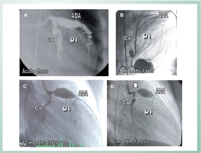
Fig. 1 -Giant diffuse left coronary artery aneurysms (1A) with a clear involution some months later (1B), albeit showing a residual giant aneurysm at the anterior descending artery (1C), which decreased progressively until 16 months following the initial treatment (1D), but developed an obstructive lesion (arrow) before the remaining aneurysm. There was also total obstruction at the right coronary artery. Cx: circumflex artery, Di: diagonal artery, IVA: anterior INTERVENTRICULAR artery
B
A
D
C
Image
Edmar Atik
KAWASAKI DISEASE: REGRESSION AND LATER STENOSIS OF GIANT CORONARY ARTERY ANEURYSMS
Arq Bras Cardiol 2007; 88(1) : e21-e22
An angiographic study on the 27th day of the disease
revealed extensive diffuse left coronary artery aneurysms with a total right coronary artery obstruction (Fig.1A). Continuing high-dose aspirin for two months beyond standardized treatment, a clear aneurysm involution by repeated angiography was observed, 3 months later (Fig.1B). This aneurysm involution occurred mostly after 11 months (Fig.1C) and 16 months (Fig.1D), with normalization in caliber of the left coronary artery, except for an isolated aneurysm in the anterior descending coronary artery that diminished but did not suffer total involution.
In this last coronary angiography, an artery stenosis (arrow) before the aneurysm finally led to the indication of myocardial revascularization by saphenous vein anastomosis to the diagonal and anterior descending arteries, with great success.
1. Atik E, Foronda A, Bustamante LNP. Doença de Kawasaki. Involução de gigantes aneurismas coronarianos após tratamento anti-inflamatório prolongado. Relato de Caso. Arq. Bras. Cardiol. 2003; 81: 265-8.
2. Chantepie A, Mauran P, Lusson JR, Vaillant MC, Bozio A. Cardiovascular complications of Kawasaki syndrome: results of a French multicenter study. Arch Pediatr. 2001; 8: 713-9.
3. Yoshikawa Y, Yagihara T, Kameda Y, Taniguchi S, Tsuda E, Kawahira T, et al. Result of surgical treatments in patients with coronary-arterial obstructive disease after Kawasaki disease. Eur J Cardiovasc Surg. 2000; 17: 515-9.
4. Oki I, Tanihara S, Ojima T, Nakamura Y, Yanagawa H. A multicenter collaborative study on the risk factors of cardiac sequelae due to Kawasaki disease: a one-year follow-up study. Acta Pediatr. 2000; 89: 1435-8.
References
This report claims our attention owing to:
1. the involution of diffuse and extensive giant coronary aneurysms in the acute period of Kawasaki disease with prolonged aspirin treatment, drawing necessary attention to a more careful clinical management in this phase because of an undoubtedly greater surgical risk at this time.
2. the slower regression of isolated giant aneurysms that require careful follow-up with suitable anticoagulation therapy and an indication to surgery at an appropriate time in order to prevent a total coronary obstruction.
Potential Conflict of Interest
No potential conflict of interest relevant to this article was reported.