Case Report
Introduction
Aneurysms of aortocoronary saphenous vein grafts (SVG) are an unusual complication of coronary artery bypass grafting. Spontaneous rupture of SVG aneurysms is exceedingly rare1,2. We report a 39-year-old man with multiple coronary graft aneurysms detected 10 years after surgery with rupture of the largest into the left pleural space.
Case Report
A 39-year-old white man was admitted 10 years after internal mammary and saphenous vein coronary bypass surgery with chest pain and hematemesis. He was found to have acute non-ST segment elevation myocardial infarction and bilateral pneumonia. On cardiac auscultation, he had regular rate and rhythm with normal S1 and S2 and a II/IV systolic ejection murmur at the left upper sternal border but no carotid or abdominal bruits. Initial laboratory values on presentation were as follows: white blood cell count, 8.4 U/l; hemoglobin level, 9.6 g/dl; hematocrit, 29.9%; platelet count, 55,000/ µl; Na+, 149 mmol/l; K+, 4.4 mmol/l; serum urea nitrogen, 57 mg/dl; creatinine, 1.2 mg/dl; creatinine kinase (CK), 284 U/l (reference range, 26–190); CK-MB, 4.6 IU/l (reference range, 0.0–3.9); cardiac troponin-I (cTnI), 1.4 ng/ml (reference range, 0.0–1.0); and myoglobin, 81 IU/l (reference range, 0–70). Repeat laboratory values
after 12 hours were as follows: Na+, 137 mmol/l; K+, 4.2 mmol/l; serum urea nitrogen, 23 mg/dl; creatinine, 1.2 mg/dl; glucose, 89 mg/dl; CK, 276 U/l; CK-MB, 4.9 IU/l; cTnI, 1.4 ng/ml; and myoglobin, 79 IU/L. Coagulogram, and ECG were strictly normal. Chest x-ray showed bilateral pulmonary infiltrates, an enlarged cardiac silhouette and left pleural effusion. The day after admission he developed severe respiratory distress, requiring mechanical respiratory support. He was started on piperacillin, tazobactam and gentamicin. Doripenem was added later. Heparin was not given secondary to the hematemesis.
He underwent thoracocentesis for left pleural effusion and 900 milliliters of bloody pleural fluid were drained. He continued to drain large amounts of blood (about one liter per day) through the left chest tube during the subsequent two days and expired after unsuccessful resuscitation.
Chest CT images performed the day after admission revealed two aortocoronary saphenous vein grafts with aneurysms. There were two visible aneurysms on the left saphenous vein graft to the region of the left anterior descending coronary artery, and a large thrombosed aneurysm on a graft to the region of the posterior descending coronary artery. No aneurysmal ruptures could be assessed. An internal thoracic bypass graft was also noted that appeared unremarkable (Figure 1).
At autopsy, the heart weighed 460 grams. There were three aortocoronary bypass vein grafts. One to the first diagonal was cord-like, with proximal occlusion at the aortic anastomosis. The graft to the left anterior descending artery demonstrated an 8 cm aneurysm near the distal anastomosis (Figure 2A), which was filled with atherosclerotic debris, with a focally calcified wall. The graft to the right coronary artery showed a 5-cm aneurysm, which was adherent to the right atrium and pulmonary artery and that had ruptured, hemorrhaging into the thoracic cavity (Figure 2B). There was a posterior healed left ventricular subendocardial infarct, and a lateral healed transmural infarct in the left ventricle. There were no mural thrombi. A mammary graft to the LAD distal to the vein graft was identified and appeared patent. There was moderate dilatation of the left ventricle. The right ventricle was 5 mm thick at the posterior wall, and the left ventricle was 14 mm thick in the anterior wall, with thinning in the lateral wall in the area of the infarct. There was no significant calcification of the valves and no vegetation. The right and left lungs weighted 560 and 410g respectively. The parenchyma of both lungs was congested and focally atelectatic.
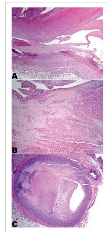
Microscopic sections of saphenous graft aneurysm to the right coronary artery demonstrated pseudoaneurysm
Key words
Coronary aneurysm; myocardial revascularization; cardiac surgery; heart rupture; coronary artery bypass.
Aortocoronary saphenous vein graft (SVG) aneurysms are rare, and are usually asymptomatic and detected incidentally. Spontaneous rupture of SVG is rare and imaging data are few. We report on a 39-year old man who was admitted to the hospital with hematemesis 10 years after aortocoronary bypass surgery. CT images revealed 3 aortocoronary SVG aneurysms, but failed to detect any rupture. His subsequent death due to rupture of SVG aneurysm was documented at autopsy, illustrating the need for aggressive treatment of symptomatic coronary graft aneurysms.
Multiple Aneurysms of Aortocoronary Saphenous Vein Grafts with
Fatal Rupture
Fábio R. Távora, Jean Jeudy, Allen P. Burke
University of Maryland, Department of Pathology, University of Maryland, Department of Radiology, Thoracic Imaging – Baltimore, MD - USA
Mailing address: Fabio Távora •
University of Maryland Medical School - 22 South Greene Street, Room NBW43 - Baltimore, MD, USA 21201
E-mail: [email protected]
Manuscript received July 24, 2006; revised received December 18, 2006; accepted January 17, 2007.
Case Report
Távora et al Saphenous aneurysm with rupture
Arq Bras Cardiol 2007; 88(5) : e104-e107
as mediastinal masses with compression to the adjacent structures or can be visualized after work-up for myocardial infarction4. CT and MRI are useful in determining aneurysm size, patency, and effect on nearby structures5.
There have been four reported cases of ruptured saphenous vein aneurysms1-3,6. Davey et al reported CT localization of the aneurysm with repeat imaging demonstrating leakage of contrast into surrounding tissue, without pinpointing the exact location of rupture2. Dimitri et al reported CT imaging with identification of aneurysm later confirmed by coronary bypass angiography3. The other two cases reported did not have CT images available and one of the ruptures was secondary to balloon angioplasty1,6.
Non-surgical interventions in the treatment of saphenous aneurysms have been attempted. Dimitri et al reported a case of successful coil embolization of a leaking saphenous aneurysm bypass to the right coronary artery that was followed by rapid wound healing3. The use of polytetrafluoroethylene-covered stents can be challenging in relatively large aneurysms and unusual anatomical locations can make the procedural technique risky. Successful long term outcome has been formation with fibrin, hemorrhage (Figure 3A) into adjacent
fat and the thymus gland, calcification of the graft wall, and adherent right atrial wall. Myocardial sections revealed diffuse interstitial fibrosis and myocyte hypertrophy. There were healed areas of infarction on the lateral wall (transmural; Figure 3B) and left posterior wall (subendocardial) of the left ventricle. A section of saphenous vein graft to the first diagonal demonstrated a cord-like vessel with near total occlusion. Sections of the saphenous vein graft to the LAD showed atheromatous plaque formation with near total occlusion by calcified fibroatheroma (Figure 3C). Distal to the occlusion, the lumen was narrowed by severe calcified atherosclerotic plaque. Sections of vein graft to LAD showed aneurysmal dilation, lipid-rich atherosclerotic plaque, and intraplaque hemorrhage.
Discussion
Saphenous grafts aneurysms are rare and detection is usually incidental. Failure to diagnose early in the course has led to several complications that included bleeding3, myocardial infarction4 and death2. They may also present
Fig. 1 - A - Contrast enhanced axial CT demonstrating aneurysmal dilatation of a left saphenous vein graft arising from the aorta and inserting on left anterior descending coronary artery (arrowhead). Large pleural collections and compressive atelectasis also noted (asterisk). B - Large thrombosed aneurysm of a right saphenous vein graft which originates from the aorta and inserts on posterior descending coronary artery (arrowhead). C - Coronal multiplanar reformatted (MPR) image showing the left saphenous vein graft aneurysm (asterisk). D - Coronal image demonstrating thrombosed aneurysm of the right saphenous vein graft (asterisk).
Case Report
Távora et al
Saphenous aneurysm with rupture
Arq Bras Cardiol 2007; 88(5) : e104-e107
described, as well as the formation of a new aneurysm after six months of the procedure7. In cases such as the one reported by Guerios et al8, where the aneurysm is located on the native artery without high risk of rupturing, a more conservative approach may be attempted, avoiding the direct approach or closure of the aneurysm.
The underlying cause of death was severe coronary atherosclerosis, which in this young man was accelerated or premature, suggesting a genetic component to his cardiovascular risk. The immediate cause of death was spontaneous rupture of a vein graft, which is a very rare occurrence2. Saphenous vein bypass graft aneurysms are in themselves infrequent, and may on occasion compress surrounding structures5.
The current report demonstrates the utility of CT in detecting saphenous vein graft aneurysms and is the first death due to rupture documented at autopsy. Because a bleeding site was not identified, the usefulness of imaging in detecting the potentially fatal complication of rupture and hemothorax remains to be shown. Therefore, awareness of the potential complication of rupture should prompt immediate consideration of non-invasive treatments, including percutaneous stenting, to prevent complications
Fig. 2 -A - Gross photograph of aneurysmal dilatation and total occlusion by atherosclerotic debris in saphenous graft aneurysm to left anterior descending artery. Metallic probe at proximal native artery. B - Gross photograph of ruptured aneurysm of graft to right coronary artery with adhesion to right pulmonary artery and surrounding hemorrhage.
Fig. 3 -A - Low power view of the saphenous vein graft aneurysm to the right coronary artery showing dissection plane (right to left) with disruption of the intima and of the media, fibrin thrombus formation and hemorrhage (original magnification, 20x). B - Low power view of left ventricular lateral wall section showing fibrosis (healed infarct) and focal myocyte hypertrophy (original magnification, 20x). C - Low power view of distal left anterior descending artery cross section showing near total occlusion by calcified fibroatheroma (original magnification, 12.5x).
of leakage and hemorrhage.
Potential Conflict of Interest
No potential conflict of interest relevant to this article was reported.
Case Report
References
1. Drummer E, Furey K, Hollman J. Rupture of a saphenous vein bypass graft during coronary angioplasty. Br Heart J. 1987; 58: 78-81.
2. Davey P, Gwilt D, Forfar C. Spontaneous rupture of a saphenous vein graft. Postgrad Med J. 1999; 75: 363-4.
3. Dimitri WR, Reid AW, Dunn FG. Leaking false aneurysm of right coronary saphenous vein graft; successful treatment by percutaneous coil embolisation. Br Heart J. 1992; 68: 619-20.
4. Taliercio CP, Smith HC, Pluth JR, Gibbons RJ. Coronary artery venous bypass graft aneurysm with symptomatic coronary artery emboli. J Am Coll Cardiol. 1986; 7:435-7.
5. Kalimi R, Palazzo RS, Graver LM. Giant aneurysm of saphenous vein graft
to coronary artery compressing the right atrium. Ann Thorac Surg. 1999; 68:1433-7.
6. Murphy JP, Jr., Shabb B, Nishikawa A, Adams PR, Walker WE. Rupture of an aortocoronary saphenous vein graft aneurysm. Am J Cardiol. 1986; 58: 555-7.
7. Bosmans JM, Claeys MJ, Dilling D, Vrints CJ. Unsuccessful long-term outcome after treatment of a vein graft false aneurysm with a polytetrafluoethylene-coated Jostent. Catheter Cardiovasc Interv. 2000; 50: 105-8.
8. Guerios EE, Bueno RR, Andrade PM, Nercolini DC, Pacheco AL, Tarastchuk JC, et al. Aneurysm of the left main coronary artery. Arq Bras Cardiol. 2000; 75 (6):531-6.
Távora et al Saphenous aneurysm with rupture