Several patients who presented with paralytical poliomyelitis developed, after many years of neuro-logic and functional stability, a progressive worsen-ing of clinical symptoms1,2, thus constituting a new
syndrome comprising fatigue, muscular atrophy, and pain. It is called “post polio syndrome” (PPS)3,4. Acute
anterior poliomyelitis (AAP) is a viral disease5
, which is characterized by headache, fever, pharyngitis, as well as signs of advanced meningeal irritation, fol-lowed by a lower motor neuron syndrome located to the spinal cord and consisting of an assymetric flaccid palsy of spinal muscles, and predominantly in the lower limbs6,7
. Following the advent of immuni-zation, the overall incidence of AAP declined drasti-cally worldwide, and a portion of patients who had previously developed polio during the 1940’s and 50’s are now currently presenting with the delayed, tar-dive effects of poliomyelitis1
. These delayed clinical
signs is probably related to the excess of physical ac-tivity during the phase of relative stability7.
CASE
A 40 years man, tax auditor, reported that at five years of age presented with clinical picture of AAP that subse-quently resulted in upper right limb and lower left limb pa-resis, difficulty walking and in the carrying out of some ba-sic daily activities. Normally returned to his activities and later enrolled in a regular exercise program. After three decades of clinical stability, he began referring weakness, muscle aches, and cramps in muscle groups not previously affected during his first bout of polio. In August of 2005, was diagnosed with PPS. The neurological examination showed assymetrical paresis in upper and lower limb as well as absent tendon reflex and except the flexor muscles of the hand. Myofasciculations and general twitching were also noted in the scapular region bilaterally, as well as in the braquial biceps, braquial triceps and quadriceps muscle groups. Figures 1 and 2 show the patient with marked right
POST-POLIOMYELITIS SYNDROME
Case report
Marco Antonio Orsini Neves
1, Mariana Pimentel de Mello
2,
Viviane Vieira dos Santos
3, Osvaldo J. M. Nascimento
4,
Reny de Souza Antonioli
5, Gabriel Rodrigues de Freitas
6, Marcos R.G. de Freitas
7ABSTRACT - The post-polio syndrome (PPS) is an entity characterized for an episode of muscular weakness and/or abnormal muscular fatigue in individuals that had presented acute polio years before. We report the case of PPS in a patient, 40 years, that thirty-five years after had had paralytic poliomyelitis, developed new symptoms of fatigue, muscular atrophy, dyspnea, difficulties in deambulation and muscular and joint pain. The electromyographic findings revealed injuried neurons of the anterior horn of the marrow and reinnervation after muscular tests.
KEY WORDS: post-poliomyelitis syndrome, paralytic poliomyelitis, neuromuscular diseases.
Síndrome pós-polio: relato de caso
RESUMO - A síndrome pós-polio (SPP) é entidade caracterizada por um episódio de fraqueza muscular e/ ou fadiga muscular anormal em indivíduos que apresentaram poliomielite aguda anos antes. Relatamos o caso de SPP em um paciente, 40 anos, que trinta e cinco anos após haver tido poliomielite paralítica, de-senvolveu novos sintomas de fadiga, atrofia muscular, dispnéia, dificuldades na deambulação e dores arti-culares e musarti-culares. Os achados eletromiográficos revelaram acometimento dos neurônios da ponta an-terior da medula e reinervação após a realização de testes musculares.
PALAVRAS-CHAVE: síndrome pós-polio, poliomielite paralítica, doenças neuromusculares.
Neuromuscular Disease Outpatient Division, Department of Neurology of Universidade Federal Fluminense, Niteroi RJ, Brazil (UFF): 1 Associ-ate Professor of Clinical Neurology - FESO (Teresópolis) and Voluntary Assistant of the Department of Neurology (Neuromuscular Diseases Division) - UFF; 2
Undergraduate in Physical Therapy and Trainer of Neurology Rehabilitation Division; 3
Graduate in Physical Therapy and Voluntary Assistant of the Department of Neurology (Neuromuscular Diseases Division) - UFF ; 4Chair of Neurology - UFF; 5Undergraduate in Physical Therapy - FESO; 6Chair of Neurology - UFF; 7Chair of Neurology and Chief of Staff, Neurology Department - UFF.
Received 15 September 2006, received in final form 8 December 2006. Accepted 26 February 2007.
Dr. Marco Antonio Orsini Neves - Rua Professor Miguel Couto 322 / 1001 - 24230-240 - Niterói RJ - Brasil. E-mail: [email protected]
Arq Neuropsiquiatr 2007;65(2-B) 529
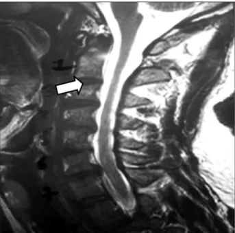
deltoid atrophy, and in practically all of the left lower limb muscle groupings, respectively. Tables 1 and 2 show various degrees of paresis in diverse muscle groups8. An electroneu-romyogram revealed decreased insertional activity in prox-imal muscles of the upper limbs as well as of the right scap-ular group. The recruitment pattern observed in affected muscles was of the incomplete type, thus indicating chron-ic reinnervation (old lesion). A magnetchron-ic resonance imaging (MRI) of the cervical spinal cord showed spinal (medullary) atrophy in the presence of a left postero lateral herniated disk at the C6/C7 level (Fig 3).
DISCUSSION
PPS is defined as the delayed development of a neuromuscular syndrome, at least 15 years following the stabilization of a previous bout of clinically evident poliomyelitis4. The mean interval between AAP and the
first signs and symptoms of PPS is roughly around 35
years9
. It is a slowly progressive disease2
, usually with an insidious, subacute onset, and resulting in some-times important restricion of daily activities10.
The precise etiology is unknown, although innu-merable theories have been proposed, including im-munopathogenic mechanisms, neuronal aging, and viral reactivation11.The most plausible hypothesis
would be excessive metabolism demand on surviv-ing, giant-sized motor units, which in turn would be brought on by equally excessive use of muscles over the years, thus resulting in a reduction in axo-nal “sprouting” in muscle fibers12,13. The giant-sized
motor units would develop during the AAP phase2,
with the goal of maximum reinnervation of previ-ously dennervated muscle fibers in order to maintain adequate function7
.Although this process is clinically efficient, neuronal overload ensues4.
Table 1. Degree of paresis in diverse muscle groups (MRC-up-per limbs)
Muscles Left side Right side
C5 – Elbow flexors 5 4
C6 – Wrist extensors 5 5
C7 – Elbow extensors 3 2
C8 – Fingers flexors 5 5
T1 – Finger abductors 4 4
Table 2. Degree of paresis in diverse muscle groups (MRC-low-er limbs).
Muscles Left side Right side
L2 – Hip flexors 3 4
L3 – Knee extensors 2 4
L4 – Ankle dorsiflexors 0 5
L5 – Long toe extensors 0 5
S1 – Ankle plantar flexors 0 5
Fig 1. Marked right deltoid atrophy. Fig 2. Atrophy in the left lower limb muscles.
530 Arq Neuropsiquiatr 2007;65(2-B)
The process of distal wasting of motor units pro-duces gaps in the neuromuscular junction, this prob-ably being the main cause of of fatigue, while muscle fibre dennervation causes the weakness “per se” en-countered in PPS. Certain risk factors for the devel-opment of PPS have been identified and include the degree of severity of AAP, advanced age of onset of AAP, number of permanent deficits present after recovery, female gender, recent weight gain, and a greater amount of physical activity during stability14.
Patients who develop PPS have a history of severe, extensive paralysis at the time of the original bout of poliomyelitis14. The clinical manifestations most
commonly found are muscular weakness, fatigue and pain3,10. Fatigue is the most incapacitating symptom,
occurring in almost 90% of patients9,15
. It is defined as a profound exhaustion that worsens with mini-mal physical activity. Patients with fatigue usually feel best during morning hours, growing worse as the day progresses2. Fatigue has been characterized
as a decrease in the tolerability as well as resistance to exercise, which improves upon rest2,9. The new-
onset weakness, which may be accompanied by at-rophy, mostly affects muscles previously affected by AAP, sometimes even extending to muscle groups un-affected by the original disease9,11.It is usually
pro-gressive and assymetrical, proximal or distal2,11.Other
signs that signal muscle involvement include: fascicu-lations, cramps, and high serum creatine phophate kinase levels7
.
Pain is a common complaint, and is usually the one that dominates the clinical picture and can be of either muscular or articular origin2. Pain is also
associ-ated with excessive activity15
, more commonly in the lower limbs and back in walking patients and in the upper limbs in wheelchair-bound or crutch wielding patients16. Patients referring pain usually limit their
day-to-day activities, which can lead to disuse atro-phy11. Other clinical symptoms less commonly found
include respiratory insufficiency or breathing difficul-ty in general, sleep disturbances, intolerance to cold, dysarthria, dysphagia and joint deformities5,9.
The principal diagnostic criteria are: proven past history of poliomyelitis, a period of complete or par-tial clinical recovery followed by a period of clinical stability of at least 15 years, gradual or rarely sud-den onset muscular weakness or abnormal fatigue; and finally, the exclusion of other neurologic and orthopedic causes17
. Eletroneuromyographic studies are useful for identifying previous neuronal loss due to AAP 2,5
, and also to exclude other neurologic dis-eases, though it does not distinguish PPS from other,
assymptomatic patients that present with subclinical (AAP) polio13
.
The goal of treatment is to provide the patient with the principles and means of self-sufficiency and to effect change in lifestyle, so as to reduce ex-cessive metabolic demands on muscle7
, and should include methods for energy conservation, regular periods of rest, weight loss, use of ortheses, and ex-ercise in moderate amounts2,7.The key to treatment
is to discover an equilibrium between activity and rest, preventing excessive muscle use and the subse-quent deterioration that comes with it. The physical limitations caused by the recent appearance of new motor symptoms added to the restrictions caused previously by AAP lead to a drastic reduction in day to day activity7, as well as to psychological
repercus-sions caused by an unexpected and acute disability2
. Many patients, like the one included in our study, received motivation to practice exercise and to make use of extreme compensatory methods during years in order to maintain their daily functions. These com-pensation methods consisted of utilizing muscles to their maximum capacity, a greater caloric consump-tion for the realizaconsump-tion of baseline activities, and the use of ligaments for stability which used to lead to hypermobility. However, these compensations were the cause of microtraumas and microcontusions of joints and ligaments, leading to overuse and ultimate exhaustion of neuromuscular motor units in an al-ready constrained system7
.
Patients who lose half (50%) of their motor neu-rons are still able to maintain clinically normal mus-cular activity/function18
, therefore subclinical involve-ment of certain muscle groups may be present. Many patients may concentrate excess weight on a limb up until then thought to be unaffected by disease, thus new onset weakness can involve apparently un-affected limbs7.
The aging process, with gradual loss of neurons mainly after age 60, may be a contributing factor to PPS19. The superposition of this process in an already
constrained motor system of limited motor units will result in a significant decrease of force13
.However, this process does not fully explain the clinical aspects found in patients under 50 years of age20. The signs
Arq Neuropsiquiatr 2007;65(2-B) 531
that benefits may bring upon by such activities may be potentialized and that the deleterious effects of excessive use can be avoided20
.
Despite the fact that there is no specific medical treatment for PPS, clinical trials using human intrave-nous imunoglobulyne and pyridostigmine have been conducted21-23
.
Kaponides et al. obtained impressive results while evaluating possible changes in motor power, physical performance and overall quality of life in patients daignosed with PPS. Of a total of 14 patients studied, all related improvement in quality of life, however without noticeable improvement in motor power nor physical performance. The results suggest that intra-venous immunoglobulyne may produce substantial clinical effects resulting in improvement in overall life quality. Such an effect may be the result of a reduction in the inflammatory process of the central nervous system. Further controlled studies as well as random ones are needed, however, since the placebo effect may have influenced the outcome.21
Gonzales et al. conducted a randomized, double-blind clinical study in patients treated with intrave-nous immunoglobulyne for PPS. A number of 142 patients were selected from four teaching hospitals in Stockholm, Sweden. The patients were divided into two groups. The experimental group, consisting of 73 patients, received intravenous immunoglobu-lyne during 3 consecutive days, repeated after three months. The control group, consisting of 69 patients, received a placebo. A questionnaire with the objec-tive of evaluating life quality was added to an evalu-ation of motor power. The results indicated that pa-tients who received intravenous immunoglobulyne had an increase in muscle strength when compared to the control group. Life quality did not vary be-tween the two groups. The authors believe that the immunoglobulyne may be an option to to supportive treatment for patients with PPS.22
Trojan et al. conducted a double-blind, random-ized and controlled study with the object of evalu-ating the effects of pyridostigmine on life quality, motor power, fatigue, and blood levels of IGF-1 in126 patients with SPP during a six-month period. The
re-sults did not show any difference between the two groups23.
Acknowledgement – The authors wish to thanks Dr
Peter Salem Jr. for the English version.
REFERENCES
1. Kidd D, Howard RS, Williams AJ, Heatley FW, Panayiotopoulos CP, Spencer GT. Late functional deterioration following paralytic polio-myelitis. Q J Med 1997;90:189-196.
2. Trojan DA, Cashman NR. Post-poliomyelitis syndrome. Muscle Nerve 2005;31:6-19.
3. Halstead LS. Post-polio syndrome: definition of an elusive concept. In Munsat TL (Ed). Post-polio syndrome. Boston: Butterworth-Heine-mann; 1991:23-38.
4. Dalakas MC, Elder G, Hallett M, et al. A long-term follow-up study of patients with post-poliomyelitis neuromuscular symptoms. N Engl J Med 1986;314:959-963.
5. Owen RR. Postpolio syndrome and cardiopulmonary conditioning, In Rehabilitation Medicine-Adding Life to Years [Special Issue]. West J Med 1991;154:557-558.
6. Oliveira ASB. Síndrome pós-poliomielite: aspectos neurológicos. Rev Neurociências 2002;10:31-34.
7. Smith LK, Kelly C. A síndrome pós-polio. In Umphred DA (Ed). Reabi-litação neurológica. 4.Ed. São Paulo: Manole; 2004:608-626.
8. Medical Research Council. Aids to the examination of the peripheral nervous system. 4.Ed. Edinburgh: WB Saunders, 2000:1-2.
9. Jubelt B, Cashman NR. Neurological manifestations of the post-polio syndrome. Crit Rev Neurobiol 1987;3:199-220.
10. Halstead LS, Rossi D. Post-polio syndrome: clinical experience with 132 consecutive outpatients. Birth Defects 1987;23:13-26.
11. Jubelt B, Agre JC. Characteristics and management of postpolio syn-drome. JAMA 2000;284:412-414.
12. Wiechers DO, Hubbell SL. Late changes in the motor unit after acute poliomyelitis. Muscle Nerve 1981;4:524-528.
13. Cashman NR, Maselli R, Wollman RL, Roos R, Simon R, Antel JP. Late denervation in patients with antecedent paralytic poliomyelitis. N Engl J Med 1987;317:7-12.
14. Klingman J, Chui H, Corgiat M, Perry J. Functional recovery. A major risk factor for the development of postpoliomyelitis muscular atrophy. Arch Neuro1 1988;45:645-647.
15. Berlly MH, Strauser WW, Hall KM. Fatigue in post-polio syndrome. Arch Phys Med Rehabil 1991;72:115-118.
16. Smith LK, McDermott K. Pain in post-poliomyelitis: addressing causes versus treating effects. In Halstead LS, Wiechers DO (Eds). Research and clinical aspects of the late effects of poliomyelitis. White Plains, NY: March of Dimes Birth Defects Foundation; 1987:121-134. 17. Halstead LS. Assessment and differential diagnosis for post-polio
syn-drome. Orthopedics 1991;14:1209-1217.
18. Lin KH, Lim YW. Post-poliomyelitis syndrome: case report and review of the literature. Ann Acad Med Singapore 2005;34:447-449.
19. Campbell MJ, McComas AJ, Petito F. Physiological changes in aging muscles. J Neurol Neurosurg Psychiatry 1973;36:174-182.
20. Chang CW, Huang SF. Varied clinical patterns, physical activities, mus-cle enzymes, electromyographic and histologic findings in patients with post-polio syndrome in Taiwan. Spinal Cord 2001;39:526-531. 21. Kaponides G, Gonzalez H, Olsson T, Borg K. Effect of intravenous
im-munoglobulin in patients with post-polio syndrome: uncontrolled pi-lot study. J Rehabil Med 2006;38:138-140.
22. Gonzales H, Sunnerhagen KS, Sjoberg I, Kaponides G, Olsson T, Borg K. Intravenous immunoglobulin for post-polio syndrome: a random-ized controlled trial. Lancet Neurol 2006;5:493-500.