Arquivos Brasileiros de Cardiologia - Volume 85, Nº 5, November 2005
Coronary Artery Aneurysm One Year and Five
Months After Sirolimus-Eluting Stent Placement
Mailing Address: George Ximenes Meireles • Rua Sena Madureira, 1265/102 - 04021-051 - São Paulo, SP - Brazil
E-mail: [email protected] Received on 12/07/04 • Accepted on 05/04/05
Luciano Abreu, George César Ximenes Meireles, Antonio Artur Forte,
Marcos Sumita, Jorge Hayashi, José Solano
Departamento de Cardiologia do Hospital Stella Maris - São Paulo, SP - Brazil
A 52-year-old man with diagnosis of post-infarction unstable angina. Coronary angiography revealed 90% luminal obstruction in the middle third of the right coronary artery and 90% in the marginal branch of the circumflex artery. After the administration of clopidogrel 300 mg associated with acetylsalicylic acid, the patient underwent the implantation of a sirolimus-eluting stent
Randomized studies with the use of drug-eluting stents have demonstrated an inhibition of neointimal hyperplasia in the majority of patients1,2. With the increasing use of
these stents, information on their long-term effect is extremely important. The presence of a coronary aneurysm one year and five months after the implantation of a sirolimus-eluting stent is of utmost interest due to the lack of previously reported case reports.
C
ASE
R
EPORT
The patient was a 52-year-old male admitted with a history of very intense oppressive precordial pain at rest. He presented risk factors for coronary disease, systemic arterial hypertension and was a smoker; he also used captopril 12.5 mg twice a day. An electrocardiogram and enzyme measurements were carried out and an acute myocardial infarction was diagnosed, with no upper unleveling of the ST segment. During hospital stay, he presented precordial pain and post-infarction angina was diagnosed. At the physical examination he presented arterial pressure of 120/70 mmHg and cardiac frequency of 80 bpm. Cardiopulmonary auscultation was normal.
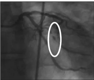
The electrocardiogram showed a sinusal rhythm, with no abnormalities. Thorax X-ray showed a cardiothoracic index of 0.5, elongated aorta and normal pleuropulmonary fields. The patient underwent a coronary angiography by the Sones technique, which disclosed a luminal obstruction of 90% in the middle third of the right coronary artery (RCA) and 90% in the first marginal branch (MG) (fig. 1) of the circumflex artery (CA). The venticulography showed a slight lower lateral hypocontractility.
The patient was medicated with clopidogrel 300 mg and acetylsalicylic acid (AAS) 200 mg on the same day the angiography was carried out, and on the following day, after a pre-dilation with a Wordpass balloon-catheter (Johnson&Johnson-Cordis) 2.0 x 20 mm, he underwent the implantation of a sirolimus-eluting stent (CYPHER; Johnson&Johnson-Cordis) 2.5 x 18 mm in the lesion at the MG branch with success (fig. 2). The stent was released with a 12 ATM pressure and the right femoral artery (RFA) was the access via used.
The use of the Philips System for the quantitative angiographic analysis of the measurements of the left marginal branch disclosed, at the pre-implantation assessment, a reference diameter of 2.50 mm, a percent
Case Report
Case Report
Case Report
Case Report
Case Report
Case Report
Arquivos Brasileiros de Cardiologia - Volume 85, Nº 5, November 2005 of stenosis in diameter of 81.01%, minimal luminal
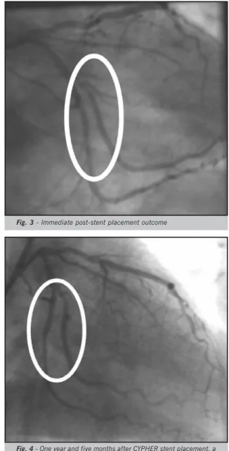
diameter of 0.47 mm and lesion extension of 8.21 mm. After the implant, the reference diameter was 2.50 mm and the minimal luminal diameter was 2.54 mm (fig. 3).
Post-procedure laboratory assessment showed levels of creatine phosphokinase of 60 U/L with a 5U/L MB fraction and 227 U/mL lactate dehydrogenase. The RFA introductory device was removed 4 hours after procedure completion and a manual compression was performed for 15 minutes. There was no post-compression bleeding or hematoma.
During hospital stay, the patient evolved with no precordial pain and no electrocardiographic or enzymatic alterations were obser ved. Two days after stent implantation, he was released from the hospital with a clopidrogel prescription of 75 mg/day for 60 days and AAS 200 mg from the release date on, indefinitely.
Thirty-six days after the first stent placement, a Bx Velocity stent (Johnson&Johnson-Cordis) 4.0 x 23 mm was successfully implanted at the RCA lesion.
The patient presented a new episode of precordial pain
one year and five months after the implantation of the sirolimus-eluting stent. He then underwent a new coronary angiography, which disclosed the presence of a coronary aneurysm in the marginal branch, in the intra-stent proximal portion, with no luminal obstruction in both stents (fig. 4).
D
ISCUSSION
The formation of a coronary artery aneurysm, defined as a dilation of the coronary artery that exceeds 1.5 times the reference diameter of the adjacent coronary segments that are angiographically normal3, has been reported after
coronary angioplasty, directed coronary atherectomy, and laser angioplasty at a frequency that varies from 2 to 10%4. At the STRESS3 study, a the presence of a coronary
artery aneurysm was observed in 3.9% of the patients submitted to Palmaz-Schatz stent implant.
Stent placement associated to anti-inflammatory agents has been correlated to an increased frequency of coronary aneurysms. Rab et al5 have reported the
occurrence of coronary artery aneurysms in 32% of patients submitted to stent placement, when corticoid drugs and colchicine were administered after the implant. These investigators concluded that the aneurysm formation was probably increased due to the concomitant use of anti-inflammatory agents.
The new drug-eluting stents approved for medical practice, such as paclitaxel6 and sirolimus1,2, are capable
of inhibiting neointimal hyperplasia. Sirolimus acts specifically at the G1 phase of the cell cycle. Due to its early action on the cell cycle, sirolimus can block cell proliferation with late vascular sequelae potential2.
Additionally, sirolimus stimulates apoptosis and reduces inflammation7. Thus, drug-eluting stents can lead to the
formation of coronary aneurysms and among the late post-implantation findings, the incomplete apposition of the stent filaments could be the responsible mechanism.
The late incomplete apposition of the stent filaments, obser ved during the follow-up with intracoronary ultrasound, is defined as a separation of one or more stent filaments from the intima, with no overlap on the lateral branch and evidence of blood flow behind the filament. It occurs in 4 to 5% of non-eluting stents and at an unknown frequency in drug-eluting ones. Many hypothesis have been formulated to explain its origin, such as positive regional vascular remodeling, plaque regression, late dissolution of the thrombotic material captured by the stent filaments, cell necrosis, apoptosis, and allergic reaction to sirolimus8.
At the RAVEL1, SIRIUS2, E-SIRIUS3 and C-SIRIUS4
studies, the presence of aneurysms was not reported, in patients evaluated by angiography and intra-coronary ultrasound six to eight months after sirolimus-eluting stent placement.
Recently, 13 patients with incomplete apposition were evaluated by intra-coronary ultrasound, six to twelve Fig. 1 - Obstructive lesion of 90% at the marginal branch of the
circumflex artery
Fig. 2 - Sirolimus-eluting stent placement (CYPHER; Johnson & Johnson/ Cordis) 2.5 x 18 mm in the lesion site at the left marginal branch
Arquivos Brasileiros de Cardiologia - Volume 85, Nº 5, November 2005
months after sirolimus-eluting stent placement and it was observed that the area of incomplete apposition did not show alterations during this period, except in one patient in whom the presence of a coronary aneurysm was observed during the follow-up period of twelve months; the condition was probably pre-existing at the time of the stent placement and failed to be detected due to the fact that it was filled with a thrombus. Based on the fact that sirolimus elution of the stent filaments remains for only six weeks, with a half-life of sirolimus in tissue for 60 hours, it is unlikely that sirolimus itself cam induce long-term alterations of the vessel wall. The possibility that this aneurysm was probably present since the first evaluation, having been masked by a thrombus, was then raised9.
Regarding the present case, a possible explanation for the aneurysm formation, which is unrelated to use of a sirolimus-eluting stent, is that, being an acute myocardial infarction with no upper unleveling of the ST segment, there was a spontaneous dissection that was not visible through the angiography, which triggered the acute coronary syndrome. The presence of a dissection has been observed by intra-coronary ultrasound prior to Palmaz-Schatz stent placement, and the image did not disappear entirely immediately after the implant. The angiography was repeated six months later and the presence of an intra-stent aneurysm was observed. The coronary ultrasound disclosed that the same dissection presented continuity with the coronary aneurysm, suggesting that the dissection was responsible for the aneurysm formation10.
A limitation observed in the present study was the lack of an intra-coronary ultrasound, which is the only way to detect the incomplete stent apposition.
No other therapeutic measures were applied in this case, as patients with coronary artery aneurysm post-percutaneous coronary intervention have shown a benign clinical course, with no association with an increase in mortality, acute myocardial infarction or artery rupture3.
Fig. 3 - Immediate post-stent placement outcome
Fig. 4 - One year and five months after CYPHER stent placement, a new coronariography disclosed an intra-stent coronary aneurysm in the left marginal branch
R
EFERENCES
1. Morice MC, Serruys PW, Souza JE et al. A randomized comparison
of a sirolimus-eluting stent with a standard stent for coronar y revascularization (RAVEL). N Engl J Med 2002;346:1773-80.
2. Moses JW, Leon M, Popma JJ et al. Sirolimus-Eluting Stents versus
Standard Stents in Pacientes with Stenosis in a Native Coronary Artery (SIRIUS). N Engl J Med 2003;349:315-23.
3. Slota PA, Fischman DL, Savage M, Rake R, Goldberg S. Frequency
and Outcome of Development of Coronary Artery Aneurysm After
Intracoronar y Stent Placement and angioplasty.Am J Cardiol
1997;79:1104-5.
4. Bal ET, Plokker HWT, Van de Berg EMJ, Ernst SMPG, Mast EG, Ascoop
CAPL. Predictability and prognosis of PTCA - induced coronary artery aneurysms. Cathet Cardiovasc Diagn 1991;22:85-8.
5. Rab ST, King SB, Roubin GS, Carlin S, Hearns JA, Douglas JS.
Coronary artery aneurysms after stent placement: a suggestion of altered vessel wall healing in the presence of anti-inflammatory
agents.J Am Coll Cardiol 1991;18:1524-8.
6. Grube E, Silber S, Hauptmann KE et al. Six- and twelve-month
results from a Randomized, double-blind trial on a slow-release placlitaxel-eluting stent for de novo coronary lesions. Circulation 2003;107:38-42.
7. Roque M, Cordon-cardo C, Fuster V. Modulation of apoptosis,
proliferation and p27 expression in a porcine coronary angioplasty model. Atherosclerosis 2000;153:315-22.
8. Mintz GS, Shah VM, Weissman NJ. Regional remodeling as the cause
of late malapposition. Circulation 2003;107:2660-3.
9. Degertekin M, Serruys PW, Tanabe K et al. Long-term follow-up of
incomplete stent apposition in patients who received sirolimus-eluting stent for de novo coronar y lesions. Circulation 2003;108:2747-50.
10. Voigtländer T, Rupprecht HJ, Stähr P, Nowak B, Kupferwasser I, Meyer J. Development of a coronary aneurysm 6 months after stent implantation assessed by intracoronary ultrasound. Am. Heart J 1996;131:833-4.