Arquivos Brasileiros de Cardiologia - Volume 85, Nº 5, November 2005
Case Report
Case Report
Case Report
Case Report
Case Report
Case Report
Mechanism of Cardiac Resynchronization Therapy
by Real-time Three-dimensional
Echocardiography in Patients with Heart Failure
Mailing Address:Marcelo Luiz Campos Vieira • R u a C a r d o s o d e M e l o , 4 6 3 / 2 1 - 0 4 5 4 8 - 0 0 2 - S ã o P a u l o , S P - B r a z i l
E-mail: [email protected] Received on 05/29/05 • Accepted on 07/01/05
Marcelo Luiz Campos Vieira, Prasad V. Maddukuri, Robert S. Phang, Natesa G. Pandian,
Wilson Mathias Júnior, José A. F. Ramires
Instituto do Coração do Hospital das Clínicas - FMUSP e Tufts University - New England Medical Center São Paulo, SP, Brazil - Boston, MA, EUA
We report the case of a 66-year-old man with heart failure NYHA class IV treated with biventricular pacing for cardiac resynchronization. The patient was evaluated by real-time three-dimensional transthoracic echocar-diography before and 48 hours after pacemaker
implan-Cardiac resynchronization using biventricular pacing (CRT) has been recommended as an adjuvant therapy for patients with symptomatic, advanced heart failure and under optimized drug therapy.1,2 Indication for CRT
takes into account clinical aspects, the presence of severe left ventricular contractile dysfunction, and either electrical or mechanical dyssynchrony. We report a patient with drug-refractory heart failure submitted to real-time three-dimensional transthoracic echocardiography (3D echo) for assessment of ventricular dyssynchrony and who progressed satisfactorily during hospitalization.
C
ASE
REPORT
A sixty-six-year-old male patient with history of orthotopic heart transplantation for ischemic cardiomyopathy complicated by graft vasculopathy, in 1999, was admitted with decompensated heart failure. At presentation, he complained of dyspnea at rest, paroxysmal nocturnal dyspnea, and right hypochondrium pain.
On physical examination, the patient showed regular heart rate of 106 bpm, respiratory rate of 20 breaths/ min, blood pressure of 112/66 mmHg, axillar y temperature of 36.6 oC, abdominal bloating with liver
edge palpable up to 8 cm from the right costal margin and weight gain of 12 kg in the previous two months, despite optimization of oral drug therapy. Cardiovascular
examination revealed jugular venous distention up to the mandibular angle, evidence of ventricular gallop due to a third heart sound (S3), and grade 3/6+ holosystolic
murmur loudest at the ventricular apex. Resting ECG showed sinus tachycardia, right bundle-branch block (QRS duration = 143 ms).
Arquivos Brasileiros de Cardiologia - Volume 85, Nº 5, November 2005
tachycardia, and widened QRS complex. Biventricular pacing was performed endocardially via de coronary sinus, according to the cardiac venous anatomy. A left ventricular electrode was implanted in the lateral wall. Real-time three-dimensional transthoracic echocardiography was performed (Philips Sonos 7500, Andover, USA) before and 48 hours after biventricular pacing (Table I). Three-dimensional echocardiographic analysis was performed in workstation using a special software (TomTec Imaging Systems, 4D CardioView software 1.3, Munich, Germany). The patient was discharged clinically improved three days after pacing and is currently under outpatient follow-up.
D
ISCUSSION
Biventricular pacing for cardiac resynchronization in patients with advanced heart failure has therapeutic benefits, resulting in clinical and laborator y improvement. This is due to enhanced intra- and inter ventricular synchrony, as well as increased ventricular filling time1. Clinical and functional
improvement following biventricular pacing is evidenced by increased LVEF and oxygen consumption (VO2), enhanced quality of life (Minnesota score), decreased mitral regurgitation, reduced number of hospital admissions, lower serum neurohormone levels, and decreased QRS complex duration2-6.
CRT is currently indicated for symptomatic patients despite drug therapy optimization when they present NYHA functional class IV, QRS complex duration > 130 ms, LBBB morphology, LVEF < 35% (as shown on echocardiography by Simpson’ method), and left ventricular diastolic diameter > 55 mm (Degree of recommendation IIa, level of evidence A).7 However, about
30% of the patients with ventricular dyssynchrony seen on ECG do not benefit from CRT5. Hence, electrical
dyssynchrony on ECG alone seems to be inadequate to identify patients who will respond to treatment. It should be also noted that CRT is costly, in addition to presenting complication rates of up to 8%2. Therefore,
electrome-chanical inter- and/or intraventricular dyssynchrony should be observed for CHF patients to be indicated for CRT.
Electromechanical ventricular asynchrony may be evidenced by using radiopharmaceuticals, nuclear magnetic resonance or echocardiography. Echocar-diography is a simple, highly reproducible and easy-to-perform technique that provides information not only for CRT indication, but also for drug therapy optimization and clinical follow-up. Accordingly, echocardiography provides data regarding atrioventricular, inter- and intraventricular synchrony and contributes to CRT optimization through optimization of atrioventricular interval and its relationship to diastolic filling. Also, during clinical follow-up it allows the determination of LVEF and yields information on LV negative remodeling by the calculation of ventricular volumes, ventricular mass index, mass/volume ratio, and ventricular sphericity index.
Different echocardiographic modalities have been used for the assessment of ventricular synchrony. From conventional techniques, such as M-mode and pulsed Doppler, to more advanced modalities, such as tissue Doppler, strain and strain rate, plus 3D real-time echo, they can provide information on ventricular synchrony. Using M-mode, LV septal-to-posterior wall motion delay is measured. Patients considered most likely to respond to CRT are those with a delay >130 ms8. Pulsed Doppler
provides information on cardiac synchrony by measuring the ventricular pre-ejection period (intraventricular synchrony) and ventricular filling time (atrioventricular synchrony), and also the differences between the onset of the QRS complex and the pulmonary flow and the beginning of the QRS complex and the aortic flow (interventricular synchrony)5. Tissue Doppler and strain
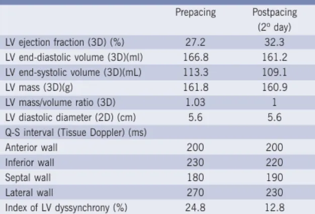
imaging are reliable indicators of regional ventricular contraction, making echocardiographic analysis more accurate and close to reality. The use of tissue Doppler allows measurement of the Q-S interval, which represents the time elapsed between the beginning of the QRS complex and peak of the S-wave (ventricular contraction) on the four LV walls and on the free wall of RV. The QS interval is usually measured at basal segments (at atrioventricular ring level) of the four LV walls and the RV free wall. Then, the greatest difference between left ventricle QS intervals and also right ventricle QS intervals is taken. In the case of this patient, only left ventricle QS intervals were measured, because that was the hospital protocol. Prior to CRT, the greatest difference of left ventricle QS intervals was 90 ms, and after CRT, 40 ms (Table I), showing intraventricular synchrony improvement.
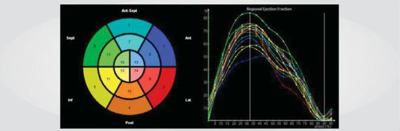
Three-dimensional echo allows calculation of LV dyssynchrony index (SDI), corresponding to the standard deviation of the mean time of systolic contraction for each of the 16 left ventricular segments (fig. I), with normal
Prepacing Postpacing (2º day) LV ejection fraction (3D) (%) 27.2 32.3 LV end-diastolic volume (3D)(ml) 166.8 161.2 LV end-systolic volume (3D)(mL) 113.3 109.1
LV mass (3D)(g) 161.8 160.9
LV mass/volume ratio (3D) 1.03 1 LV diastolic diameter (2D) (cm) 5.6 5.6 Q-S interval (Tissue Doppler) (ms)
Anterior wall 200 200
Inferior wall 230 220
Septal wall 180 190
Lateral wall 270 230
Index of LV dyssynchrony (%) 24.8 12.8
3D - real-time three-dimensional transthoracic echocardiography; 2D - two-dimensional transthoracic echocardiography; LV - left ventricle; Q-S - interval between the beginning of Q-wave on the ECG and the peak of S-wave on tissue Doppler
Table I - Real-time three-dimensional transthoracic echocardiographic and tissue Doppler studies before
and two days following biventricular pacing
Arquivos Brasileiros de Cardiologia - Volume 85, Nº 5, November 2005
value < 8%. SDI may also be calculated taken into account only 12 ventricular segments, leaving out the 4 apical segments. In this patient, SDI was 24.8% pre-CRT and 12.8% post-pre-CRT, characterizing great improvement in left ventricular synchrony (Table I).
SDI calculation enables not numerical determination of ventricular dyssynchrony, but also identification of the site of greatest dyssynchrony, where the left electrode should ideally be placed. In this case, pre-CRT synchrony pattern is quite heterogeneous, but the greatest conduction delay was found on the lateral wall (Fig. 2, red line), where the left ventricular electrode was implanted. Implantation of left electrode in a site other than that where the greatest conduction delay occurs and the ensuing ventricular contraction may be one of the mechanisms that account for failure to appropriate response following CRT.
By determining SDI immediately after pacemaker implantation, three-dimensional echo provides an insight into the mechanism involved in ventricular resynchronization therapy. In the reported case, an increase in LVEF (from 27.2% to 32.3%) 48 hours following CRT was noted, with maintenance of ventricular
Fig. 1 - Regional left ventricular contraction in a 16-segment model of the left ventricle using real-time 3D echocardiogram. Normal volunteer
Fig. 2 - Regional left ventricular contraction in a patient who underwent resynchronization by biventricular pacing before and 48 hours following the procedure (Post-CRT) using real-time 3D echocardiogram. A great improvement in left ventricular dyssynchrony is noted after the procedure
Pre-CRT Post-CRT
volumes, ventricular mass index, LV diastolic diameter and LV mass/volume ratio. Therefore, the mechanism associated with the improvement in LV contractile performance was enhanced left intraventricular synchrony, as shown by lower SDI 48 hours after CRT. A further characteristic of this patient was the presence of prolonged QRS and RBBB morphology, as with most Brazilian patients with Chagas’ disease. Larger studies are needed to determine whether CRT may be a promising treatment for this group of patients with CHF class III and IV (NYHA) and RBBB.
The use of real-time three-dimensional echocardiogram has advantages regarding CRT, namely: to allow real-time, simultaneous, fast analysis of all ventricular segments, facilitate and standardize communication between cardiac electrophysiologists or surgeons who perform biventricular pacing, enable quantification of pre-implantation ventricular dyssynchrony (dyssynchrony index) imme-diately after the procedure and during clinical follow-up. Its disadvantages, however, include the high cost of 3D echocardiography equipment, the need of cardiologists trained in and familiar with the technique, and the limited quality of images acquired for three-dimensional analysis.
Arquivos Brasileiros de Cardiologia - Volume 85, Nº 5, November 2005
C
ONCLUSION
The use of three-dimensional echocardiography allowed evaluation of cardiac resynchronization after biventricular pacing in this patient with RBBB and CHF class II and IV
REFERENCES
1. Yu CM, Chau E, Sanderson, Fan K et al. Tissue Doppler echo-cardiographic evidence of reverse remodeling and improved synchronicity by simultaneously delaying regional contraction after biventricular pacing therapy in hear t failure. Circulation 2002;105:438-45.
2. Bristow MR, Saxon LA, Boehmer J et al. Cardiac resynchronization therapy with and without an implantable defibrillator in advanced chronic heart failure. N Eng J Med 2004;350: 2140-50.
3. Abraham WT, Fisher WG, Smith AL et al. Cardiac resynchronization in chronic heart failure. N Engl J Med 2002;346:1845-53.
4. Cazeau S, Leclercq C, Lavergne T et al. Effects of multisite biventricular pacing in patients with heart failure and intraventricular conduction delay. N Engl J Med 2001;344:873-80.
5. Lane RE, Chow AW, Chin D et al. Selection and optimization of biventricular pacing: the role of echocardiography. Heart 2004; 90(Suppl VI): vi 10-16.
6. Bax JJ, Ansalone G, Breithardt OA et al. Echocardiographic evaluation of cardiac resynchronization therapy: ready for routine clinical use. A clinical appraisal. J Am Coll Cardiol 2004;44:1-9.
7. Gregorates G, Abrams J, Epstein AE et al. ACC- AHA- NASPE 2002 guideline update for implantation of cardiac pacemakers and antiarrhythmia devices. J Am Coll Cardiol 2002;40:1703-19.
8. Ptizalis MV, Acoviello M, Romito R et al. Cardiac resynchronization therapy tailored by echocardiographic evaluation of ventricular asynchrony. J Am Coll Cardiol 2002;40:1615-22.
MECHANISM OF CARDIAC RESYNCHRONIZATION THERAPY BY REAL-TIME THREE-DIMENSIONAL ECHOCARDIOGRAPHY IN PATIENTS WITH HEART FAILURE