Coxsackievirus B5 induced apoptosis of HeLa cells: Effects on p53 and SUMO
Rogério Gomes
a,⁎
, Renata Guerra-Sá
b, Eurico Arruda
a,caDepartment of Cell Biology, University of São Paulo School of Medicine at Ribeirão Preto, Av. dos Bandeirantes, 3900, 14049-900, SP, Brazil bFederal University of Ouro Preto, MG, Brazil
cVirology Research Center, University of São Paulo School of Medicine at Ribeirão Preto, SP, Brazil
a b s t r a c t
a r t i c l e
i n f o
Article history: Received 27 July 2009 Returned to author for revision 27 September 2009
Accepted 3 October 2009 Available online 10 November 2009
Keywords: Coxsackievirus B5 p53 SUMO Apoptosis UBC9
Coxsackievirus B5 (CVB5), a human enterovirus of the familyPicornaviridae, is a frequent cause of acute and chronic human diseases. The pathogenesis of enteroviral infections is not completely understood, and the fate of the CVB5-infected cell has a pivotal role in this process. We have investigated the CVB5-induced apoptosis of HeLa cells and found that it happens by the intrinsic pathway by a mechanism dependent on the ubiquitin–proteasome system, associated with nuclear aggregation of p53. Striking redistribution of both SUMO and UBC9 was noted at 4 h post-infection, simultaneously with a reduction in the levels of the ubiquitin-ligase HDM2. Taken together, these results suggest that CVB5 infection of HeLa cells elicit the intrinsic pathway of apoptosis by MDM2 degradation and p53 activation, destabilizing protein sumoylation, by a mechanism that is dependent on a functional ubiquitin–proteasome system.
© 2009 Elsevier Inc. All rights reserved.
The human enteroviruses of the family Picornaviridae, are distributes in 5 species:Poliovirus,Human enteroviruses A,B,CandD
(http://www.ncbi.nlm.nih.gov/ICTVdb/). A wide spectrum of human diseases is caused by over 70 different serotypes of non-polio enteroviruses, and coxsackie B viruses are among the most frequent of them (Pallansch, 1997). Among the more extensively studied coxsackie B viruses, coxsackievirus B5 (CVB5) is one of the most importantly associated with human diseases, mainly affecting the central nervous system (CNS), and the myocardium, but also in association with long-term sequelae, such as insulin-dependent diabetes mellitus and dilated cardiomyopathy (Endres et al., 2002; Moschovi et al., 2002; Pallansch, 1997).
The progression of a viral infection ultimately results from the interplay of virulence factors and the host immune system, frequently leading to inflammatory responses that contribute to tissue damage. Central to this is the fate of the infected host cell, which depends mainly on the induction of apoptotic or necrotic cell deaths (McLean et al., 2008). There is plenty of accumulated evidence that picorna-viruses interfere with apoptosis (Buenz and Howe, 2006; Hwang et al., 2007). Apoptosis is a programmed cell death, responsible for the tissue remodeling, elimination of senescent and anomalous cells, and also elimination of infected cells. It can be activated by extra cellular (extrinsic pathway) or intracellular signals (intrinsic pathway). Both pathways activate the cisteino proteases with specificity to arpartic acid (caspases) (Danial and Korsmeyer, 2004). One of the key molecules for apoptosis triggering is p53. The sequence-specific
DNA-binding transcription factor p53 has a central role in initiating cell cycle arrest and apoptosis, principally through its ability to increase the transcription of genes that control these processes (Watson and Irwin, 2006).Pampin et al. (2006) have demonstrated that Poliovirus infection induces the recruitment of p53 to promye-locytic leukemia nuclear bodies (PML NBs), p53 phosphorylation on Ser15, and activation of p53 target genes, leading to the induction of p53-dependent apoptosis. The regulation of p53 activity and stability is controlled by a series of post-translational modifications (phos-phorylation, acetylaton, methylation, ubiquitination and sumoyla-tion) (Lavin and Gueven, 2006). One mechanism of p53 regulation involves the SUMO-specific protease SUSP4 which upregulates p53 by promoting MDM2 self-ubiquitination and consequently degradation by the proteasome complex (Lee et al., 2006). Viral interference with the ubiquitination of host proteins could be beneficial to the virus, as long as it results in an environment that is more favorable for viral propagation.
We describe in this paper the mechanisms of induction of apoptosis by CVB5 in HeLa cells.
Results
Mitochondrial membrane potential change (ΔΨm) and cytochrome c release
An increase inΨm was observed byflow cytometry of cells treated with JC-1 at 4 h post-infection, indicating that CVB5 induced apoptosis of HeLa cells (Fig. 1A). Release of cytochromecfrom mitochondria to cytosol at 4 h post-infection was evidenced by immunoblot (Fig. 1B). ⁎ Corresponding author. Fax: +55 16 3602 3376.
E-mail address:[email protected](R. Gomes).
0042-6822/$–see front matter © 2009 Elsevier Inc. All rights reserved. doi:10.1016/j.virol.2009.10.005
Contents lists available atScienceDirect
Virology
Fig. 1.Activation of apoptotic intrinsic pathway by CVB5 infection of HeLa cells. Percentage of cells with depolarized mitochondria detected by cytofluorimetric quantification of JC-1 staining in CVB5- and mock-infected HeLa cells. Bivariate plots of orange (FL2) versus green (FL1)fluorescence as an estimate of mitochondrial membrane potential variation (ΔΨm). Values indicate percentages of cells with depolarized mitochondria (A). Western blot analysis shows that infection of CVB5 also causes release of cytochromec(Cyt C) from mitochondria to cytosol (B), and that procaspase 9 is cleaved into caspase 9 (C). Increase on caspase 3 activity was also observed by FACS analysis in CVB5 infected cells (D). Al those events were observed at the time of 4 h post-infection.
Taken together, these results indicate that CVB5 induces depolariza-tion of mitochondria and cytochromecrelease to the cytosol.
Caspases 3 and 9
Western blot assay showed the cleavage of procaspase 9 into caspase 9, noted at 4 h post-infection (Fig. 1C). This caspase 9 activation came along with an increase in caspase 3 activity detected by FACS also at 4 h post-infection of HeLa cells by CVB5 (Fig. 1D).
p53 nuclear aggregation
In mock-infected HeLa cells, p53 staining was observed as a slight diffuse signal throughout the cell. In contrast, in CVB5-infected cells the p53 labelling was concentrated in dots within the nucleus. This also occurred in HeLa cells treated with actinomycin D, an inductor of apoptosis, as well as in Vero cells infected with CVB5 (Fig. 2A). Possibly these nuclear aggregates could be a result of accumulation of p53 on transcription sites of proapoptotic genes. No colocalization of p53 nuclear aggregates with PML NB was observed on confocal images (Fig. 2B). The detection by western blot of the p53 ubiquitin ligase, MDM2, showed a decrease during the time course of CVB5 infection, better observed 4 h post-infection, as compared to mock infected cells (Fig. 2C). The reallocation of p53 to nucleus could be a consequence of MDM2 degradation which causes the release of p53 to activation of proapoptotic genes.
SUMO and UBC9
In non-infected HeLa cells the SUMO E2 ligase UBC9 is localized as an aggregate next to the nucleus, but at 4 h post-infection this distribution is greatly altered, and appears dispersed throughout the
cell (Fig. 3A). Although the virus infection induced UBC9 dispersion throughout the cell, the immunoblot assay indicated that the total amount of UBC9 remained apparently constant during the experiment (Fig. 3B). Similar to the findings with UBC9, SUMO is distributed around the nucleus of non-infected HeLa cells, while in CVB5 infected ones smaller aggregates of it were seen scattered throughout the cell (Fig. 3A).
Inhibition of proteasome delays p53 nuclear aggregation during CVB5 infection
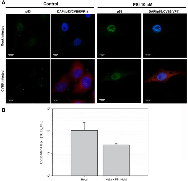
We have found that CVB5 progeny production in HeLa cells was reduced in the presence of the proteasome inhibitors MG132 and PSI in a dose- and time-dependent manner (data not shown). Here we found that in cells incubated with 10μM PSI, CVB5 infection did not induce p53 nuclear doted aggregates observed in non-infected cells (Fig. 4A). The absence of those aggregates could be a consequence of a partial reduction of CVB5 replication in the presence of PSI (Fig. 4B), or the nuclear aggregation of p53 could be dependent on a fully functional proteasome.
Discussion
Several mechanisms of apoptosis induced by picornaviruses have been documented (Buenz and Howe, 2006). For example, protease 3C induces apoptosis in cells infected by poliovirus (Barco, Feduchi, and Carrasco, 2000) and enterovirus 71 (Li et al., 2002). Likewise, the Theiler's virus L protein interferes with nucleocytoplasmic trafficking (Delhaye, van Pesch, and Michiels, 2004), which could alter the subcellular localization of proteins essential to the cell homeostasis, such as nuclear factor-κB (NF-κB). Interestingly, CVB3 VP2 capsid protein induces apoptosis via interaction with a CD27 cytoplasmic
Fig. 3.CVB5 infection of HeLa cells causes conspicuous redistribution of SUMO-1 and UBC9. Mock- and CVB5-infected HeLa cells (MOI = 10) werefixed at 4 h post-infection and stained with antibodies to SUMO-1 and UBC9 by immunofluorescence. DAPI was used to stain nuclei and images were taken by confocal microscopy (A). Western blot shows that UBC9 amounts were kept apparently constant during the infection (B).
binding protein named Siva, thus triggering activation of procaspase 3 (Henke et al., 2000; Martin et al., 2004). The UPS is important for CVB3 replication, since proteasome inhibitors block the phosphorylation of CVB3-induced extracellular signal-regulated kinase (ERK) through the inhibition of degradation of mitogen-activated protein kinase (MAPK) phosphatase-1 (MKP-1), resulting in reduced CVB3 replica-tion (Wong et al., 2007).
The best-known mechanisms of immortalization of HeLa and Vero cells involve degradation or inactivation of p53 (Ahuja, Saenz-Robles, and Pipas, 2005; Scheffner and Whitaker, 2003). In HeLa cells, the human papillomavirus (HPV) oncoprotein E6 binds to p53 and stimulates its degradation by the UPS (Scheffner et al., 1990), while in Vero cells the large T antigen of the polyomavirus SV40 binds to the DNA-binding surface of p53, inhibiting its transcription activating function (Ahuja, Saenz-Robles, and Pipas, 2005; Chen and Hahn, 2003). The differences between mechanisms of immortalization might explain why the staining for p53 in HeLa cells showed amounts generally reduced in comparison to the levels observed in Vero cells. However, CVB5 infection was found to cause striking alteration in p53 distribution in both cell lineages, mainly characterized by the formation of aggregates in the nucleus, probably equivalent to p53 docking sites for transcription of proapoptotic genes. While a CVB5
product responsible for this effect has not yet been identified, this finding strongly indicates that CVB5-induced apoptosis is p53-dependent.
A wide spectrum of intrinsic and extrinsic stressors stabilize and activate p53, affecting it by phosphorylation, acetylation, ubiquitination, methylation and sumoylation (Lavin and Gueven, 2006). Such post-translational modifications modulate levels of p53 and its trafficking between cell compartments, enabling induction of cell cycle arrest, senescence or apoptosis, thus controlling the ultimate fate of the cell. HDM2 (human double minute-2) is a homologue of MDM2 (mouse double minute-2), which is a p53-specific E3 ligase responsible for the regulation of p53, whose levels are also regulated by the UPS (Honda, Tanaka, and Yasuda, 1997). Remarkably, the HDM2 gene is one of the target genes whose transcription is activated by p53, and its product regulates p53 by a feedback loop. The regulation of p53 by HDM2 and a proposed model for how it can be affected by CVB5 infection are summarized inFig. 5. CVB5 infection of HeLa cells causes deregulation of protein sumoylation, which in turn leads to the availability of free HDM2 for ubiquitination and degradation by the proteasome, (Buschmann et al., 2000). HDM2 reduction leads to increased levels of p53, which is then made available for nuclear translocation, stimulating the induction of apoptosis.
Fig. 4.Inhibition of proteasome abrogates p53 nuclear aggregation in CVB5-infected cells. Mock- and CVB5-infected HeLa cells (MOI = 10) were incubated with or without 10μM PSI,
The present study found that SUMO-1, whose localization appeared sharply perinuclear in control HeLa cells, was markedly redistributed in CVB5 infected cells, appearing as dots dispersed throughout the cell. Similarly, the SUMO-1 conjugating enzyme UBC9, normally found concentrated at a restricted region near the nucleus, was redistributed all over the cell upon CVB5 infection. These results strongly indicate that CVB5 infection interferes with, and perhaps destabilizes protein sumoylation in host cells. It has been shown that other viruses, such as human cytomegalovirus, herpes simplex virus and adenovirus, interfere with sumoylation of host proteins (Boggio and Chiocca, 2005; Boggio and Chiocca, 2006). For example, the protein Gam1 of avian adenovirus interferes with sumoylation, causing degradation of UBC9 by a mechanism still not understood (Chiocca, 2007). This interference with sumoylation caused by Gam1 leads to the disassembly of PML-NB, a structure that seems to have an anti-virus function (Everett and Chelbi-Alix, 2007), thus helping adenovirus replication (Colombo et al., 2002).
CVB5 infected cells had diminished levels of HDM2, which could result from the transcriptional/translational shut-off of protein synthesis, or from the HDM2 cleavage by a CVB5 protease.
In the light of the importance of the UPS for the regulation of p53 activity, and considering that UPS activity is intimately intertwined with the efficient replication of another coxsackie B virus, CVB3 (Gao et al., 2008; Luo et al., 2003; Si et al., 2005; Si et al., 2007; Szalay et al., 2006), it is noteworthy that in the present study a functional proteasome was found to be essential for the CVB5-induced nuclear translocation/aggregation of p53 in HeLa cells. It should be pointed out that in the presence of a proteasome inhibitor that caused a lesser than 10-fold reduction in CVB5 titers, there was a virtually complete
abrogation of aggregation of p53 in the nucleus of HeLa cells. Therefore, it is reasonable to say that a functional proteasome is essential for the p53-dependent CVB5-induced apoptosis.
Alternatively, the stabilization of p53 in HeLa cells could be related to the transcriptional/translational shutoffs of protein synthesis caused by CVB5, which could in turn reduce levels of the protein E6 of HPV in HeLa cells and the large T antigen of SV40 in Vero cells, thus leading to the stabilization of p53 levels. However, even in the presence of higher levels of p53, a unifying mechanism must exist for the nuclear translocation and for the conspicuous and apparently targeted aggregation of p53 in the nucleus, both in HeLa and Vero cells infected by CVB5. In summary, CVB5 infection triggers HeLa cell death by apoptosis, which is associated with nuclear aggregation of p53 and redistribution of SUMO, by a mechanism that is dependent on a functional ubiquitin–proteasome system.
Materials and methods
Virus, cell culture, viral infections
CVB5 was originally a gift from Dr. Roger M. Loria (Virginia Commonwealth University, Richmond, Virginia, USA) and further propagated in the laboratory by standard protocols. HeLa and Vero cells were grown in Dulbecco's modified minimal essential medium with Eagle's salts (DMEM) supplemented with 10% fetal bovine serum (FBS), to 80–90% confluence prior to virus infection. Cells were infected with CVB5 (MOI = 10) for 1 h at 4 °C for virus adsorption. Cells were then washed with PBS and cultured in DMEM supple-mented with 2% FBS for the indicated periods of time.
Fig. 5.Model of p53 activation: HDM2 stability is sustained by a small ubiquitin-like modifier (SUMO), which is attached to the target at the same lysine residues used for ubiquitination, thus blocking HDM2 degradation (Ia), leaving p53 available for ubiquitination (Ib) and degradation by the proteasome (Ic). Exposure of the cell to CVB5 infection would reduce sumoylation of HDM2 (IIa), leading to its ubiquitination (IIb) and proteasomal degradation, (IIc), ultimately causing p53 stabilization (IId) and its availability for nuclear translocation and activation of proapoptotic genes (IIe). Small red circles represent SUMO; small green squares represent ubiquitin, small pink circles represent cytochromec.
Antibodies and reagents
Rabbit polyclonal anti-UBC9 (H-81) and anti-p53 (FL-393), goat polyclonal anti-SUMO-1 (N-19) and mouse monoclonal anti-MDM2 (SMP14) were all obtained from Santa Cruz (California). Both mouse monoclonal anti-caspase 9 and anti-cytochrome c were obtained from BD Pharmingen (California). Mouse monoclonal anti-enterovi-rus was obtained from Dako (place). The mitochondrial potential sensors JC-1 and secondary antibodies conjugated with Alexa 488 and 594 were obtained from Molecular Probes® (Oregon) and secondary antibodies conjugated with HRP were obtained from Zymed (Cali-fornia). The proteasome inhibitors PSI (Z-LLe-(OBut)-Ala-Leu-H) was
obtained from Biomol® International (Exeter).
Caspase 3 assay
HeLa cells (106cells/ml) were collected at the indicated periods of
times. The rhodamine-labelled peptide substrate (Z-DEVD)2-Rh 110 (Calbiochem, Germany) was added to afinal concentration of 50μM, followed by incubation for 10 min at 37 °C. Cells were then washed in PBS and resuspended in 300μl of PBS for FACS analysis. The cellular rhodamine 110 greenfluorescence (515–545 nm) was measured with excitation by a 488 nm argon laser on a FACScanflow cytometer (Becton Dickinson). A total of 104cells per sample were analyzed with
the WinMID® software.
JC1 assay
Changes in the mitochondrial membrane potential (ΔΨm) consequent to mitochondrial dysfunction were evaluated using JC-1. The accumulation of this compound in the mitochondrial matrix is controlled by theΨm and its absorption/emission spectra is such that color emission changes from green to orange as theΨm increases (Nicholls and Ward, 2000). CVB5 infected and control cells were collected and washed with PBS, then resuspended in 300μl of PBS and incubated with 10μg/ml of JC-1 for 10 min at 37 °C before analysis by flow cytometry. A total of 104cells per sample were analyzed using
the WinMID® software.
Mitochondria enrichment
CVB5- or mock-infected HeLa cells were mechanically harvested by scraping in PBS, centrifuged at 500×gfor 5 min, the pellets were resuspended in MB buffer (210 mM mannitol, 70 mM sucrose, 1 mM EDTA, 10 mM HEPES pH 7.5) supplemented with of a cocktail of protease inhibitors [phenylmethylsulfonylfluoride (PMSF), 1 mM; leupeptine, 1μg/ml; aprotinin, 6.8 mg/ml; benzamidine, 1 mM], and then disrupted by several passages through a 25G1 needlefitted onto a 5-ml syringe. The suspensions obtained were centrifuged at 500×g
for 5 min at 4 °C and the supernatants were further centrifuged at 10,000×g for 30 min at 4°C, in order to obtain a pellet of heavy membranes (HM) mitochondrial-enriched fraction, which was sup-plemented with 1% Triton X-100. The supernatant was further centrifuged at 100,000×gfor 1 h at 4°C to obtain a supernatant of thefinal soluble cytosolic fraction (S), and a pellet of light membranes, which was discarded. The fractions HM and S were stored at−70° until further analysis.
Immunoblotting
Proteins separated by sodium dodecyl sulphate-polyacrylamide gel electrophoresis (SDS-PAGE) were transferred onto nitrocellulose membranes, which were then blocked for 1 h with 3% nonfat dry milk in 10 mM Tris-HCl pH 7,5 with 0.1% Tween 20. Blots were incubated for 3 h with the appropriate primary antibody, followed by incubation for 1.5 h with an adequate peroxidase-labelled secondary antibody.
Immunoreactive bands were visualized by ECL Western Blotting Detection Reagents (Amersham Biosciences®).
Immunofluorescence
Cell culture monolayers grown on 12 mm glass coverslips inside the wells of 24-well dishes were infected with CVB5 andfixed at 4 h postinfection in 2% paraformaldehyde for 20 min, rinsed three times with PBS, permeabilized with 0.3% Triton X-100 in PBS for 15 min, and then blocked with 1% BSA in PBS for 1 h. Incubation with the primary antibody diluted in PBS containing 1% BSA was done for 60 min, followed by washing ten times with PBS, incubated with secondary antibody for 45 min in 1% BSA/PBS, washed 10 times with PBS, and mounted in Flourmount-G ® (Southern Biotech, place), all done at room temperature. DNA was stained with DAPI (4′ ,6-diamidino-2-phenylindole) and images were acquired using a Leica TCSP5 confocal scanner microscope.
Acknowledgments
We acknowledge thefinancial supports from CNPq (Scholarships for EA and RG, numbers 310825/2006-7 and 151212/2007-4, respectively) and FAPESP (Grant Numbers 95/09692-2—research funding for EA; 03/02531-1—scholarship for RG; and 04/08868-0 to establish a confocal microscopy facility). We thank Maria Lucia Silva for technical support.
References
Ahuja, D., Saenz-Robles, M.T., Pipas, J.M., 2005. SV40 large T antigen targets multiple cellular pathways to elicit cellular transformation. Oncogene 24 (52), 7729–7745. Barco, A., Feduchi, E., Carrasco, L., 2000. Poliovirus protease 3C(pro) kills cells by
apoptosis. Virology 266 (2), 352–360.
Boggio, R., Chiocca, S., 2005. Gam1 and the SUMO pathway. Cell Cycle 4 (4), 533–535. Boggio, R., Chiocca, S., 2006. Viruses and sumoylation: recent highlights. Curr. Opin.
Microbiol. 9 (4), 430–436.
Buenz, E.J., Howe, C.L., 2006. Picornaviruses and cell death. Trends Microbiol. 14 (1), 28–36.
Buschmann, T., Fuchs, S.Y., Lee, C.G., Pan, Z.Q., Ronai, Z., 2000. SUMO-1 modification of Mdm2 prevents its self-ubiquitination and increases Mdm2 ability to ubiquitinate p53. Cell 101 (7), 753–762.
Chen, W., Hahn, W.C., 2003. SV40 early region oncoproteins and human cell transformation. Histol. Histopathol. 18 (2), 541–550.
Chiocca, S., 2007. Viral control of the SUMO pathway: Gam1, a model system. Biochem. Soc. Trans. 35 (Pt. 6), 1419–1421.
Colombo, R., Boggio, R., Seiser, C., Draetta, G.F., Chiocca, S., 2002. The adenovirus protein Gam1 interferes with sumoylation of histone deacetylase 1. EMBO Rep. 3 (11), 1062–1068.
Danial, N.N., Korsmeyer, S.J., 2004. Cell death: critical control points. Cell 116 (2), 205–219.
Delhaye, S., van Pesch, V., Michiels, T., 2004. The leader protein of Theiler's virus interferes with nucleocytoplasmic trafficking of cellular proteins. J. Virol. 78 (8), 4357–4362.
Endres, A.S., Helms, T., Steinfuhrer, S., Meisel, H., 2002. Transient broca aphasia in an elderly man caused by coxsackievirus B5. J. Neurol. 249 (9), 1318–1319. Everett, R.D., Chelbi-Alix, M.K., 2007. PML and PML nuclear bodies: implications in
antiviral defence. Biochimie 89 (6-7), 819–830.
Gao, G., Zhang, J., Si, X., Wong, J., Cheung, C., McManus, B., Luo, H., 2008. Proteasome inhibition attenuates coxsackievirus-induced myocardial damage in mice. Am. J. Physiol. Heart Circ. Physiol. 295 (1), H401–H408.
Henke, A., Launhardt, H., Klement, K., Stelzner, A., Zell, R., Munder, T., 2000. Apoptosis in coxsackievirus B3-caused diseases: interaction between the capsid protein VP2 and the proapoptotic protein siva. J. Virol. 74 (9), 4284–4290.
Honda, R., Tanaka, H., Yasuda, H., 1997. Oncoprotein MDM2 is a ubiquitin ligase E3 for tumor suppressor p53. FEBS Lett. 420 (1), 25–27.
Hwang, H.Y., Kim, J.Y., Lim, J.Y., Chung, S.K., Nam, J.H., Park, S.I., 2007. Coxsackievirus B3 modulates cell death by downregulating activating transcription factor 3 in HeLa cells. Virus Res. 130 (1-2), 10–17.
Lavin, M.F., Gueven, N., 2006. The complexity of p53 stabilization and activation. Cell Death Differ. 13 (6), 941–950.
Lee, M.H., Lee, S.W., Lee, E.J., Choi, S.J., Chung, S.S., Lee, J.I., Cho, J.M., Seol, J.H., Baek, S.H., Kim, K.I., Chiba, T., Tanaka, K., Bang, O.S., Chung, C.H., 2006. SUMO-specific protease SUSP4 positively regulates p53 by promoting Mdm2 self-ubiquitination. Nat. Cell Biol. 8 (12), 1424–1431.
Luo, H., Zhang, J., Dastvan, F., Yanagawa, B., Reidy, M.A., Zhang, H.M., Yang, D., Wilson, J. E., McManus, B.M., 2003. Ubiquitin-dependent proteolysis of cyclin D1 is associated with coxsackievirus-induced cell growth arrest. J. Virol. 77 (1), 1–9.
Martin, U., Nestler, M., Munder, T., Zell, R., Sigusch, H.H., Henke, A., 2004. Characterization of coxsackievirus B3-caused apoptosis under in vitro conditions. Med. Microbiol. Immunol. 193 (2-3), 133–139.
McLean, J.E., Ruck, A., Shirazian, A., Pooyaei-Mehr, F., Zakeri, Z.F., 2008. Viral manipulation of cell death. Curr. Pharm. Des. 14 (3), 198–220.
Moschovi, M., Sterpi, P., Youroukos, S., Tzortzatou-Stathopoulou, F., 2002. Encephalitis and myocarditis in a child with acute lymphoblastic leukemia: role of coxsack-ievirus B5? Pediatr. Hematol. Oncol. 19 (3), 205–210.
Nicholls, D.G., Ward, M.W., 2000. Mitochondrial membrane potential and neuronal glutamate excitotoxicity: mortality and millivolts. Trends Neurosci. 23 (4), 166–174. Pallansch, M.A., 1997. Coxsackievirus B epidemiology and public health concerns. Curr.
Top. Microbiol. Immunol. 223, 13–30.
Pampin, M., Simonin, Y., Blondel, B., Percherancier, Y., Chelbi-Alix, M.K., 2006. Cross Talk between PML and p53 during Poliovirus Infection: Implications for Antiviral Defense, Vol. 80, pp. 8582-8592.
Scheffner, M., Whitaker, N.J., 2003. Human papillomavirus-induced carcinogenesis and the ubiquitin-proteasome system. Semin. Cancer Biol. 13 (1), 59–67.
Scheffner, M., Werness, B.A., Huibregtse, J.M., Levine, A.J., Howley, P.M., 1990. The E6 oncoprotein encoded by human papillomavirus types 16 and 18 promotes the degradation of p53. Cell 63 (6), 1129–1136.
Si, X., McManus, B.M., Zhang, J., Yuan, J., Cheung, C., Esfandiarei, M., Suarez, A., Morgan, A., Luo, H., 2005. Pyrrolidine dithiocarbamate reduces coxsackievirus B3 replication through inhibition of the ubiquitin–proteasome pathway. J. Virol. 79 (13), 8014–8023.
Si, X., Wang, Y., Wong, J., Zhang, J., McManus, B.M., Luo, H., 2007. Dysregulation of the ubiquitin-proteasome system by curcumin suppresses coxsackievirus B3 replica-tion. J. Virol. 81 (7), 3142–3150.
Szalay, G., Meiners, S., Voigt, A., Lauber, J., Spieth, C., Speer, N., Sauter, M., Kuckelkorn, U., Zell, A., Klingel, K., Stangl, K., Kandolf, R., 2006. Ongoing coxsackievirus myocarditis is associated with increased formation and activity of myocardial immunoprotea-somes. Am. J. Pathol. 168 (5), 1542–1552.
Watson, I.R., Irwin, M.S., 2006. Ubiquitin and ubiquitin-like modifications of the p53 family. Neoplasia 8 (8), 655–666.
Wong, J., Zhang, J., Si, X., Gao, G., Luo, H., 2007. Inhibition of the extracellular signal-regulated kinase signaling pathway is correlated with proteasome inhibitor suppression of coxsackievirus replication. Biochem. Biophys. Res. Commun. 358 (3), 903–907.