GENETIC DIVERSITY AND ANTIFUNGAL SUSCEPTIBILITY TESTING OF
TRICHOSPORON ASAHII ISOLATED OF INTENSIVE CARE UNITS PATIENTS
Rosana Bellan de Oliveira Silva1,2; Ana Marisa Fusco-Almeida2; Marcelo Teruyuki Matsumoto2; Lilian Cristiane Baeza3; Tatiane Benaducci2; Maria José Soares Mendes-Giannini2*
1Instituto Adolfo Lutz, Rio Claro, SP, Brasil; 2Departamento de Análises Clínicas, Faculdade de Ciências Farmacêuticas,
Universidade Estadual Paulista, Araraquara, SP, Brasil; 3Universidade Estadual de Maringá, Maringá, PR, Brasil
Submitted: December 10, 2007; Returned to authors for corrections: February 08, 2007; Approved: July 05, 2008.
ABSTRACT
Trichosporon asahii is an opportunistic pathogen, associated with a high mortality rate in
immunocompromised patients. In this study, ten isolates, recovered from oral cavity and urine of patients in Intensive Care Units (ICU) over six months, were identified by classical and molecular methods, typed by RAPD and tested in vitro for susceptibility to fluconazole, itraconazole, 5-flucytosine and amphotericin B. A total agreement between the identification of Trichosporon sp by PCR based on sequences of the Internal Transcribed Spacer Regions (ITS) and on the sequences of small-subunit (SSU) ribosomal DNA (rDNA) was found. Randomly amplified of polymorphic DNA (RAPD), with primers P6 and M13, was used to determine the genomic profiles. The dendogram analysis indicated that almost all strains showed similarity >0.9 among them and all strains were multidrug-resistant. This study brings new results on the identification and genotyping of T. asahii isolated from Brazilian ICU patients and information about their antifungal drugs susceptibility.
Key-words: Trichosporon, PCR, RAPD, susceptibility, antifungal drugs
*Corresponding Author. Mailing address: Laboratório de Micologia Clínica, Departamento de Análises Clínicas, Faculdade de Ciências Farmacêuticas, UNESP, Rua Expedicionários do Brasil, 1621, CEP 14801-902, Araraquara, SP, Brasil. Tel.: +55 (16) 33016556 / Fax: +55 (16) 33016547. E-mail: giannini@fcfar.unesp.br
INTRODUCTION
In the past few decades, a worldwide increase in the incidence of fungal infections has been observed, as well as the recognition of innocuous yeasts, previously thought incapable of harming the human body, as pathogens (38). The emergence of less common but medically important fungal pathogens has contributed to the rate of morbidity and mortality, especially in the increasingly expanding population of immunocompromised patients. Various groups have stressed the importance of newopportunistic fungal pathogens as causes of life-threateninginfections (1,14). Among them, deep-seated trichosporonosis is a lethal opportunistic infection occasionally found in immunocompromised patients, particularly those who are neutropenic due to cytotoxic therapy for hematological malignancies.
Over the past decade, the taxonomy of the genus
Trichosporonhas been subjected to extensive revision on the
basis of moleculardata, and the previously named T. beigelii
(or T. cutaneum)corresponds, in the most recent classification,
to six differentspecies: T. asahii, T. asteroides, T. cutaneum,
T. inkin, T. mucoides, and T. ovoides (15,16).
Trichosporon is found in the commensal mycobiota of the
skin surface, mainly in the genito crural and perianal areas of homosexuals (47). This yeast is found in the normal skin, nails and mouth of healthy individuals (28,35,58), as well as, in Brazil,
T. asahii was also recovered from rice and cassava used in
fermented beverages (45).
Trichosporon infections are rare but have been associated
immunocompromised patients with a pattern similar to candidiasis (6,19,29,30,43,58). Rodrigues et al. (43) described twenty-two cases of nosocomial infection caused by T. asahii, detected during a period of six years (1999-2005) in a Brazil hospital. Recently, a case was reported of systemic Trichosporon
cutaneum infection in a 3-year-old infant with Wilms’ tumor (5).
In the same hospital, an oropharyngeal secretion sample was collected from an AIDS patient with oral lesions and
Trichosporonpullulans was isolated(34).
At the moment, Trichosporon asahii is the most clinically important pathogenic yeast in the genus Trichosporon, as this species causes both deep-seated infection and summer-type hypersensivity pneumonitis (9,15,17,28,49,52). Sugita et al. (49) have suggested that T. asahii is common environmental pathogen, and Martins-Diniz et al. (25) have also encountered this genus in both biotic (professional health) and abiotic sources (areas of hospital) with prevalence around 30% among yeast isolated. Pini et al. (39) have found massive air contamination with T. asahii in the ward where neutropenic patients were staying and the corridor immediately outside.
Trichosporon infection is associated with high morbidity
and mortality and is difficult to treat, frequently involving resistance to 5-fluorocytosine, azoles and amphotericin B (10,41). Walsh et al. (57) noted that blood isolates were more resistant to amphotericin B than skin isolates. The optimal antifungal therapy for these infections is unclear. There is growing evidence that azole drugs have good activity against Trichosporon spp. (3,11,36) and that combined administration of fluconazole and amphotericin B may be superior to either drug used alone against invasive infection (2,18). Meyer et al. (28) report a case of chronic disseminated T. asahii infection in a child with leukemia, where a cure was achieved after treatment with itraconazole.
The therapies are only effective if the disease is detected at an early stage, and, therefore, early diagnosis is an important factor in the successful management of patients with disseminated trichosporonosis. Unfortunately, difficulties in the identification of these microorganisms lead to delays in treatment and post-mortem diagnosis (30,31).
Species identification and molecular typing have become critical elements of nosocomial fungal outbreak investigations. The studies should include the origins of nosocomial infection, genetic comparison between invasive or noninvasive isolates, and comparison of the genotypes and susceptibilities of the isolates (53).
Many investigations of the intraspecies diversity and epidemiology of pathogenic fungi using random amplified polymorphic DNA (RAPD) analysis, hybridization with specific probes such as CA3, and multilocus enzyme electrophoresis (MLEE) have been reported, but there are only a few reports for
Trichosporon species (12,48).
Sugita et al. (51) have sequenced the internal transcribed spacer (ITS) region and describe a rapid PCR-based approach
for detection of all species of emerging yeasts of the
Trichosporon genus. The PCR product length alone identified
all the isolates, the primers TRF (forward) and TRR (reverse) being chosen to align with regions were not conserved in other medically important yeasts (50). Their data indicate that the PCR detection system is useful for identifying these yeasts.
In Brazil there are few survey on these yeasts (30,34,43). In this study we examined the genetic diversity and biochemical characteristics of T.asahii strains recovered from clinical specimens (oral cavity and urine) from patients in Intensive Care Units (ICU), as well as the correlation between the drug susceptibility profile and genetic changes detected by RAPD (primer M13 and 6).
MATERIALS AND METHODS Yeast isolates
All isolates were recovered from clinical specimens (two from oral cavity and eight from urine) from patients in Intensive Care Units (ICU), collected from April to September 2001. The isolates were subcultured on Sabouraud glucose agar (SGA). Isolates were identified by API 20C AUX (BioMérieux, France) strip in accordance with the manufacturer’s instructions and by evaluation of their micromorphological features, on cornmeal Tween 80-agar. The interpretation of the data was based on Kurtzman and Fell (21).
Oligonucleotide primers and PCR amplification
The internal transcribed spacer (ITS) regions were amplified with primers ITS1 (5' - TCC GTA GGT GAA CCT GCG G - 3') and
ITS4 (5' - TCC TCC GCT TAT TGA TAT GC - 3’), which were
designed from conserved regions to amplify the 18S rDNA, 25S and 5,8S regions of ribosomal DNA (51). The primers, TRF (forward) (5' – AGA GGC CTA CCA TGG TAT CA- 3') and TRR (reverse) (5' –TAA GAC CCA ATA GAG CCC TA- 3'), were chosen because they would specifically amplify only
Trichosporon species and to align with regions which were not
conserved in other medically important yeasts (50). These PCR primers correspond to nucleotides 154 to 173 and 354 to 335 of
Saccharomyces cerevisiae SSU rDNA. For both PCRs, the
amplification reactions were performed in 25 μL sterile distilled water containing 1 μM of each primer, 3.0 μL of template DNA solution (1.5 μg/mL), 1.5 mM MgCl2, 2.5U of Taq polymerase,
products were run on 2.0% agarose gel in 1 × TBE (90 mM Tris, 90 mM boric acid, 2 mM EDTA pH 8) buffer for 150 min at 150 V, stained with 0.5 μg/mL ethidium bromide (GIBCO), and visualized under UV light, with the aid of Image Master VDS (PHARMACIA BIOTECH), and photographed.
RAPD analysis
Genomic DNA was extracted and purified by a slight modification of the method of Lasker et al. (22). RAPD was carried out with primer 6 (5’-d[CCC GTC AGC A]-3’), (Amersham Pharmacia Biotech) and M13 (5'- d [GAG GGT GGC GGT TCT]-3’), in a total volume of 25 μL, using the Ready-To-Go RAPD Kit in accordance with the manufacturer’s instructions. The following cycle conditions were used for primer 6: initial denaturation at 95ºC for 5 min, followed by 45 cycles of denaturation at 95ºC for 1 min, annealing at 36ºC for 1 min and amplification at 72ºC for 2 min, with a final extension at 72ºC for 10 min. The cycle used for primer M13 was: initial denaturation at 94ºC for 3 min, followed by 40 cycles of denaturation at 94ºC for 20 s, annealing at 50ºC for 1 min, and amplification at 72ºC for 20 s with a final extension at 72ºC for 6 min. Amplification products were separated by electrophoresis on 2% agarose gels in 1X TBE buffer (0.1M Tris, 0.09 M boric acid, 0.001 M EDTA [pH 8.4] at 150V for 2.5 h, stained with 0.5 mg of ethidium bromide per mL of deionized water for 30 min., and then visualized under UV light and photographed. Patterns were analyzed both by visual comparison and by the Image Master VDS Software (Pharmacia Biotech). The computer program GelCompar II version 2.0 was used to determine the genetic relationship of the isolates. Similarity coefficients were calculated by the Dice Algorithm and cluster analysis was performed by an UPGMA algorithm (46).
In vitro antifungal susceptibility testing
The susceptibility testing followed the Clinical and Laboratory Standards Institute (CLSI) recommendations for microdilutionprocedures, but included modifications described previously (7,44). Briefly, in the susceptibility test, RPMI 1640 supplemented with 2% glucose was inoculated with 2x10⁵ CFU/
mL, in the flat-bottomed wells of microplate cultures trays. All microplates were wrappedwith film sealer to prevent the medium from evaporating, attachedto an electrically-driven wheel inside the incubator, agitatedat 350 rpm, and incubated at 35ºC for 48 h. Candida parapsilosis(ATCC 22019) was used as the control strain.The antifungal agents used in the study were: amphotericinB (AMB) (Sigma Aldrich Quimica S.A.), 5-fluorocytosine(5FC) (Sigma Aldrich), fluconazole (FCZ) (Pfizer S.A.) and itraconazole (ITZ) (Janssen S.A.). AMB, FCZ, and ITZ were dissolved in 100% dimethyl sulfoxide (Sigma Aldrich) and 5FC in sterile distilled water. All drug stocksolutions were frozen at -70ºC as 100x stocks untilused.The MICs were determined spectrophotometrically as follows: after 48h. incubation, the
optical density of eachwell of the microtiter plate was read with a microplate reader(Bio-Rad) at 490 nm. ForAMB, the MIC endpoint was defined as the lowest drug concentrationexhibiting a reduction in growth of 90% or more, compared withthe control. For 5FC and the azole drugs, theMIC endpoint was defined as the concentration producing 50%inhibition (13). The breakpoints MICs was based on CLSI M27-A2 (7) reference microdilution method described for Candida sp. Experiments were repeated in duplicate, and MICs with one dilution higher were found and accepted as described by CLSI (7).
RESULTS Identification
All isolates presented membranousand finely cerebriform, white to cream-colored colonies. Initial microscopic examination of portionsof these colonies, as well as slide cultures, demonstrated roundto oval, budding yeast-like cells and true hyphae forming cylindricalarthroconidia.They grew on SGA at 30 and 35ºC but not at 45ºC. The overall micro- and macroscopic appearancewas consistent with that for members of the genus
Trichosporon and morphological, physiological, and
biochemical features were as described for T. asahii.
In vitro antifungal susceptibility testing
The MICs of the four antimicrobial agents for the quality controlorganisms were consistently within 2 or 3 dilutionsof each other. No differences were observed between MICs obtainedby the CLSI reference procedure and those achieved by the modifiedmethod. The MICs of four antifungal drugs for
the T. asahii isolates, obtained by the CLSI broth microdilution
method, are presented in Table 1. As indicated, the isolates had reduced susceptibility in vitro to all drugs, showing 4-6 times higher MICs to itraconazole and 3-4 times to 5-flucytosine compared of MICs breakpoint values. FCZ exhibited the best activity in vitro against the majority of the isolates (90%), with MIC of 16 μg/mL and only one isolate with 32 μg/mL. Three isolates have MIC value for AMB above 2 μg/mL.
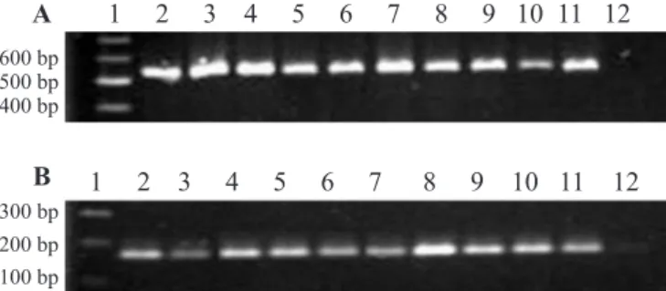
DNA fragment
Both primers for the ITS region and TRR/TRF amplified all DNA isolates, producing distinct DNA fragments of approximately 530 bp and 170 pb, respectively, as shown in Fig. 1.
RAPD analysis
only high-intensity bands were used. Each primer generated between six and nine bands for an individual isolate.
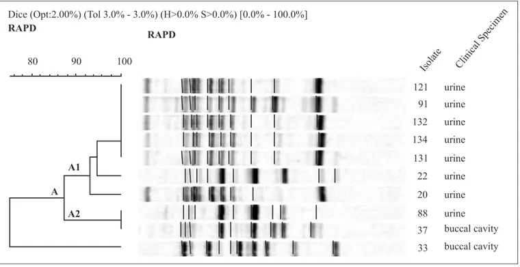
Among the 10 isolates analyzed by primer 6,various RAPD profiles were observed with molecular weights of bands between 170 and 1100 bp. All visible and definite bands were included in the analysis (Fig. 2). Dendrogram analysis obtained with this random primer showed a high homogeneity among the isolates from different body sites, with five RAPD profiles. Only one group (A) was generated with a coefficient of similarity greater than 85%. The largest (A1) contained seven isolates from urine, one grouped in a cluster (5 isolates) and two isolates with high degree (>90%). The subgroup A2 grouped in a cluster contained two isolates from oral cavity and urine. Subgroups A1 and A2 were related by similarity coefficient above 85%, suggesting these strains are closely related to each other. The isolate 33 from oral cavity has a low similarity coefficient of 70% in relation to the other groups, suggesting different strains.
For the primer M13 (Fig. 3), four different profiles were observed. One group was generated by a coefficient of similarity higher that 80%. Two clusters were formed in the subgroup A1, the first with five isolates from urine and the second containing three isolates, two from urine and one from oral cavity. In this last cluster, the three isolates suggest colonization of different sites with the same strain. The subgroup A2 and the isolate 22
from urine also presented a high degree of similarity with subgroup A1.
DISCUSSION
The total agreement between the identification of
Trichosporon sp by PCR based on sequences of the Internal
Transcribed Spacer Regions (ITS) and on the sequences of small-subunit (SSU) ribosomal DNA (rDNA) aligned by the primers TRF/TRR indicates that T. asahii can be readily identified by these two methods, as shown by Sugita et al. (51).
T. asahii has been isolated from various types of clinical
specimens from immunocompromised patients, including blood, skin biopsy, and urine specimens (9,17,19,29,32,43,52), but few were isolated from patients without immunological disorders (20). Factors that enhance mucosal colonization and subsequent invasion of Trichosporon species include broad-spectrum antibiotic treatment and scratches in mucosal barriers (6,24,56). Despite the increasing frequency and severity of trichosporonosis, data on the antifungal susceptibility of
Trichosporon spp. are limited and recommendations for in vitro
testing of this fungus are not included in the guidelines of the National Committee for Clinical Laboratory Standards (27). There are few data available regarding T. asahii susceptibility to antifungal drugs in Brazil. The emergence of T. asahii isolates with reduced susceptibility to common antifungal drugs is a matter for concern. In our study, all isolates had reduced susceptibility to AMB, 5-FC and azoles. ITZ and FCZ exhibited MICs of 2 to 8 and 16 to 32 μg/ml, respectively. The first one corresponds to resistant isolates (≤ 1) and for the FCZ intermediate susceptibility (16-32 μg mL-1) based on CLSI
breakpoints for Candida sp (7). For AMB, the controversy is
Table 1. Isolation sites and antifungal susceptibility test results
for T. asahii isolates recovered from patients in an Intensive
Care Unit.
Isolate MIC (mg/liter)
a
FLZ ITZ 5FC AMB
20 urine 16 8 32 2
22 urine 32 4 32 2
33 oral cavity 16 2 32 4
37 oral cavity 16 2 16 2
88 urine 16 2 16 4
91 urine 16 4 32 2
121 urine 16 2 16 2
131 urine 16 2 16 4
132 urine 16 2 16 2
134 urine 16 2 16 2
ATCC 22019
(type strain) 2-8 0,06-0,25 0,12-0,5 0,25-1 Susceptibility
breakpointsb ≤ 8 ≤ 0.125 ≤4 >2c
aAbbreviations: FLZ, fluconazole, ITZ, itraconazole, 5FC,5-fluorocytosine, AMB, amphotericin B; bThe breakpointsare based on NCCLS M27-A2 reference microdilution method described by
Candida sp.; cEspinell-Ingroff (8).
Figure 1. Agarose gel electrophoresis of PCR products of T.
asahii isolates recovered from clinical specimens (oral cavity
greater, because we did not have MICs values that discriminated susceptible and resistant yeasts strains. Recently, Espinel-Ingroff (8) discussed the MIC value of 2 μg mL-1 as the
breakpoint to filamentous fungus. Wolf et al. (58) reported the
isolation of multidrug-resistant T. asahii from infected patients who exhibited risk factors and had developed either superficial infections or invasive infections while in ICU. Thus, the pathogenesis of Trichosporon infection in the ICU setting may
Figure 2. Dendrogram of T. asahii obtained from RAPD molecular patterns with primer 6.
be similar to that of the more commonly observed Candida
infection. Paphitou et al. (36) results confirmed the previous observation of Perparim et al. (37) as well as of other authors (26,33,54,55) that the azoles, in general, appear to be more active
in vitro against this pathogen than to AMB. Recently, a
47-year-old man with diagnosed acute myeloblastic leukemia and non-insulin-dependent diabetes mellitus developed T. asahii
fungemia and the in vitro antifungal susceptibilities showed high caspofungin and amphotericin B MICs values (16 μg/ml, and > 32 μg/ml, respectively) while fluconazole, itraconazole, and voriconazole exhibited low MICs (4 μg/ml, 0.5 μg/ml, and 0.015 μg/ml, respectively) (4). In another patient, a neutropenic individual with acute myeloid leukaemia experienced a breakthrough infection of T. asahii despite posaconazole treatment (42). Although there are no established breakpoints to define the antifungal susceptibility of Trichosporon spp, our study suggests the FCZ may be used to treat patients with trichosporonosis. McGinnis et al. (26), as well as in our study, also showed that T. asahii strains exhibited reduced susceptibility to AMB (MICs, 1 to 4 mg/liter), but they were susceptible to ITZ and FCZ (MICs=0.125 to 1 and 0.5 to 4 mg/ liter, respectively). On the other hand, the FCZ MICs for two
T.asahii isolates recovered from esophageal biopsy specimens
of two patients were found to be relatively high and these isolates were also resistant to nystatin. Lemes et al. (23) verified that the isolates of T. asahii were 5FC susceptible, ITZ resistant and some isolates were resistance to FCZ. The in vitro resistance to antifungal drugs may not be critical in immunocompetent patients, who may overcome the infection when the precipitating cause (e.g., a catheter) is removed, even if only partial inhibition of fungal growth is achieved through the use of antifungal drugs. However, immunocompromised patients may be dependent on the action of fungicidal drugs, so that infection with multidrug-resistant Trichosporon species may be catastrophic in this population.
RAPD analysis with primers 6 and M13 generated a similar dendrogram structure. In general, there was, in both dendrograms, formation of groups containing strains with high similarities (>0.8) among them associated with high MICs. The exception was strain 33 recovered from oral cavity, which showed minor similarity (around 0.7) with primer 6, but high (> 0.85) with primer M13. Moreover, RAPD analysis suggested that the isolates the different body sites were similar, suggesting a common origin. In addition, these patients were hospitalized in ICU between April and December, revealing a possibly clonal source of infection.
In conclusion, a rapid and reliable identification of T. asahii
was achieved by molecular techniques. In addition, RAPD can be used to differentiate genotypes and reveal the correlation among them, drug susceptibility and the origin of a strain. The higher correlation among genotypes and the higher MICs of these strains offer potential risks of exogenous nosocomial
infection (e.g. to catheter). In addition, increasing use of antifungal drugs in the ICU may lead to the selection and isolation of more resistant species in the future.
ACKNOWLEDGEMENTS
This investigation was financially supported by the Brazilian Organizations, CNPq (Processo n. 400141/1998-1), FCF-UNESP and Instituto Adolfo Lutz.
RESUMO
Diversidade genética e suscetibilidade a antifúngicos
de Trichosporon asahii isolado de pacientes de
Unidades de Terapia Intensiva
Trichosporon asahii é um patógeno oportunista que
apresenta altos índices de mortalidade em pacientes imunocomprometidos. No presente trabalho, dez cepas foram isoladas da cavidade bucal e urina de pacientes internados na Unidade de Terapia Intensiva (UTI) por seis meses. Todos os isolados foram identificados por métodos clássicos e moleculares, tipados por RAPD e testados in vitro quanto à sensibilidade ao fluconazol, itraconazol, 5-fluorocitosina e anfotericina B. Houve concordância total entre a identificação
de Trichosporon sp por PCR baseado na seqüência da região
ITS (Internal Transcribed Spacer) e na seqüência da subunidade menor do DNA ribossômico (rDNA).Os perfis genéticos foram determinados por RAPD utilizando dois iniciadores P6 e M13. A análise do dendrograma mostrou que a maioria das amostras apresentou alta similaridade entre elas (>0.9) e todas foram multidrogas resistentes. Este estudo traz novos resultados em relação à identificação e genotipagem de isolados brasileiros de T. asahii em pacientes internados em UTI, bem como dados sobre o perfil de sensibilidade aos antifúngicos.
Palavras-chave:Trichosporon, PCR, RAPD, sensibilidade às drogas antifúngicas, drogas antifúngicas.
REFERENCES
1. Anaissie, E.J. (1992). Opportunistic mycoses in the immunocompromised host: experience at a cancer center and review. Clin. Infect. Dis., 14, 43-53.
2. Anaissie, E.J.; Hachem, R.; Karyotakis, N.C.; Gokaslan, A.; Kignani, M.C.; Stephens, L.C.; Tin, U.C. (1994). Comparative efficacies of amphotericin B, triazoles, and combination of both as experimental therapy for murine trichosporonosis. Antimicrob. Agents
Chemother., 38, 2541-2544.
3. Arechavala, A.I.; Bianchi, M.H.; Robles, A.M.; Santiso, G.; Negroni, R. (2007). Identification and susceptibility against fluconazole and albaconazole of 100 yeasts’ strains isolated from vaginal discharge.
4. Bayramoglu, G.; Sonmez, M.; Tosun, I.; Aydin, K.; Aydin, F. (2008). Breakthrough Trichosporonasahii fungemia in neutropenic patient with acute leukemia while receiving caspofungin. Infection, 36, 68-70.
5. Carvalho, A.M.R.; Melo, L.R.B.; Moraes, V.L.; Neves, R.P. (2008). Invasive Trichosporoncutaneum infection in an infant with wilms’ tumor. Braz. J. Microbiol., 39, 59-60.
6. Chan, R.M.; Lee, P.; Wroblewski, J. (2000). Deep-seated trichosporonosis in an immunocompetent patient: a case report of uterine trichosporonosis. Clin. Infect. Dis., 31, 621.
7. Clinical and Laboratory Standards Institute. (2002). Reference method for broth dilution antifungal susceptibility testing of yeasts. Approved standard document M27-A2. CLSI, Wayne, PA, USA. 8. Espinel-Ingroff, A. (2008). Mechanisms of resistance to antifungal
agents: Yeasts and filamentous fungi. Rev. Iberoam. Micol., 25, 101-106.
9. Fagundes Junior, A.A.; Carvalho, R.T.; Focaccia, R.; Fernandez, J.G.; Araújo, H.B.N.; Strabelli, T.M.V.; Kopel, L.; Lage, S.G. (2008).
Trichosporon asahii an emerging etiologic agent of fungal infection
and colonization in heart failure patients in intensive care unit: case report and literature review. Rev. Bras. Ter. Intensiva, 20, 106-109. 10. Fleming, R.V.; Walsh, T.J.; Anissie, E.J. (2002). Emerging and less common fungal pathogens. Infect. Dis. Clin. North Am., 16, 915-933.
11. Fournier, S.; Pavageau, W.; Feuillhade, M.; Deplus, S.; Zagdanski, A.M.; Verola, O.; Dombret, H.; Molina, J.M. (2002). Use of voriconazole to successfully treat disseminated Trichosporon asahii
infection in a patient with acute myeloid leukaemia. Eur. J. Clin.
Infect. Dis., 21, 892-896.
12. Fujita, S.I.; Senda, Y.; Nakaguchi, S.; Hashimoto, T. (2001). Multiplex PCR using internal transcribed spacer 1 and 2 regions for rapid detection and identification of yeast strains. J. Clin. Microbiol., 39, 3617-3622.
13. Ghannoum, M.A.; Ibrahim, A.S.; Fu, Y.; Shafiq, M.C.; Edwards, J.E. Jr.; Criddle, R.S. (1992). Susceptibility testing of Cryptococcus
neoformans: a microdilution technique. J. Clin. Microbiol., 30,
2881-2886.
14. Girmenia, C.; Pagano, L.; Martino, B.; D’Antonio, D.; Fancir Specchia, G.; Melillo, L.; Buelli, M.; Pizzarelli, G.; Venditti, M.; Martino, P.; Gimema Infection Program. (2005). Invasive infections caused by Trichosporon species and Geotrichum capitatum in patients with hematological malignancies: a retrospective multicenter study from Italy and review of the literature. J. Clin. Microbiol., 43, 1818-1828.
15. Guého, E.; de Hoog, G.S.; Smith, M. (1992). Neotypification of the genus Trichosporon. Antonie Leeuwenhoek, 61, 285-288. 16. Gueho, E.; Improvisi, L.; de Hoog, G.S.; Dupont, B. (1994).
Trichosporon on humans: a practical account. Mycoses, 37, 3-10.
17. Itoh, T.; Hosokawa, R.; Kodhdera, U.; Toyazaki, N.; Asada, Y. (1996). Disseminated infection with Trichosporonasahii. Mycoses, 39, 195-199.
18. Kamberi, P.; Asturo, T.; Takayoshi, T.; Massaru, N. (1998). Efficacy of amphotericin B and azoles alone and in combination against disseminated trichosporonosis in neutropenic mice. Chemoterapy, 44, 55-62.
19. Kataoka-Nishimura, S.; Akyama, H.; Saku, K.; Dkashiwa, M.; Mori, S.; Tanikawa, S.; Sakanaki, H.; Onozawa, Y. (1998). Invasive infection due to Trichosporon cutaneum in patients with hematologic malignancies. Cancer, 82, 484-487.
20. Kim, Y.J.; Kim, S.I.; Kim, Y.R.; Park, Y.M.; Park, Y.J.; Kang, M.W. (2007). Successful treatment of septic shock with purpura fulminans caused by Trichosporon asahii in an immunocompetent patient.
Ann. Clin. Lab. Sci., 37, 366-369.
21. Kurtzman, C.P.; Fell, J.W. (1998). The yeasts: a taxonomic study. Elsevier, Amsterdam.
22. Lasker, B.; Page, L.S.; Lott, T.J.; Kobayashi, G.S. (1992) Isolation, characterization, and sequencing of Candidaalbicans repetitive element 2. Gene, 116, 51-57.
23. Lemes, R.M.S.; Colombo, A.L.; Resende, M.A. (2004). Perfil de susceptibilidade de Trichosporon spp. à anfotericina B e azólicos. Proceedings of the Brazilian Congress of the Mycology 4rd ed., Ouro Preto, p. 139.
24. Lyman, C.A.; Garrett, K.F.; Pizzo, P.A.; Walsh, T. (1994). Response human polymorphonuclear leukocytes and monocytes to
Trichosporonbeigelli: host defense against an emerging opportunistic
pathogen. J. Infect. Dis., 170, 1557-1565.
25. Martins-Diniz, J.N.; Silva, R.A.; Miranda, E.T.; Mendes-Giannini, M.J. (2005). Monitoring of airborne fungus and yeast species in a hospital unit. Rev. Saúde Pública, 39, 398-405.
26. McGinnis, M.R.; Pasarel, L.; Sutton, D.A.; Fothergill, A.W.; Cooper, C.R.; Rinaldi, M.G. (1998). In vitro activity of voriconazole against selected fungi. Med. Mycol., 36, 239-242.
27. Metin, D.Y.; Hilmioglu-Polat, S.; Hakim, F.; Inci, R.; Tumbay, E. (2005). Evaluation of the microdilution, Etest and disk diffusion methods for antifungal susceptibility testing of clinical strains of
Trichosporon spp. J. Chemother., 17, 404-408.
28. Meyer, M.H.; Letscher-Bru, V.; Waller, J.; Lutz, P.; Marcellin, L.; Herbrech, R. (2002). Chronic disseminated Trichosporon asahii
infection in a leukemic child. Clin. Infect. Dis., 15, 22-25. 29. Mirza, S.H. (1993). Disseminated Trichosporonbeigelli infection
causing skin lesions in a renal transplant patient. J. Infect., 27, 67-70. 30. Moretti-Branchini, M.L.; Fukushima, K.; Schreiber, A.Z.; Nishimura, D.; Papaiordanou, P.M.; Trabasso, P.; Tanaka, R.; Miyaji, M. (2001).
Trichosporon species in bone marrow transplanted patients. Diag.
Microbiol. Infect. Dis., 39, 161-164.
31. Nagai, H.; Yamakami, Y.; Hashimoto, A.; Tokimatsu, I.; Nasu, M. (1999). PCR detection of DNA specific for Trichosporon species in serum of patients with disseminated trichosporonosis. J. Clin.
Microbiol., 37, 694-699.
32. Nakajima, M.; Sugita, T.; Mikami, Y. (2007). Granuloma associated with Trichosporonasahii infection in the lung: Unusual pathological findings and PCR detection of Trichosporon DNA. Med. Mycol., 18, 1-4.
33. Nenoff, P.; Oswald, U.; Haustein, U.F. (1999). In vitro susceptibility of yeasts for fluconazole and itraconazole. Evaluation of a microdilution test. Mycoses, 42, 629-639.
34. Neves, R.J.; Cavalcanti, M.A.Q.; Chaves, G.M.; Magalhães, O.M.C. (2002). Trichosporon pullulans (Lidner) diddens & lodder isolated from the oral cavity of AIDS patient. Braz. J. Microbiol., 33, 241-242.
35. Nucci, M.; Pulcheri, W.; Spector, N.; Bueno, A.P.; Bacha, P.C.; Caiuby, M.J.; Derossi, A.; Costa, R.; Morais, J.C.; de Oliveira, H.P. (1995). Fungal infections in neutropenic patients. A 8-year prospective study.
Rev. Inst. Med. Trop. Sao Paulo, 37, 397-406.
36. Paphitou, N.I.; Ostrosky-Zeichner, L.; Paetznick, V.L.; Rodriguez, J.R.; Chen, E.; Rex, J.H. (2002). In vitro antifungal susceptibilities of
Trichosporon species. Antimicrob. Agents Chemother., 46, 1144-1146.
37. Perparim, K.; Nagai, H.; Hashimoto, A.; Goto, Y.; Tashiro, T.; Nasu, M. (1996). In vitro susceptibility of Trichosporon beigelli to antifungal agents. J. Chemother., 8, 445-448.
38. Pfaller, M.A.; Diekema, D.J. (2004). Rare and emerging opportunistic fungal pathogens: concern for resistance beyond
Candidaalbicans and Aspergillusfumigatus. J. Clin. Microbiol.,
42, 4419-4431.
39. Pini, G.; Faggi, E.; Donato, R.; Fanci, R. (2005). Isolation to
Trichosporon in a hematology ward. Mycoses, 48, 45-49.
41. Quindos, G.; Ruesga, M.T.; Martin-Mazuelos, E.; Salesa, R.; Alonso-Vargas, R.; Carrillo-Munoz, A.J.; Brena, S.; San Milla, R.; Ponton, J. (2004). In vitro activity of 5-fluorocytosine against 1021 spanish clinical isolates of Candida and other medically important yeasts.
Rev. Iberoam. Micol., 21, 63-69.
42. Rieger, C.; Geiger, S.; Herold, T.; Nickenig, C.; Ostermann, H. (2007). Breakthrough infection of Trichosporonasahii during posaconazole treatment in a patient with acute myeloid leukaemia. Eur. J. Clin.
Microbiol. Infect. Dis., 26, 843-845.
43. Rodrigues, Gda. S.; de Faria, R.R.; Guazzelli, L.S.; Oliveira, Fde. M.; Severo, L.C. (2006). Nosocomial infection due to Trichosporonasahii: clinical revision of 22 cases. Rev. Iberoam. Micol., 23, 85-89. 44. Rodríguez-Tudela, J.L.; Martín-Díez, F.; Cuenca-Estrella, M.; Rodero,
L.; Carpintero, Y.; Gorgojo, B. (2000). Influence of shaking on antifungal susceptibility testing of Cryptococcus neoformans: a comparison of the NCCLS standard M27A medium, buffered yeast nitrogen base, and RPMI-2% glucose. Antimicrob. Agents
Chemother., 44, 400-404.
45. Schwan, R.F.; Almeida, E.G.; Souza-Dias, M.A.; Jespersen, L. (2007). Yeast diversity in rice-cassava fermentations produced by the indigenous Tapirapé people of Brazil. FEMS Yeast Res., 7, 966-972. 46. Sneath, P.H.A.; Sokal, R.R. (1973) Numerical taxonomy: the
principles and practice of numerical classification. W.H. Freeman
and Company, San Francisco.
47. Stenderup, A.; Schonheyder, H.; Ebbesen, P.; Melbye, M. (1986). White piedra and Trichosporon beigelii carriage in homosexual men.
J. Med. Vet. Mycol., 24, 401-406.
48. Sugita, T.; Ichikawa, T.; Matsukura, M.; Sueda, M.; Takashima, M.; Ikeda, R.; Nishikawa, A.; Shinoda, T. (2001). Genetic diversity and biochemical characteristics of Trichosporon asahii isolated from clinical specimens, houses of patients with summer-type hypersensitivity pneumonits, and environmental materials. J. Clin.
Microbiol., 39, 2405-2411.
49. Sugita, T.; Nishikawa, A.; Ichikawa, T.; Ikeda, R.; Shinoda, T. (2000). Isolation of Trichosporon asahii from environmental materials. Med.
Mycol., 38, 27-30.
50. Sugita, T.; Nishikawa, A.; Shinoda, T. (1998). Identification of
Trichosporon asahii by PCR based on sequences of the internal
transcribed spacer regions. J. Clin. Microbiol., 36, 2742-2744. 51. Sugita, T.; Nishikawa, A.; Shinoda, T. (1998). Rapid Detection of
species of the opportunistic yeast Trichosporon by PCR. J. Clin.
Microbiol., 36, 1458-1460.
52. Takamura, S.; Oono, T.; Kanzaki, H.; Arata, J. (1999). Disseminated trichosporonosis with Trichosporon asahii. Eur. J. Dermatol., 9, 577-579.
53. Toscano, C.M.; Jarvis, W.R. (1999). Emerging issues in nosocomial fungal infections. Curr. Infect. Dis., 1, 347-361.
54. Uchida, K.; Nishiyama, Y.; Okota, N.; Yamaguchi, H. (2000). In
vitro antifungal activity of a novel lipopeptide antifungal agent,
against various fungal pathogens. J. Antibiot. (Tokyo), 53, 1175-1181.
55. Uzun, O.; Arikan, S.; Docagoz, S.; Sancak, B.; Unai, S. (2000). Susceptibility testing of voriconazole, fluconazole, itraconazole and amphotericin B against yeast isolates in a Turkish University Hospital and effect of time of reading. Diag. Microbiol. Infect. Dis., 38, 2101-2107.
56. Walsh, T.J.; Lee, J.W.; Melcher, G.P.; Navarro, E.; Bacher, J.; Callender, D.; Reed, K.D.; Wu, T.; Lopez-Berestein, G.; Pizzo, P.A. (1992). Experimental Trichosporon infection in persistently granulocytopenic rabbits: implications for pathogenesis, diagnosis, and treatment of an emerging opportunistic mycosis. J. Infect. Dis., 28, 121-133.
57. Walsh, T.J.; Melcher, G.P.; Rinaldi, M.G.; Lecciones, J.; McGough, D.A.; Kelly, P.; Lee, J.; Callender, D.; Rubin, M.; Pizzo, A. (1990).
Trichosporon beigelli, an emerging pathogen resistant to amphotericin
B. J. Clin. Microbiol., 28, 1616-1622.
58. Wolf, D.G.; Falk, R.; Hacham, M.; Theelen, B.; Boekhout, T.; Scorzetti, G.; Shapiro, M.; Block, C.; Salkin, I.; Polacheck, I. (2001). Multidrug-resistant Trichosporon asahii infection of nongranulocytopenic patients in three intensive care units. J. Clin.