ANTIFUNGAL SUSCEPTIBILITY PROFILE OF TRICHOSPORON ISOLATES: CORRELATION BETWEEN CLSI AND ETEST METHODOLOGIES
Raquel M.L. Lemes1; Juliana P. Lyon2*; Leonardo M. Moreira3; Maria Aparecida de Resende 1
1
Departamento de Microbiologia, Instituto de Ciências Biológicas, Universidade Federal de Minas Gerais, Belo Horizonte, MG,
Brasil; 2Instituto de Pesquisa e Desenvolvimento da Universidade do Vale do Paraíba, São José dos Campos, SP, Brasil; 3
Departamento de Engenharia Biomédica da Universidade Federal de São João Del Rei, São João Del Rei, MG, Brasil.
Submitted: April 08, 2009; Returned to authors for corrections: December 03, 2009; Approved: February 18, 2010.
ABSTRACT
The aim of the present study was to evaluate the antifungal susceptibility profile of Trichosporon species
isolated from different sources employing the Clinical and Laboratory Standards Institute (CLSI) method
and E-test method. Thirty-four isolates of Trichosporon spp. and six CBS reference samples were tested
for their susceptibility to Amphotericin B, 5-flucytosine, Fluconazole, Itraconazole, Voriconazole and
Terbinafine. All species showed high Minimun Inhibitory Concentrations (MIC) for Itraconazole and
susceptibility to Fluconazole, The comparison among the results obtained by the CLSI method and E-test
revealed larger discrepancies among 5-flucytosine and Itraconazole. The present work provides
epidemiological data that could influence therapeutic choices. Furthermore, the comparison between
different methodologies could help to analyze results obtained by different laboratories.
Key Words:Trichosporon spp., antifungal drugs, CLSI, E-test
INTRODUCTION
Systemic fungal infections occur with increasing
frequency in hospitalized patients. Although Candida species
account for the majority of fungal systemic diseases, the
number of yeasts that can cause infection continues to increase.
In recent years, several reports of trichosporonosis have
appeared (3, 6, 11, 16) Trichosporon infections are associated
with a wide spectrum of clinical manifestations, ranging from
superficial cutaneous involvement in immunocompetent
individuals to severe systemic disease in immunocompromised
patients (19).
Trichosporonosis is mainly caused by six species: T.
asahii and T. mucoides are causative agents of deep-seated
infection, T. cutaneum and T. asteroides cause superficial
infections, and T. ovoides and T. inkin are involved in white
piedra of the head and genital area, respectively (4, 17).
Kustimur et al. (6) reported the first case of disseminated
infection due to T. asteroides in an intensive care patient.).
Carvalho et al. (2) reported a systemic infection involving T.
cutaneum in a child with Wilm’s tumour and Neves et al. (9)
isolated T. pulullans from the oral cavity of an HIV positive
patient.
The increase in the incidence and morbidity of fungal
infections has caused interest in the development of new
appropriate therapeutics (13). The CLSI method (8) for
antifungal susceptibility testing includes the genera Candida
and Cryptococcus, but not Trichosporon spp. According to
Arikan and Hasçelik (1), it is unknown whether there is any
species-related variation in the antifungal susceptibility profile
of Trichosporon isolates. Additionally, the concordance
between the methodologies used to delineate antifungal
susceptibility profiles has not been extensively accessed for
Trichosporn spp.
The present study was undertaken to determine the in vitro
antifungal susceptibility profile of different species of
Trichosporon isolates by using the CLSI reference method and
E-test. The epidemiological data obtained could be helpful in
the development of therapeutic strategies. Besides, the
comparison between different methodologies is important to
analyze results obtained by different laboratories.
MATERIAL AND METHODS
Isolates
Thirty-four isolates belonging to the collection of the
laboratory of Mycology of the department of Microbiology of
the Federal University of Minas Gerais were tested. The
isolates were obtained from different sources: urine (12),
oropharynx (8), blood (4), nail (3) skin (3), hair (1),
bronchoalveolar lavage (1), fingers (1) and environment (1).
The isolates were previously identified using morphological,
physiological and biochemical proofs according with Kurtzman
and Fell (4). The tests employed for identification were
macroscopic appearance of the giant colony, microscopic
features, susceptibility to cycloheximide, growth temperature,
diazonium Blue color reaction, urease production and
carbohydrate and nitrogen assimilation profiles. Six CBS
reference samples (CENTRAALBUREAU VOOR
SCHIMMELCULTURES, Baarn, The Netherlands) were
included in susceptibility testing: T. asahii (CBS – 2479), T.
coremiiforme (CBS -2482), T. asteroides (CBS - 3481), T.
inkin (CBS - 5585), T. mucoides (CBS - 7625) and T. ovoides
(CBS – 7556). All the strains were maintained at 4° C on
Sabouraud Dextrose Agar (SDA, Difco, Detroit, MI, USA)
with 300 µg/ml of chloramphenicol and BHI broth (Brain
Heart Infusion, Difco), until susceptibility tests were carried
out. Transfers were done at 3-month intervals. The isolates
were stored for three months.
Antifungal susceptibility testing
CLSI method- Susceptibility testing was performed
according to the M27-A2 document of the CLSI (8).
Amphotericin B (Sigma Chemical Co., St. Louis, USA),
5-flucytosine (Hoffman La Roche, Bale, Switzerland),
Fluconazole (Pfizer São Paulo, Brazil), Itraconazole (Janssen
Pharmaceutica, São Paulo, Brazil), Voriconazole (Pfizer, São
Paulo, Brazil), and Terbinafine (Novartis Biociências S.A., São
Paulo, Brazil) were obtained as reagent grade powders from
their respective manufacturers. Dilutions were made in RPMI
1640 medium (Sigma, St Louis, Mo, USA) buffered to pH 7
with 0,165M (3[N-morpholino] propanesulfonic acid) buffer
(Sigma). The inoculum was prepared in a concentration of 1-5
x 106 cells/ml. The final concentration of the inoculums was 1.0
× 103 to 1.5 × 10³ cell/ml. The final concentration of the
antifungal agents was 0.03 to 16 µg/ml for Itraconazole, 0.12 to
64.0 µg/ml for Amphotericin B, Voriconazole, Terbinafine,
and Fluconazole and 0.25 to 128.0 g/ml for 5-flucytosin.
Trays were incubated at 35°C and MIC (Minimum Inhibitory
Concentration) endpoints were read after 48h of incubation.
Drug free and yeast controls were included.
Following incubation, the MICs of Fluconazole,
Terbinafine, Voriconazole and Itraconazole were read as the
lowest concentration at which prominent decrease in turbidity
relative to the growth control was observed (decrease of 80%
in turbidity). For Amphotericin B and 5-flucytosine, MIC was
considered as the complete inhibition of growth. Quality
control was ensured by testing the CLSI recommended strain
C.parapsilosis ATCC 22019. The isolate was considered to be
susceptible if the MIC value was 2-8µg/ml for Fluconazole, ≤
0,125µg/ml for Itraconazole, 8-16µg/ml for 5-flucytosine, and
≤1µg/ml for Amphotericin B, as suggested by Wolf et al (19)
and ≤1µg/ml for Voriconazole, as suggested by Pfaller et al
(10). The isolate was considered to be susceptible if the MIC
Candida albicans by Ryder et al (13).
E-test
All the samples were tested by E-test for susceptibility
profile. The test was performed according to the
manufacturer’s instructions (AB Biodisk, Solna, Sweden).
Briefly, the inoculum concentration was adjusted to a 0.5
McFarland standard for Candida species (1-5 x 10⁶ cell/ml).
Then, 0.5 ml of this suspension was inoculated onto plates
containing RPMI 1640 agar (1.5%) with 2% glucose using a
cotton swab. After a period of 15 minutes, the E-test strips
were applied. The antifungal drugs Amphotericin B,
Itraconazole, Fluconazole and 5-flucytosine were tested. The
plates were incubated at 35° C and read after 24 and 48 hours.
RESULTS
MICs of Voriconazole, Terbinafine, Amphotericin B, 5 -
flucytosine, Fluconazole and Itraconazole for six CBS
Trichosporon spp. strains are represented in Table 1.
Amphotericin B, 5 - flucytosine, Fluconazole and Itraconazole
were tested by both, CLSI and E-test methods.
Table 1. Minumun inhibitory concentration ( g/ ml) for CBS (Centraalbureau Voor Schimmelcultures) strains obtained by CLSI and E-test methodologies
AMB 5-FC FLU ITR TER VOR
T. asahii (CBS 2479)
CLSI 0.25 2 32 2 0.03 0.5
E-test 0.25 8 32 1 - -
T. asteroides (CBS 3481)
CLSI 1 32 0.5 0,5 1 0.5
E-test 0.125 32 0.75 0.75 - -
T. coremiiforme (CBS 2482)
CLSI 0.5 0.5 1 2 2 1
E-test 0.38 0.75 256 0.75
T. inkin (CBS 5585)
CLSI 64 2 64 64 8 4
E-test 32 0.006 256 32 - -
T. mucoides (CBS 7625)
CLSI 0.12 64 0.5 2 0.25 0.25
E-test 0.38 32 8 0.75 - -
T. ovoides (CBS 7556)
CLSI 0.125 2 2 2 2 0.25
E-test 0.25 0.38 0.38 0.38 - -
- = Test not performed
AMB= Amphotericin B; 5-FC= 5 flucytosine; FLU = Fluconazole; ITR = Itraconazole; TER= Terbinafine; VOR= Voriconazole.
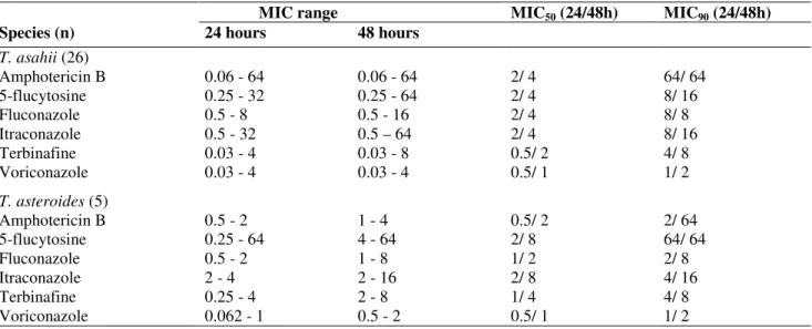
Results shown in table 2 regarding MIC50 and MIC90 (MIC
for 50 % and 90 % of the strains tested respectively) reveal that
T. mucoides was susceptible to Amphotericin B (1/ 1µg/ ml),
Terbinafine (0.5/ 1µg/ ml), and Voriconazole (0.25/ 0.5 µg/ml),
while the other species were resistant to these drugs (8, 11, 16)
All species showed resistance to Itraconazole and susceptibility
to Fluconazole, according to the parameters suggested by Wolf
et al (16). T. ovoides showed susceptibility to Fluconazole and
5-flucytoosine only.
The samples of Trichosporon spp. were submitted to
comparison between the two methods (CLSI and E-test). Only
by the CLSI method (2 µg/ ml) and susceptibility to this drug
by E-test (0,75 µg/ ml). When the samples were compared for
5-flucytosine, two (T. mucoides, MIC50/ 90 > 250 / > 32 µg/ ml
and T. asahii, MIC50/ 90 16/ >32 µg/ ml) showed resistance in
both methods. Regarding Itraconazole, two isolates showed
susceptibility by the E-test and resistance by CLSI method. The
parameters for resistance and susceptibility are those proposed
by Wolf et al (16) and Pfaller et al (8).
Table 2. In vitro antifungal susceptibility profile of 34 isolates of Trichosporon spp
MIC range MIC50 (24/48h) MIC90 (24/48h)
Species (n) 24 hours 48 hours
T. asahii (26)
Amphotericin B 0.06 - 64 0.06 - 64 2/ 4 64/ 64
5-flucytosine 0.25 - 32 0.25 - 64 2/ 4 8/ 16
Fluconazole 0.5 - 8 0.5 - 16 2/ 4 8/ 8
Itraconazole 0.5 - 32 0.5 – 64 2/ 4 8/ 16
Terbinafine 0.03 - 4 0.03 - 8 0.5/ 2 4/ 8
Voriconazole 0.03 - 4 0.03 - 4 0.5/ 1 1/ 2
T. asteroides (5)
Amphotericin B 0.5 - 2 1 - 4 0.5/ 2 2/ 64
5-flucytosine 0.25 - 64 4 - 64 2/ 8 64/ 64
Fluconazole 0.5 - 2 1 - 8 1/ 2 2/ 8
Itraconazole 2 - 4 2 - 16 2/ 8 4/ 16
Terbinafine 0.25 - 4 2 - 8 1/ 4 4/ 8
Voriconazole 0.062 - 1 0.5 - 2 0.5/ 1 1/ 2
MIC: Minimum Inhibitory Concentration
MIC90: Minimum Inhibitory concentration for 90% of the isolates
MIC50: Minimum Inhibitory concentration for 50% of the isolates
T. ovoides and T. mucoides were not included in this table due to the small number of isolates tested
DISCUSSION
The comparison of the susceptibility profile of CBS
reference samples with the data available in the literature
reveals that the results obtained for T. asahii (CBS 2479) in the
present work were concordant with those obtained by Wolf et
al. (19) for Amphotericin B and Fluconazole. When we
compared the results obtained in the present work by the CLSI
method of (2µg/ml), with those obtained by Ghého et al. (4),
(0,019µg/ ml) the discrepancy among MICs obtained for
Itraconazole against T. mucoides and T. ovoides reached 100
folds the dilution. The comparisons allowed the observation of
the differences in MIC values obtained for different groups,
even considering reference samples. These differences
emphasize the importance of the standardized intra-laboratory
conduct, since results obtained in our experiment were
compatible with those obtained by Wolf et al. (19) that also
used a visual system of evaluation, while Guého et al. (4) used
an automatic system.
The presented data reveal a profile of high resistance of
the genus Trichosporon to Amphotericin B and Itraconazole,
high susceptibility to Fluconazole and moderate resistance to
5-flucytosine. Rodriguez-Tudela et al. (12) reported that the
majority of the T. asahii, T. faecali and T. coremiiforme
exhibited resistance to Amphotericin B in vitro. Li et al. (7)
reported resistance to Amphotericin B among T. asahii, T.
cutaneum and T. inkii strains. Differently, Uzun et al. (18),
analyzing the in vitro susceptibility of eight samples of
Trichosporon spp. reported that those samples were susceptible
to Amphotericin B, but susceptible to Fluconazole. Silva, et al.,
(15) also evaluate the antifungal susceptibility profile of T.
asahii clinical isolates. The isolates had reduced susceptibility
in vitro to all drugs, showing 4-6 times higher MICs to
breakpoint values. In this study, Fluconazole exhibited the best
activity in vitro against the majority of the isolates (90%), with
MIC of 16 g/ml and only one isolate with 32 g/ml. Three
isolates have MIC value for Amphotericin B above 2 g/ml.
Variable susceptibility to Amphotericin B has been
observed with samples of the genus Trichosporon, especially
among isolates obtained from immunocompromised patients.
This observation could result in a better conduct for antifungal
therapy. On the other hand, Fluconazole was effective
indicating that the azolic can be the valid option in the therapy
of this infection, commonly difficult to treat.
Resistance to Itraconazole was observed in all of the
Trichosporon species, as well as susceptibility to the
Fluconazole. Only T. mucoides was susceptible to
Amphotericin B, Terbinafine and Voriconazole, being the other
species resistant to these drugs. The results obtained in this
work with Voriconazole, when compared to those obtained by
Uzun et al. (17) were surprisingly more than 30 times higher.
On the other hand, Serena et al. (14) used a guinea pig model
of systemic trichosporonosis to demonstrate a better efficacy of
Voriconazole in comparison with Amphotericin B.
Regarding the comparison between the CLSI method and
the E-test, the greater divergence from those reported in
previous works were obtained for 5-flucytosine, while data
obtained for Amphotericin B were considerably in agreement
with other researchers. Although the number of samples
analyzed is small, the correlation between the two methods for
Amphotericin B is significant (1, 4, 19).
Trichosporon species has been increasingly involved in
systemic infections and, despite this fact, there are relatively
few studies about its antifungal susceptibility profile. Although
these data do not correlate the susceptibility "in vitro/in vivo",
the present work provides epidemiological data that could
influence therapeutic choices. Furthermore, the comparison
between different methodologies could help to analyze results
obtained by different laboratories.
ACKNOWLEDGEMENTS
This work was supported by CNPq (Conselho Nacional de
Pesquisa)
REFERENCES
1. Arikan, S.; Hascelik, G. (2002). Comparison of NCCLS microdiluition method and E-test in antifungal susceptibility testing of clinical Trichosporon asahii isolates. Diagn. Microbiol. Infect. Dis. 43,107-111.
2. Carvalho, A.M.R.; Melo, L.R.B.; Moraes, V.L.; Neves, R.P. (2008). Invasive Trichosporon Cutaneum Infection In An Infant With Wilms’ Tumor. Braz. J. Microbiol. 39, 59-60.
3. Gökahmetoglu, S.; Nedret Koc, A.; Günes, T.; Cetin, N. (2002). Case Reports. Trichosporon mucoides infection in three premature newborns. Mycoses 45, 123-125.
4. Guého, E.; Improvisi, L.; Hoog, G.S.; Dupont, B. (1994). Trichosporon on humans: a practical account. Mycoses 37, 3-10.
5. Kurtzman, C.P.; Fell, J.W. (1998). The yeasts-A taxonomic study. 4 ed. Elsevier.
6. Kustimur, S.; Kalkanci, A.; Caglar, K.; Disbay, M.; Aktas, F. (2002). Nosocomial fungemia due to Trichosporon asteroides: firstly described bloodstream infections. Diagn. Microbiol. Infect. Dis. 43, 167-170. 7. Li, H.M.; Du, H.T.; Liu, W.; Wan, Z.; Li, R.Y. (2005). Microbiological
characteristics of medically important Trichosporon species. Mycopathologia. 160, 217-224.
8. National Commitee for Clinical Laboratory Standards. (2002). Reference Method for Broth Dilution Antifungal Susceptibility Testing of Yeasts. Approved Standard M27-A2. National Commitee for Clinical Laboratory Standards, Wayne, Pa.
9. Neves, R.P.; Cavalcanti, M.A.Q.; Chaves, G.M.; Magalhaes, O.M.C. (2002). Trichosporon Pullulans (Lidner) Diddens & Lodder Isolated From The Oral Cavity Of Aids Patient. Braz. J. Microbiol. 33, 241-242. 10. Pfaller, M.A.; Diekema, D.J.; Messer, S.A.; Boyken, S.; Hollis, R.J.;
Jones, R.N. (2003). In vitro activities of Voriconazole, Pozaconazole, and four licensed systemic antifungal gents against Candida species infrequently isolated from blood. J. Clin. Microbiol. 1, 78-83.
11. Rodrigues, G.S.; Faria, R.R.; Guazzelli, L.S.; Oliveira, F.M.; Severo, L.C. (2006). Nosocomial infection due to Trichosporon asahii: clinical revision of 22 cases. Rev. Iberoam. Micol. 23, 85-89.
12. Rodriguez-Tudela, J.L.; Diaz-Guerra, T.M.; Mellado, E.; Cano, V.; Tapia, C.; Perkins, A.; Gomez-Lopez, A.; Rodero, L.; Cuenca-Estrella, M. (2005). Susceptibility Patterns and Molecular Identification of Trichosporon Species Antimicrob Agents Chemother 49, 4026–4034.
13. Ryder, N.S. (1999). Activity of terbinafine against serious fungal pathogens. Mycoses 42, 115-119.
14. Serena, C.; Gilgado, F.; Marine, M.; Pastor, F.J.; Guarro, J. (2006). Efficacy of voriconazole in a guinea pig model of invasive trichosporonosis. Antimicrob. Agent. Chemother. 50, 2240-2243. 15. Silva, R.B.O.; Fusco-Almeida, A.M.; Matsumoto, M.T.; Baeza, L.C.;
Antifungal Susceptibility Testing Of Trichosporon Asahii Isolated Of Intensive Care Units Patients. Braz. J. Microbiol. 39, 585-592. 16. Sood, S.; Pathak, D.; Sharma, R.; Rishi, S. (2006). Urinary tract infection
by Trichosporon asahii. Indian. J. Med. Microbiol. 24, 294-296. 17. Sugita, T.; Nishikawa, A.; Shinoda, T.; Kume, H. (1995). Taxonomic
position of deep-seated, mucosa associated, and superficial ioslates of
Trichosporon cutaneum from trichosporonosis patients. J. Clin.
Microbiol. 33, 1368-1370.
18. Uzun, O.; Arikan, S.; Kocagoz, S.; Sancak, B.; Unal, S. (2000). Susceptibility testing of voriconazole, fluconazole, itraconazole and amphotericin B against yeast isolates in a Turkish University Hospital and effect of time of reading. Diag. Microbiol. Infect. Dis. 38,101-107. 19. Wolf, D.G.; Hacham, M.; Theelen, B.; Boekhout, T.; Scorzetti, G.;

