online | memorias.ioc.fiocruz.br
Prevalence and clinical aspects of respiratory syncytial virus
A and B groups in children seen at Hospital de Clínicas of Uberlândia,
MG, Brazil
TFM Oliveira, GRO Freitas, LZG Ribeiro, J Yokosawa, MM Siqueira1, SAR Portes1, HL Silveira2, T Calegari2, LF Costa, OC Mantese2, DAO Queiróz/+
Laboratório de Virologia, Instituto de Ciências Biomédicas 2Faculdade de Medicina, Universidade Federal de Uberlândia, Av. Pará 1720,
Bloco 4C, 38400-902 Uberlândia, MG, Brasil 1Laboratório de Vírus Respiratórios, Instituto Oswaldo Cruz-Fiocruz, RJ, Brasil
Respiratory syncytial virus (RSV) is well recognized as the most important pathogen causing acute respiratory disease in infants and young children, mainly in the form of bronchiolitis and pneumonia. Two major antigenic groups, A and B, have been identified; however, there is disagreement about the severity of the diseases caused by these two types. This study investigated a possible association between RSV groups and severity of disease. Re-verse transcription-polymerase chain reaction was used to characterize 128 RSV nasopharyngeal specimens from children less than five years old experiencing acute respiratory disease. A total of 82 of 128 samples (64.1%) could be typed, and, of these, 78% were group A, and 22% were group B. Severity was measured by clinical evaluation associated with demographic factors: for RSV A-infected patients, 53.1% were hospitalized, whereas for RSV B pa-tients, 27.8% were hospitalized(p = 0.07). Around 35.0% of the patients presented risk factors for severity (e.g., pre-maturity). For those without risk factors, the hospitalization occurred in 47.6% of patients infected with RSV A and in 18.2% infected with RSV B. There was a trend for RSV B infections to be milder than those of RSV A. Even though RSV A-infected patients, including cases without underlying condition and prematurity, were more likely to require hospitalization than those infected by RSV B, the disease severity could not to be attributed to the RSV groups.
Key words: Respiratory syncytial virus groups - young children - disease severity
Respiratory syncytial virus (RSV) is a common cause of hospitalization and visits to the emergency rooms among neonates and young children, especially in their first years of life (Sangaré et al. 2006). It is the principal cause of bronchiolitis and pneumonia in chil-dren under six months of age (Queiróz et al. 2002, Ca-legari et al. 2005). An RSV infection does not produce substantial immunity to subsequent infection; thus, reinfections are common, with the frequency of symp-toms decreasing with age (Black 2003). Sequelae from the RSV infection have been observed, which are mostly persistent wheezing, increased airway hypereactivity or atopic asthma (Simoes 2001). Virtually all infants have been RSV infected by the age of two (Parrott et al. 1974). In the first infection, 40-50% have involvement of the lower respiratory tract, and 1-2% of previously healthy infants develop severe disease requiring hospitalization (Yun et al. 1995).
No effective vaccine is currently available for prophy-laxis, and ribavirin therapy has presented only modest benefits (Walsh et al. 1997). In addition, younger infants
Financial support: Fapemig, CNPq, UFU +Corresponding author: dqueiroz@icbim.ufu.br
J Yokosawa’s present address: NL Biotechnology LLC, Little Rock, AR, USA
Received 16 November 2007 Accepted 7 July 2008
tend to have a prolonged illness with little response to medications in general (Howidi et al. 2007). On the other hand, palivizumab, a humanized monoclonal antibody (IgG1K) directed against an epitope in the A antigenic site of the F protein of RSV, is effective in reducing both the incidence and severity of RSV infections in high-risk infants (Black 2003). Children at the greatest high-risk of RSV infection include those born prematurely and those with congenital heart disease or chronic lung conditions or immunocompromised (Welliver 2003). Prevention of infection in these infants for as long as possible is par-ticularly important (Black 2003).
prophy-lactic modalities against RSV have become available, and an understanding of such differences may have an effect on treatment strategies (Papadopoulos et al. 2004).
In temperate weather regions, RSV epidemics occur annually in the winter months, whereas in tropical ar-eas, the outbreaks are associated with the rainy season (Cane 2001), and, in semi-tropical areas, the virus cir-culates mainly during autumn (Vieira et al. 2001, Costa et al. 2006). Both groups circulate concurrently in each epidemic, and group A is the most prevalent in many of the epidemics. Several studies indicate that RSV A and B alternate in prevalence during successive years (Mufson et al. 1988, Sullender 2000). In Brazil, studies have identified the simultaneous circulation of the RSV groups in different years in the Northern (Mello & Silva 1992), Northeastern (Moura et al. 2003), Southeastern (Siqueira et al. 1991, Cintra et al. 2001) and Southern regions (Straliotto et al. 2001), but, to date, no epidemio-logical pattern for each specific RSV group has been es-tablished for Central Brazil.
Recently, we reported a study to determine the viral etiology of acute respiratory disease (ARD) in children less than five years of age in Uberlândia (Costa et al. 2006). RSV was the most frequently identified agent in acute lower respiratory infections, accounting for approxi-mately 25% of the cases. The purpose of this study was to determine the RSV group in the samples in which RSV was detected. Moreover, we attempted to establish whether there was a relationship between a specific RSV group and disease severity by analyzing clinical and demographic features of children with confirmed RSV infections.
PATIENTS, MATERIALS AND METHODS
Patients and specimens - Since April 2000, respira-tory viruses have been investigated by indirect immu-nofluorescence assay (IFA) in nasopharyngeal aspirates (NPA) of children who were less than five years old, experiencing ARD, and attended the Hospital de Clíni-cas (HC), Universidade Federal de Uberlândia (UFU), state of Minas Gerais, Southeastern Brazil. The study was approved by the ethics committees of UFU and signed consent was obtained from the parents of each child included in this study. Afterwards, a questionnaire was administered to ascertain information about clinical and demographic data, e.g., age, gender, gestational age, clinical symptoms, and risk factors such as immunode-ficiency, chronic respiratory disorders (bronchopulmo-nary dysplasia, cystic fibrosis), congenital heart disease and prematurity. Also, information on any intensive care unit (ICU) hospitalization, requirement of mechanical ventilation and clinical diagnostic criteria such as signs, symptoms, laboratory data, chest radiographs and the pathologies classified according to WHO (1994) were recorded. Hospitalization was required only for those patients presenting moderate or severe disease. Clinical symptoms were divided into: bronchiolitis, bronchitis (laryngotracheobronchitis, bronchospasm, Pertussis-like syndrome, whooping cough, and bronchitis), pneumonia (bronchopneumonia and pneumonia) and upper respira-tory tract infection (acute otitis media, rhinitis, sinusitis, flu and upper respiratory tract infection).
One milliliter of physiologic solution was instilled into the nostril of patients and NPA was collected within five days of the onset of symptoms for viral diagnosis. Nasal secretions were aspirated through a catheter into a mucus trap, which was connected to a vacuum source. The catheter was inserted into one nostril parallel to the palate. A vacuum was applied, and the catheter was slowly withdrawn with a rotating motion. Mucus from the other nostril was collected in the same manner. NPAs were processed and RSV diagnoses were per-formed according to Queiróz et al. (2002) using an IFA kit (Respiratory Panel I Viral Screening and Identifica-tion IFA, Chemicon Internacional, Temecula, CA).
RSV isolation in cell culture - NPAs were inoculated in cell culture (HEp-2 cells; ATCC CCL-23) according to the methods by Queiróz et al. (2002). To confirm the isolation of RSV, regardless of the presence of cytopathic effects, cells were removed from the flasks and tested by an IFA kit, as mentioned above. Cell culture superna-tants were used in RT-PCR. Some NPA specimens were not inoculated in cell culture and were tested directly (in natura) by RT-PCR.
RSV molecular characterization - The viral RNA was
extracted from 250 μl of NPA or 125 μl of RSV-infected
HEp-2 cells, using Trizol® (Invitrogen Corp, Carlsbad, CA). Complementary DNA synthesis and PCR amplifica-tion were carried out as previously described (Stockton et al. 1998). The primers used for group determination were generated against the N and P regions of the RSV genome, according to the methods by Stockton et al (1998). Ten mi-croliters of each RT-PCR product was separated on a 1.5% agarose gel and photographed with ImageMaster® VDS GE Healthcare, after staining with ethidium bromide. RSV strains Long (group A; ATCC VR-26) and CH18537 (group B; ATCC VR-1580), both as positive controls, and water as a negative control, were run together in each RT-PCR assay to validate the amplification process and to exclude the presence of contaminants.
Statistical analysis - Data were analyzed by using the WINKS 4.8a program (TexaSoft, Cedar Hill, TX, USA) with a statistical significance level of p < 0.05. Categori-cal variables were examined by using the Fisher’s exact test (two-tailed) with95% confidence intervals (CI). For analyses in which the dependent variable was continu-ous, non-paired Mann-Whitney was used.
RESULTS
in determining the RSV group than testing of isolates in cell culture (49.4%), although only three samples needed to be assayed by both methods.
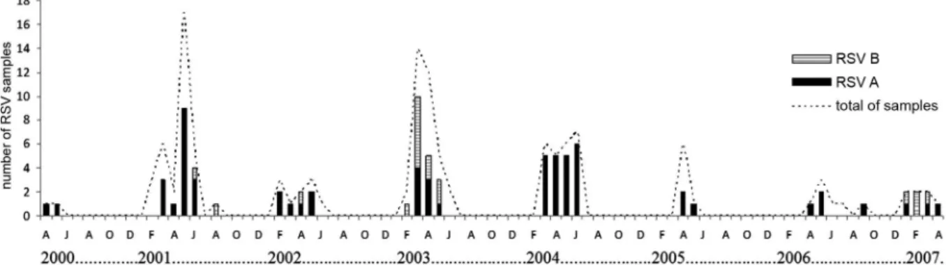
Regarding the RSV circulation by subtype, Fig. 1 shows that RSV A was detected in all seven years of the study, while RSV B was detected in only four years (2001, 2002, 2003 and 2007), when it cocirculated with RSV A. Furthermore, RSV B was more prevalent in 2003 and 2007. In general, RSV was detected from Janu-ary to June (mid-summer to late autumn) with higher incidence of cases from April to May. However, in 2001, one case was detected in August, and, in 2006, one case occurred in September.
In terms of gender, 57.8% (37/64) of the RSV A cases were males, compared with 44.4% (8/18) RSV B cases. However, the difference was not statistically significant (p = 0.42) (Table I). By comparing the age of the patients, RSV A- and B-infected patients presented similar me-dians (4.0 and 4.2 months old, respectively). For RSV A, 59.4% of the patients were < 6 months old, while for RSV B, the rate was 55.6%. However, RSV B was not detected in any patients < 1 month old (Table I).
Bronchiolitis was the clinical diagnosis more fre-quently observed in RSV A-infected children (Table II). For clinical severity, RSV B infections tended to be milder than infections with RSV A (p = 0.18), and, ex-cept in one case, all children with severe disease were
less than six months old. One fatal case was registered of an infant presenting with a RSV A-infection at her 9th day in the ICU. For both the requirement of mechanical ventilation (RSV A: 8/64; RSV B: 1/18) and of ICU hos-pitalization (RSV A: 16/64; RSV B: 3/18), no statistical difference was observed between the groups.
A total of 35.4% (29/82) of the RSV-infected patients presented with prematurity and other underlying condi-tions (RSV A: 22/64; RSV B: 7/18). Fig. 2 shows that, for the 39 hospitalized patients, 53.1% (34/64) were infected with RSV A, while 27.8% (5/18) were infected with RSV B (p = 0.07; OR 2.95; 95% CI; median age 2.1 and 3.0 months, respectively). When analyzing children without underlying conditions and/or prematurity, hospitalization occurred in 47.6% (20/42) of RSV A cases and in 18.2% (2/11) of RSV B cases (p = 0.10; OR 4.09; 95% CI). In addition, 66.7% (26/39) of the hospitalized patients were males (RSV A: 23/34; RSV B: 3/5) (Table II).
DISCUSSION
In this study, we determined the RSV group of speci-mens collected over approximately seven years from chil-dren with respiratory disease. Our results, showing that cases with RSV A predominated over cases with RSV B, are in agreement with the majority of such typing studies (Martinello et al. 2002, Savon et al. 2006). However, one study from 1992 to 1995 in Denmark, reported a predomi-nance of RSV B infections (Johansen et al. 1997).
Fig. 1: seasonal distribution of respiratory syncytial virus groups A and B detected in children attended the Hospital de Clínicas, Universidade Federal de Uberlândia, between April 2000 and first months of 2007.
TABLE I
Age and gender distribution according to RSV group of 82 patients who attended the Hospital de Clínicas, Universidade Federal de Uberlândia, with acute respiratory disease from April 2000 to 2007
RSV A RSV B
Age (months) n (%) Gender (♂) N (%) Gender (♂)
<1 9 (14.1) 5 0 (0.0) 0
1-3 18 (28.1) 11 6 (33.3) 3
3-6 11 (17.2) 7 4 (22.2) 2
6-12 10 (15.6) 6 3 (16.7) 2
≥12 16 (25.0) 8 5 (27.8) 1
Total 64 (100.0) 37 (57.8)a 18 (100.0) 8 (44.4)a
We were able to determine the subtype of approxi-mately 64% of RSV-positive specimens. Uncharac-terizeable specimens also occurred in studies by other authors (Straliotto et al. 1994, Walsh et al. 1997, Cin-tra et al. 2001). Papadopoulos et al. (2004) typed 59.0% using RT-PCR directly from NPA samples, and, in this study, using the same method and the same primers se-quences (Stockton et al. 1998), we typed 57.3%. On the other hand, when using RT-PCR in the RSV samples inoculated in cell culture, we were able to characterize 49.4% of RSV samples. This lower rate could be caused by the lack of virus infectivity or replication in the cells that could have arisen from the long period of speci-men storage in freezers at -70ºC and/or liquid nitrogen. In addition, it was shown that group B strains tend to grow more slowly in tissue culture, to produce fewer
vi-rus particles, and to lose the characteristic of syncytial formation. Therefore, these features could hamper the ability to isolate the virus from clinical specimens and, consequently, the ability to perform group determina-tion (Hall et al. 1990).
The detection of RSV B in alternating years is consis-tent with other studies (Arbiza et al. 2005, Galiano et al. 2005). Mufson et al. (1987) concluded that an infection by RSV A provides some degree of protection against the reappearance by this same group in the subsequent year. The fact that RSV B was not detected in 2005 and 2006 could be because of the few numbers of clinical specimens obtained in those years. Over 20 years ago, Anderson et al. (1985) described the existence of these two groups, the possibility of their occurrence in the same season and their worldwide distribution.
It is known that male children are more susceptible to severe disease than females (Burr et al. 1993, Queiróz et al. 2002), but the reasons are still unknown. In our study, the majority of RSV A-infected children were male. This result is consistent with that of Sangaré et al. (2006), who suggested that being female was protective against RSV hospitalization.
We also found that RSV A-infected patients tended to be slightly younger (median 4.0 vs. 4.2 years), as pre-viously observed (mean 4.6 vs. 5.1 years) (Imaz et al. 2000), and, in the current study, children less than one month of age were infected solely by the group A. In contrast, based on the mean value of ages observed, Pap-adopoulos et al. (2004) reported a predominance of RSV B infection in the youngest children.
For information on lower tract infections, our data are consistent with those of Garzon and Wiles (2002); they found that bronchiolitis was the most common clin-ical diagnosis, followed by pneumonia associated with RSV infection. Similar findings were reported by other investigators (Queiróz et al. 2002, Sangaré et al. 2006). TABLE II
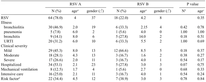
Clinical and demographic characteristics according to RSV group and patients
RSV A RSV B P value
N (%) agea gender (♂) N (%) agea gender (♂) Nb agec
RSV 64 (78.0) 4 37 18 (22.0) 4.2 8 0.35
Illness
bronchiolitis 30 (46.9) 2.0 19 6 (33.3) 2.15 4 0.42 0.78 pneumonia 5 (7.8) 6.0 2 1 (5.6) 4.0 0 1.00 1.00 bronchitis 9 (14.1) 8.0 6 5 (27.8) 14.0 2 0.18 0.51 URTI 20 (31.2) 6.0 10 6 (33.3) 5.5 2 1.00 0.69 Clinical severity
Mild 29 (45.3) 8.0 13 12 (66.6) 8.5 5 0.18 0.37 Moderate 18 (28.1) 4.3 13 3 (16.7) 1.6 2 0.38 0.27 Severe 17 (26.6) 2.0 11 3 (16.7) 4.0 1 0.54 0.17 Hospitalized 34 (53.1) 2.1 23 5 (27.8) 3.0 3 0.07 0,75 Mechanical ventilation 8 (12.5) 1.7 4 1 (5.6) 2.7 0 0.68 0.33 Intensive care 16 (25.0) 2.1 11 3 (16.7) 4.0 1 0.54 0.24 Risk factord 22 (34.4) 4.5 12 7 (38.9) 3.0 3 0.78 0.84
a: median in months; b: Fisher’s exact test; c: Mann-Whitney; d: underlying conditions and prematurity; UTRI: upper respiratory tract infection.
Papadopoulos et al. (2004), using duration of hospital-ization in the severity index, did not find significant dif-ferences between the groups. They stated, however, that the length of hospitalization is based on clinical deci-sions that cannot be controlled. Considering that includ-ing hospitalization in the severity index involves more subjective assessments, this factor was not taken into account in this study. Nevertheless, we observed that pa-tients infected by RSV A were more frequently hospital-ized than the patients with RSV B. It has been reported that RSV A was more frequently detected and caused more severe disease than RSV B strains in hospitalized infants (Coates et al. 1963). In relation to disease severi-ty, however, others have reported the opposite (Palomino et al. 2004) or even that there was a lack of evidence for differences between groups (Coggins et al. 1998, Cintra et al. 2001), which was similar to what we observed in this study. Also, it was noteworthy that most of the patients with severe symptoms were less than six months old. This observation was in accordance with the results obtained by Imaz et al. (2000) and Cintra et al (2001).
In the comparison of RSV groups with respect to se-verity of disease, group A infections tended to be slight-ly more severe than B; however, it was not statisticalslight-ly significant, maybe because of the small sample size or the subjective parameters selected. For instance, more detailed studies of RSV molecular characterization and of the immune response should be done in an attempt to better understand the association with RSV virulence and individual susceptibility.
In conclusion, we did not observe statistical differ-ences in clinical severity between the RSV groups, but we observed a trend for RSV B infection to be milder than RSV A. Even though RSV A-infected patients, in-cluding cases without underlying condition and prema-turity, were more likely to require hospitalization than those infected by RSV B, the disease severity could not to be attributed to the RSV groups.
ACKNOWLEDGEMENTS
To Dr. Dean D. Erdman (CDC), for providing some re-agents; Dr. Juan Arbiza, for useful discussions; the laborato-ries of Immunology, Molecular Biology, Parasitology, Physi-ology and Genetics, UFU, for their equipments; the health care professionals of the Hospital de Clínicas; the patients and their parents.
REFERENCES
Anderson LJ, Hierholzer JC, Tsou C, Hendry RM, Fernie BF, Stone Y, McIntosh K 1985. Antigenic characterization of respiratory syncytial virus strains with monoclonal antibodies. J Infect Dis 151: 626-633.
Arbiza J, Delfraro A, Frabasile S 2005. Molecular epidemiology of human respiratory syncytial virus in Uruguay: 1985-2001 - a re-view. Mem Inst Oswaldo Cruz 100: 221-230.
Black CP 2003. Systematic review of the biology and medical man-agement of respiratory syncytial virus infection. Respir Care 48: 209-231.
Burr ML, Limb ES, Maguire MJ, Amarah L, Eldridge BA, Layzell JC, Merrett TG 1993. Infant feeding, wheezing, and allergy: a prospective study. Arch Dis Child 68: 724-728.
Calegari T, Queiróz DAO, Yokosawa J, Silveira HL, Costa LF, Oliveira TFM, Luiz LN, Oliveira RC, Diniz FC, Rossi LMG, Carvalho CJ, Lima AC, Mantese OC 2005. Clinical-epidemiological evaluation of respiratory syncytial virus infection in children attended in a public hospital in midwestern Brazil. Braz J Infect Dis 9: 156-161.
Cane PA 2001. Molecular epidemiology of respiratory syncytial vi-rus. Rev Med Virol 11: 103-116.
Cintra OA, Owa MA, Machado AA, Cervi MC, Figueiredo LT, Rocha GM, Siqueira MM, Arruda E 2001. Occurrence and severity of infections caused by subgroup A and B respiratory syncytial vi-rus in children in southeast Brazil. J Med Virol 65: 408-412.
Coates HV, Kendrick L, Chanock RM 1963. Antigenic differences between two strains of respiratory syncytial virus. Proc Soc Exp
Biol Med 112: 958-964.
Coggins WB, Lefkowitz EJ, Sullender WM 1998. Genetic variability among group A and group B respiratory syncytial viruses in a children’s hospital. J Clin Microbiol 36: 3552-3557.
Costa LF, Yokosawa J, Mantese OC, Oliveira TF, Silveira HL, Nepo-muceno LL, Moreira LS, Dyonisio G, Rossi LMG, Oliveira RC, Ribeiro LZG, Queiróz DAO 2006. Respiratory viruses in chil-dren younger than five years old with acute respiratory disease from 2001 to 2004 in Uberlandia, MG, Brazil. Mem Inst Oswaldo
Cruz 101: 301-306.
Devincenzo JP 2004. Natural infection of infants with respiratory syncytial virus subgroups A and B: a study of frequency, disease severity, and viral load. Pediatr Res 56: 914-917.
Galiano MC, Palomo C, Videla CM, Arbiza J, Melero JA, Carballal G 2005. Genetic and antigenic variability of human respiratory syn-cytial virus (groups a and b) isolated over seven consecutive sea-sons in Argentina (1995 to 2001). J Clin Microbiol 43: 2266-2273.
Garzon LS, Wiles L 2002. Management of respiratory syncytial vi-rus with lower respiratory tract infection in infants and children.
AACN Clin Issues 13: 421-430.
Hall CB, Walsh EE, Schnabel KC, Long CE, McConnochie KM, Hil-dreth SW, Anderson LJ 1990. Occurrence of groups A and B of respiratory syncytial virus over 15 years: associated epidemio-logic and clinical characteristics in hospitalized and ambulatory children. J Infect Dis 162: 1283-1290.
Howidi M, Rajah J, Abushrar Z, Parsons H 2007. The severity of respiratory syncytial virus bronchiolitis in young infants in the United arab emirates. J Trop Pediatr 53: 22-26.
Imaz MS, Sequeira MD, Videla C, Veronessi I, Cociglio R, Zerbini E, Carballal G 2000. Clinical and epidemiologic characteristics of respiratory syncytial virus subgroups A and B infections in Santa Fe, Argentina. J Med Virol 61: 76-80.
Johansen J, Christensen LS, Hornsleth A, Klug B, Hansen KS, Nir M 1997. Restriction pattern variability of respiratory syncytial virus during three consecutive epidemics in Denmark. Apmis 105: 303-308.
Martinello RA, Chen MD, Weibel C, Kahn JS 2002. Correlation be-tween respiratory syncytial virus genotype and severity of ill-ness. J Infect Dis 186: 839-842.
Mello W, Silva C 1992. Epidemiological aspects of RSV subgroups in Belém, Brazil. Boletin Latino-americano 1: 8.
Moura FE, Borges LC, Portes SA, Ramos EA, Siqueira MM 2003. Respiratory syncytial virus infections during an epidemic period in Salvador, Brazil. Viral antigenic group analysis and descrip-tion of clinical and epidemiological aspects. Mem Inst Oswaldo
Cruz 98: 739-743.
char-acteristics of respiratory syncytial virus strains recovered from children with two consecutive infections. J Clin Microbiol 25: 1535-1539.
Mufson MA, Belshe RB, Orvell C, Norrby E 1988. Respiratory syn-cytial virus epidemics: variable dominance of subgroups A and B strains among children, 1981-1986. J Infect Dis 157: 143-148.
Palomino MMA, Larenas A J, Moraga AG, Avendano LF 2004. Se-veridad clínica de la infección respiratoria aguda baja primaria por virus respiratorio sincicial grupos A y B. Rev Chil Pediatr 75: 18-24.
Papadopoulos NG, Gourgiotis D, Javadyan A, Bossios A, Kallergi K, Psarras S, Tsolia MN, Kafetzis D 2004. Does respiratory syncy-tial virus subtype influences the severity of acute bronchiolitis in hospitalized infants? Respir Med 98: 879-882.
Park JW, Barnett DW 2002. Respiratory syncytial virus infection and the primary care physician. South Med J 95: 353-357.
Parrott RH, Kim HW, Brandt CD, Chanock RM 1974. Respiratory syncytial virus in infants and children. Prev Med 3: 473-480.
Peret TC, Hall CB, Schnabel KC, Golub JA, Anderson LJ 1998. Cir-culation patterns of genetically distinct group A and B strains of human respiratory syncytial virus in a community. J Gen Virol 79: 2221-2229.
Queiróz DAO, Durigon EL, Botosso VF, Ejzemberg B, Vieira SE, Mineo JR, Yamashita C, Hein N, Lopes CL, Cacharo AL, Stew-ien KE 2002. Immune response to respiratory syncytial virus in young Brazilian children. Braz J Med Biol Res 35: 1183-1193.
Sangaré L, Curtis MP, Ahmad S 2006. Hospitalization for respiratory syncytial virus among California infants: disparities related to race, insurance, and geography. J Pediatr 149: 373-377.
Savon C, Goyenechea A, Valdes O, Aguilar J, Gonzalez G, Palerm L, Gonzalez G, Perez Brena P 2006. Respiratory syncytial virus group A and B genotypes and disease severity among Cuban chil-dren. Arch Med Res 37: 543-547.
Simoes EA 2001. Treatment and prevention of respiratory syncytial virus lower respiratory tract infection. Long-term effects on re-spiratory outcomes. Am J Respir Crit Care Med 163: S14-17.
Siqueira MM, Nascimento JP, Anderson LJ 1991. Antigenic charac-terization of respiratory syncytial virus group A and B isolates in Rio de Janeiro, Brazil. J Clin Microbiol 29: 557-559.
Stockton J, Ellis JS, Saville M, Clewley JP, Zambon MC 1998. Mul-tiplex PCR for typing and subtyping influenza and respiratory syncytial viruses. J Clin Microbiol 36: 2990-2995.
Straliotto SM, Nestor SM, Siqueira MM 2001. Respiratory syncytial virus groups A and B in Porto Alegre, Brazil, from 1990 to 1995 and 1998. Mem Inst Oswaldo Cruz 96: 155-158.
Straliotto SM, Roitman B, Lima JB, Fischer GB, Siqueira MM 1994. Respiratory syncytial virus (RSV) bronchiolitis: comparative study of RSV groups A and B infected children. Rev Soc Bras
Med Trop 27: 1-4.
Sullender WM 2000. Respiratory syncytial virus genetic and anti-genic diversity. Clin Microbiol Rev 13: 1-15.
Vieira SE, Stewien KE, Queiróz DAO, Durigon EL, Torok TJ, An-derson LJ, Miyao CR, Hein N, Botosso VF, Pahl MM, Gilio AE, Ejzenberg B, Okay Y 2001. Clinical patterns and seasonal trends in respiratory syncytial virus hospitalizations in Sao Paulo, Bra-zil. Rev Inst Med Trop Sao Paulo 43: 125-131.
Walsh EE, McConnochie KM, Long CE, Hall CB 1997. Severity of respiratory syncytial virus infection is related to virus strain. J
Infect Dis 175: 814-820.
Welliver RC 2003. Review of epidemiology and clinical risk factors for severe respiratory syncytial virus (RSV) infection. J Pediatr 143: S112-117.
WHO - World Heath Organization 1994. International Classification of Diseases, 10th Review, ICD-10, Available from: http://www. who.int/classifications/icd/en/.