Construction and expression of aspartic protease from
Onchocerca volvulus
*
as ompA fusion protein in a mutant strain of
Salmonella typhimurium
Abbas Jolodar
1and David J. Miller
21
Department of Biochemistry, School of Veterinary Medicine, Shahid Chamran University, Ahwaz- Iran.
2
Department of Biochemistry and Molecular Biology, James Cook University of North Queensland,
Townsville, Australia.
Abstract
Two constructions in pHS164vector were designed to permit expression of OV7A and OV4A inserts encoding the N-terminal and C-terminal portion of an aspartic protease fromOnchocerca volvulus, respectively. A novel 39 kD protein ompA-OV7A fusion protein was stably expressed as ompA fusion in a modified strain of Salmonella typhimurium strain SL5000 and E.coli strain JM109. Expression of the fusion protein in bacterial strains harboring the constructs were evaluated by western blotting.E.coli and Salmonella lysates were fractionated by 10% SDS-PAGE gel and then immobilized to nitrocellulose membrane by electroblotting. Primary polyclonal antibody generated in rats against the GST-OV7A fusion protein was used in the Western blots. It remains to be seen whether the fusion protein expressedin vivo will promote effective immune response.
Key words:filarial parasite, nematode,Salmonella typhimurium. Received: March 5, 2002; accepted: March 25, 2002.
Introduction
The filarial parasiteO.volvuluscauses human oncho-cerciasis frequently leading to blindness and severe dermal as well as lymphatic pathology. Onchocerciasis affects ap-proximately 40-50 million people in equatorial Africa, Ye-men and Latin America (World Health Organization 1987). Aspartic acid proteases are a relatively small group of enzymes which includes the mammalian pepsins, chymo-sins, cathepsins and renins. Structural data indicates that the enzymes have bilobal structures comprised mainly of
β-sheets with a substrate-binding cleft containing the active site. Aspartic proteases represent attractive targets for immunoprophylaxis or the generation of protective immu-nity (McKerrowet al.,1993).
To elicit a protective immune response toO.volvulus,
antigens must be delivered and presented to the host in the correct fashion. The new generation of liveSalmonella vac-cines have been highly successful as carriers for the intrace-llular delivery of target antigens, usually on recombinant plasmids, into theSalmonellavehicle. To date, there are no publications describing the use of live vaccines for
onchocerciasis. Recently, a novel construction of the avi-rulentSalmonellastrain that expressed OvGST as a fusion to the N-terminal domain of the bacterial outer membrane protein-A with promising results was described (Catmullet al.,1999). Protein fusions of this kind are believed to en-hance the immune response due to presentation of the for-eign antigen to the outer surface of the bacteria. This initial study demonstrated that the application of this delivery sys-tem to O.volvulus antigens can induce antigen specific Th-1 like cellular immune responses (Yanget al.,1990). This approach may also be useful in the study of other dis-eases for which intracellular antigen delivery and Th-1 cellular immune responses appear to be important for pro-tection.
In this study we have made novel modifications to the mutantSalmonella strain that has expressedOnchocerca
protease as fusion to only a small region of the N-terminal domain of the bacterial outer membrane protein-A (ompA) for enhancement of cell-mediated immune responses in an-imal models.
Materials and Methods
Bacterial strains and plasmid pHS164 structure
A modified strain of S.typhimurium derived from LT2 was identified with a r- m+ phenotype, designated Send correspondence to Abbas Jolodar. Department of Clinical
Chemistry, Bernhard Nocht Institute for Tropical Medicine, Bernhard-Nocht-Str. 74, 20359 Hamburg, Germany. E-mail: jolodara@yahoo. com.
strain SL5000. This strain ofSalmonella not only allows the introduction of foreign DNA because it is restriction negative but also modifies the DNA because it remains modification positive. Thus SL5000 was used as an inter-mediate host to generateSalmonella compatible plasmid DNA from plasmids initially derived fromE.coli.The bac-terial strains were cultured in Luria broth (LB) and on LB agar. The media was supplemented with 50 µg/mL of ampicillin. The vector pHS164 (5448 bp) was used for the expression of foreign antigens as fusions with the N-terminal region of theShigellaouter membrane protein A (ompA; Pistor and Hobom, 1990). The outer membrane proteins (omp) ofShigella dysenteriae,which can be used as carrier proteins to present foreign peptide epitopes on the bacterial cell surface, has been used to construct a group of enterobacterial surface expression vectors for foreign genes protein (Lang, 2000 ). Secretory signals are located at the N-terminal region of ompA and therefore fusion proteins that include this signal peptide should be directed to the outer membrane of theSalmonellabacteria (Beheret al.,
1980). The plasmid can be chemically induced by IPTG. The presence of ampicillin resistant factor (ampr) in this construct permitted selection of transformants on LB agar containing ampicillin.
DNA sequence analysis
Sequences were obtained by the dideoxy-method (Sangeret al.,1977) using T7 Sequenase kits (Pharmacia). The sequencing procedures was performed on double stranded, denatured plasmid DNA using the pHS164 se-quencing primer.
Screening of recombinant clones using PCR
Colony picks were taken and resuspended in 12µL water which was boiled for 5 min and then the cell debris was pelleted by a 5 min centrifugation at approximately 11,000 g. 10µL of the lysis solution was added to 5µL of PCR master mix resulting in a solution containing 1 x syn-thesis buffer, 2.5 mM MgCl2,200 mM of each dNTP, 0.5 U Taq DNA polymerase and 5 pmoles of each primer. Primers specific for inserts in conjunction with the vector forward sequencing primer were used.
Preparation of protein samples
Following induction, bacterial cells were recovered from 1 mL of culture by centrifugation at 12,000xgfor 30 s. The supernatant was discarded and the cell pellet
resuspended in 0.1 mL of distilled water after washing once in 0.5 mL of ice cold 50 mM Tris-HCl pH 7.4. The protein samples were mixed with 25µL of 5 x SDS reducing load-ing buffer (60 mM Tris-HCl pH 6.8/25% glycerol/2% SDS/ 14.4 mM 2-β mercaptoethanol/0.1% bromophenol blue) and chromosomal DNA sheared by sonication. After centrifugation at 12,000xgfor 5 min, the supernatants were incubated for 2 min in boiling water prior to loading on the gel. Prestained molecular weight markers were used to es-tablish the apparent size of proteins
Western blot analysis
After SDS-PAGE, the proteins were transferred electrophoretically to nitrocellulose membrane using a BioRad minitrans-Blot apparatus. In a gel holder a piece of Whatman paper saturated in transfer buffer was placed on a pre-soaked Scotch-Brite pad and then the cathode side of the pre-equilibrated gel was located on the paper. A piece of Hybond-C membrane (Amersham), was cut to the same size as the gel and placed directly onto the anode side of the gel, removing air bubbles by rolling a glass pipette over the membrane. The gel-membrane sandwich was completed by overlaying saturated filter paper and scotch brite on top of the membrane before closing the gel holder. Electroblotting was carried out by applying a constant voltage of 80 V for 1 h at 4 °C.
The blocked membrane was probed with primary an-tibody in blocking buffer followed by rinsing the mem-brane three times in PBS/0.1% Tween-20 (v/v) for 5 min with gentle agitation. The membrane was transferred to an appropriate dilution of secondary antibody conjugate in blocking buffer and incubated for 1 h at room temperature. The enhanced chemiluminescence (ECL) method (Mathewset al,1985) was used to detect secondary anti-body-HRP conjugates, following the manufacture’s (Amersham) instructions.
Results
Two strategies were devised to clone the OVAP N-terminus and C-terminus (Jolodaret al.,1998) in frame with the ompA N-terminus in the pHS164 expression vec-tor.The summary of constructions are listed in Table I.
Construction of pHS-OV4A and pHS-OV7A
The cDNA OV4A clone encodes the OVAP C-ter-minus and includes the 364 bp 3’untranslated region. An aliquot of the OV4A clone in pGEM-T vector was digested
Table I- Summary for subcloning in pHS164.
Construction Termini Base pair Amino acid Domain Vector
pHS-OV7A Eco RI 706 235 N-terminal pHS164
with ApaI, cutting the plasmid construct once only, within the vector polylinker, resulting in a 5GGCC overhang. The 3’ recessed ends of ApaI were filled using Klenow frag-ment. The linearised pGEMT-OV4A plasmid was then di-gested with NsiI to generate a cohesive 5’end. The OV4A fragment was gel-purified and directionally subcloned into pHS164 that had 5’NsiI and 3’SmaI ends. Although the pHS-OV4A construct contained 62 bp of pGEM-T vector polylinker at the 5-end of OV4A its sequence did not en-code any stop codons to disrupt the expression process. The pHS-OV7A construct was designed to express the OV7A (Jolodaret al.,1997) insert encoding the N-terminal por-tion of the aspartic protease from O.volvulus. The subcloning procedure of the OV7A fragment from pUC18 to pHS164 was facilitated by digestion withEcoRI. The 3’ recessed ends of the OV7A fragment were end filled using Klenow fragment to allow ligation with the blunt ends of StuI-digested pHS164 vector (Figure1). Recombinant plas-mids were transformed intoE.coliJM109 and bacteria con-taining the plasmids were isolated and plasmid prepared from these for DNA sequencing and for transformation into
Salmonella.The predicted open reading frame was deter-mined by analyzing the sequences, particularly around the cloning site. Putative recombinants were screened by PCR using primers specific for OV7A in conjunction with the vector forward sequencing primer followed by the DNA se-quencing. This method allowed the presence of one or more copies of insert and their orientation to be determined. The plasmids were introduced intoS.typhimuriumSL5000 by the Lederberg and Cohen method (Lederberget al.,1974); however, due to the low efficiency of transformation into
Salmonella, 5 to 50µg of plasmid was used per transforma-tion resulting in no more than 100 transformants.
Detection of ompA fusion proteins by western blotting
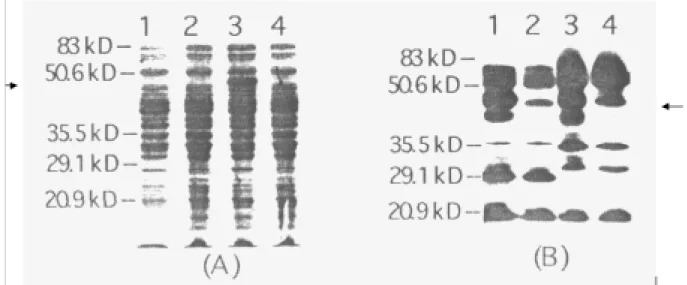
Liquid cultures of pHS-OV7A transformants were grown to an OD600 nm of 0.5 before addition of IPTG to a final concentration of 0.2 mM to induce ompA transcrip-tion. After 2 h of induction theE.coliandSalmonella bacte-ria were lysed and insoluble debris was separated from the soluble protein. The supernatant recovered from the bacte-rial lysates was further fractionated by SDS-PAGE and the results are illustrated in Figure 2. Comparisons of IPTG in-duced verses uninin-duced pHS-OV7A and the pHS164 non-recombinant control lysates revealed a novel 39 kD protein in the induced extract of pHS-OV7A. This novel
Figure 1- The subcloning strategy used to generate the pHS-OV7A con-struct with an open reading frame of OV7A continuous with the 5’ end of the OmpA gene in the pHS164 vector.
Figure 2- Expression of ompA-OV7A fusion protein as determined by SDS-PAGE and Western blotting. (A) Protein from the pHS-OV7ASalmonella
protein was similar to the size predicted for the ompA-OV7A protein, a fusion approximately 13 kD and 26 kD. There were no discernible differences between the pHS-OV4A and non-recombinant vector lysates. Due to the hydrophobic nature of this portion of the OVAP protein it is possible that the fusion product was insoluble, but it is also possible that the level of expression of the ompA-OV4A fusion protein was too low to allow visual-ization by Coomassie staining. Expression of the fusion protein in bacterial strains harboring the constructs was evaluated by western blotting.E.coli andSalmonella ly-sates fractionated by 10% SDS-PAGE gel were immobi-lized to nitrocellulose membrane by electroblotting. Primary polyclonal antibody used in the Western blots was generated in rats against the GST-OV7A fusion protein. Detection of the primary antibody was with a HRP-conju-gated goat anti-rat immunoglobulin (DAKO, High Wycombe, Bucks, UK), combined with a peroxidase in-duced chemiluminescence (ECL) detection system. It should be noted that OV4A includes approximately 80 amino acids of the OV7A. Thus, this antiserum is also likely to recognize OV4A.
Discussion
It has been suggested that the aspartic proteases which are predicted to be secreted by parasite may be valu-able for immunoprophylaxis against onchocerciasis (Lizotte-Waniewskiet al.,2000). Vaccine efficacy of re-combinant cathepsin D aspartic protease fromSchistosoma japonicum(Verityet al.,2001) andHaemonchus contortus
(Longbottom et al., 1997) has been reported. Immuno-localisation experiments performed by the authors (Jolodar
et al., unpublished data) indicated that the recombinant OV7A protein encoding an aspartic protease was predomi-nantly associated with the hypodermis and inner membrane zone of the uterine wall of adult females. The patterns of immunolabelling were highly specific when sections were probed with antibodies to the recombinant protein.
The expression of OV7A as a fusion with ompA was similar in both strains of bacteria and a single 39 kDa pro-tein of equal intensity was detected. This indicated that the ompA-OV7A fusion protein was stably expressed in two hosts with different genetic backgrounds. However, the ompA-OV4A fusion protein in the lysates generated from IPTG induced bacteria transformed with the pHS-OV4A construct was not detected by the methods used here. It is possible that the polyclonal antibody generated to GST-OV7A did not contain any idiotypes that recognized OV4A epitopes or the ompA-OV4A protein was absent. The literature implies that the omp is an important outer membrane protein partially responsible for membrane inte-grity. They all have a common structural motif of aβ-barrel that is composed of a variable number of transmembrane
β-strands connected at the periplasmic side with short turns
and at the outside with long surface-accessible loops (Hobomet al.,1995; Lang H. 2000).
This study demonstrated that the recombinant Salmo-nellaconstruct pHS-OV7A expressed wellin vitro. It re-mains to be seen whether the recombinant Salmonella
construct pHS-OV7A expressedin vivowill promote an ef-fective immune response. To determine this there will have to be a number of vaccine trials in animal models. Several of these animal models for onchocerciasis are recognized by the WHO and include mice, jirds, birds, cattle and chim-panzees. Whether the immune response to OV7A, and per-haps OV4A, delivered bySalmonella elicits the desirable Th-1 like immune response in these models and or offers protection is a question I hope will be answered positively in the near future.
References
Beher MG, Schnaitman CA and Pugsley AP (1980) Major heat-modifiable outer membrane protein in gram-negative bacteria; comparison with the ompA protein of Escherichia coli. J. Bacteriol. 143:906-913.
Catmull J, Wilson ME, Kirchhoff LV, Metwali A and Donelson JE (1999) Induction of specific cell-mediated immunity in mice by oral immunization with Salmonella expressing On-chocerca volvulus glutathione S-transferase. Vaccine 17:31-39.
Hobom G, Arnold N and Ruppert A (1995) OmpA fusion proteins for presentation of foreign antigens on the bacterial outer membrane. Dev. Bio.l Stand. 84:255-262.
Jolodar A and Miller DJ (1997) Preliminary characterization of an Onchocerca volvulus aspartic protease. Int. J. Parasitol. 27:1087-1090.
Jolodar, A and Miller DJ (1998) Identification of a novel family of non-lysosomal aspartic proteases in nematodes. Biochim. Biophys. Acta 1382:13-16.
Lang H (2000) Outer membrane proteins as surface display sys-tems. Int J Med Microbiol. 290:579-585.
Lederberg MM and Cohen SS (1974) Transformation of Salmo-nella typhimurium by plasmid deoxyribonucleic acid. J. Bacteriol. 119:1072-1074.
Lizotte-Waniewski M, Tawe W, Guiliano DB, LuW, LiuJ, Wil-liams SA and Lustigman S (2000) Identification of potential vaccine and drug target candidates by expressed sequence tag analysis and immunoscreening of Onchocerca volvulus larval cDNA libraries. Infect Immun . 68:3491-3501. Longbottom D, Redmond DL, Russell M, Liddell S, Smith WD
and Knox DP (1997) Molecular cloning and characterisation of a putative aspartate proteinase associated with a gut mem-brane protein complex from adult Haemonchus contortus. Mol Biochem Parasitol. 88:63-72.
Mathews JA, Batki A, Hynds RJC, and Kricka LJ (1985) Enhanced chemioluminescent method for the detection of DNA dot-hybridization assay. Anal. Biochem.
151:205-209.
Pistor S and Hobom G (1990) ompA-Haemagglutinin fusion pro-teins for oral immunization with live attenuated Salmonella. Res. Microbiol. 141:879-881.
Sanger F, Nicklen S and Coulson AR (1977) DNA sequencing with chain termination reaction inhibitors. Proc. Natl. Sci. USA 74:5463-5467.
Verity CK, McManus DP and Brindley PJ. (2001). Vaccine effi-cacy of recombinant cathepsin D aspartic protease from
Schistosoma japonicum. Parasite Immunol 23:153-162.
World Health Organization (1987) 2. Distribution, prevalence, and socioeconomic effects. In: WHO committee on oncho-cerciasis (Third report). World Health Organization: Ge-neva, pp. 8-21.