of Gene Expression in
Salmonella
Typhimurium
Inês de Jesus de Almeida e Silva
Dissertation presented to obtain the Ph.D degree in Biology
Instituto de Tecnologia Química e Biológica | Universidade Nova de Lisboa
Oeiras,
December,
Inês de Jesus de Almeida e Silva
Dissertation presented to obtain the Ph.D degree in Biology
Instituto de Tecnologia Química e Biológica
Universidade Nova de Lisboa
Oeiras, December, 2012
Ribonucleases in the Control of Gene
Financial Support from Fundação para a Ciência e Tecnologia (FCT) – Ph.D: grant - SFRH / BD / 43211 / 2008.
Work performed at:
Control of Gene Expression Laboratory Instituto de Tecnologia Química e Biológica
Av. da República (EAN)
2781-901 Oeiras – Portugal Tel: +351-21-4469548
Supervisor :
Professora Doutora Cecília Maria Pais de Faria de Andrade Arraiano – Investigadora
Coordenadora, Instituto de Tecnologia Química e Biológica, Universidade Nova de Lisboa.
(Head of the Laboratory of Control of Gene Expression, where the work of this
Dissertation was performed)
Co-supervisor :
Doutora Sandra Cristina de Oliveira Viegas – Investigadora Pós-Doutorada, Instituto de
Tecnologia Química e Biológica, Universidade Nova de Lisboa.
(Post-doc Fellow in the Laboratory of Control of Gene Expression, where the work of this
Dissertation was performed)
President of the Jury :
Doutora Claudina Amélia Marques Rodrigues Pousada – Professora Catedrática
Convidada do Instituto de Tecnologia Química e Biológica da Universidade Nova de
Examiners:
Professor Doutor Iñigo Lasa Uzcudun – Head of Microbial Biofilm Research Group,
Instituto de Agrobiotecnología, Pamplona (Principal Examiner).
Professor Doutor Arsénio do Carmo Sales Mendes Fialho – Professor Associado com
Agregação, Instituto Superior Técnico, Universidade Técnica de Lisboa (Principal
Examiner).
Professor Doutor Miguel Agostinho Sousa Pinto Torres Fevereiro – Investigador
Principal do Instituto Nacional de Investigação Agrária e Veterinária - INIAV
Professora Doutora Isabel Maria Godinho Sá Nogueira – Professora Associada com
"
Every experiment proves something. If it doesn’t prove what you
wanted it to prove, it proves something else."
during these last four years:
- Instituto de Tecnologia Química e Biológica (ITQB) for giving me the opportunity to develop my work during these last years in such a good
environment and with such good conditions. I also would like to thank the past
and present ITQB directors, namely Professor Doutor José Artur Martinho
Simões and Professor Doutor Luís Paulo Rebelo,for all their efforts to maintain ITQB as an excellence institute even in these “crisis” moments.
- Fundação para a Ciência e Tecnologia (FCT) for the essential financial support.
- My Supervisor Professora Cecília Maria Arraiano for giving me the opportunity to work in her lab, for believe in my capacities, for encouraging me
to do the PhD in her lab, for providing excellent work conditions and for all the
support during these last four years. Thank you also for giving me the chance to
attend to several meetings both in Portugal and abroad where I could present and
discuss my work with so many different “scientific people”.
- My Supervisor and good friend Sandra Viegas. Thank you for your support, guidance, for all the knowledge that you transmitted me, for your
companionship, for always have the right words even in the worst situations.
Thank you for being always a wonderful friend. I am sure that my scientific and
personal life would not be the same without you.
- All my past and present colleagues from CMA Lab… thank you for your support during these important four years of my life. In special, I would like to
thank to Sandra, Margarida, Susana, Andreia, Cátia, Rute, Ricardo and
special moments we spent both in and outside the lab. A special thank to my
“office mates” and very good friends Sandocha, Margarette and Suzette for your positive vibes and for your support during these last years of my life. Also, a
special and grateful thank to Andreia Aires our compass in the lab. Thank you for all your help, support and for being always so helpful and positive!
- Franscisco García-del Portillo and Álvaro Ortega from Centro Nacional de Biotecnología, Consejo Superior de Investigaciones Científicas (CNB-CSIC),
Madrid, Spain for their collaboration and precious help in the final step of this
work. I hope that we can continue our collaboration.
- All my friends that during these four years helped me to overcome the difficult moments. Especially:
Joana and Ana, my “sisters”, thank you for your support not only during these four years but also along the last ten years. Thank you for your friendship;
without you my life would never be the same. Thank you for all the wonderful
moments during our weird stay in Göttingen; you were essential to my sanity
during those five months. Danke!
To my dear friend Catarina thank you for all your support, patience and all the perfect moments we passed. I miss you!
- Jónsi, Georg, Kjartan, Orri, Eddie, Jeff, Matt, Stone, Mike, Glen, David, Rita,
especially during THE endless moving, and for looking after Pedro when I am
not at home.
- A special and profound acknowledgement to my grandparents Celina, Avelino and Amália for all your teachings. Even if they had nothing to do with science, they were really important for me in these last years. To my grandfather António thank you for your support, guidance and love. I will always miss you…
- To my special friends Sebastião and Belchior for the relaxing moments and for your unconditional love.
- To my brother Eduardo and my sister Mónica thank you for all the support and for giving me two of the most precious things of my life. To my nephews António and Mariana thank you for being part of my life!
- To my dear mum Luísa and my dear dad José thank you for all the support, unconditional love, friendship… Thank you for telling me always the best words
in the right moments. Your advices are precious for me. I have no words to thank
you for everything. You are for sure the best parents in the world! This
achievement is also yours!
- And last but not least… To my boyfriend and best friend Pedro Mendes for being always by my side. Thank you for your support, understanding, strength… thank you for being always so interested in my work even if you don’t
understand anything about that. Thank you for being my soulmate, my
ABSTRACT
... XIX
RESUMO
... XXV
LIST OF PUBLICATIONS
... XXXI
DISSERTATION OUTLINE
... XXXV
ABBREVIATIONS
... XLI
CHAPTER 1
... 1
CHAPTER 2
... 65
CHAPTER 3
... 109
CHAPTER 4
... 151
CHAPTER 5
... 187
RNAs are important effectors in the process of gene expression. In
bacteria, the levels of the transcripts have to be rapidly adjusted in response to
constantly changing environmental demands. The cellular concentration of a
given RNA is the result of the balance between its synthesis and degradation.
RNA degradation is a complex process encompassing multiple pathways.
Ribonucleases are the enzymes that directly process and degrade RNA
transcripts, regulating their cellular amounts. The rate at which RNA decay
occurs depends on the availability of ribonucleases and their specificities
according to the sequence and/or the structural elements of the RNA molecule.
Several other factors modulate RNA degradation, namely polyadenylation, which
plays a multifunctional role in RNA metabolism. Additionally, small non-coding
RNAs are crucial regulators of gene expression, and can directly modulate the
stability of their mRNA targets. In many cases this regulation is dependent on
Hfq, an RNA binding protein which can act in concert with polyadenylation
enzymes and is often necessary for the activity of the sRNAs.
The first objective of this Doctoral work was to study the importance of
the endoribonucleases RNase III and RNase E on the regulation of the sRNA
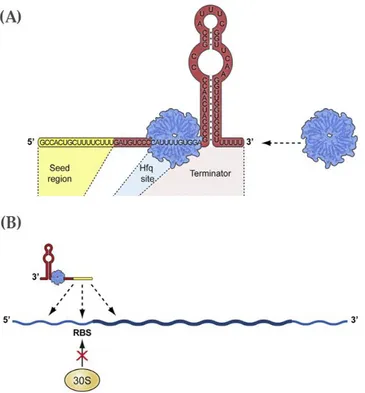
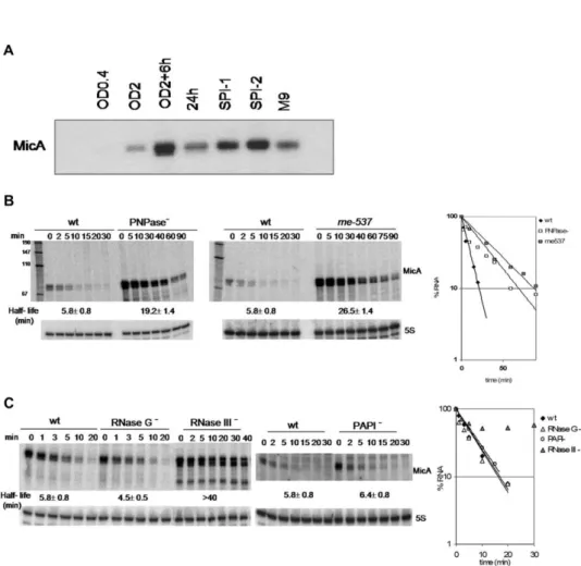
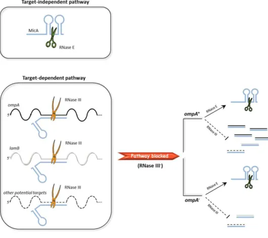
MicA in Salmonella Typhimurium. MicA is a trans-encoded sRNA, which
down-regulates porin expression in stationary-phase. Its main targets in Salmonella are
the outer membrane protein OmpA and the LamB maltoporin. In a previous
study, we have analyzed the expression profile of MicA under different growth
conditions and used distinct Salmonella RNase mutants to evaluate the
contribution of each of these enzymes in the turnover of this sRNA. In order to
further analyse the role of the two main Salmonella endoribonucleases, RNase III
and RNase E, in the degradation of MicA, both enzymes were overexpressed and
the two RNases and the in vitro activity of both enzymes using MicA as a
substrate, revealed that both endoribonucleases are involved in the control of
MicA sRNA levels. However, MicA was shown to be cleaved by RNase III when
it is in complex with its targets, while RNase E was the responsible for the control
of free-MicA levels in the cell. This study allowed us to propose a model for MicA
degradation in which RNase III and RNase E perform a different role in the decay
of this sRNA.
In the second part of this Dissertation the aim was to examine the role of
SraL sRNA in S. Typhimurium. SraL was originally identified in E. coli, and was
afterwards detected in Salmonella, particularly in late stationary phase of growth.
SraL has been little studied and its biological function was not known at the
beginning of this study. Several approaches are currently available to determine
the biological role of the sRNAs. In the present work, a proteomic analysis of
Salmonella strains expressing different SraL levels in the cell enabled the detection
of several SraL putative targets. The majority of the changes were detected in
proteins involved in several pathways of carbohydrates metabolism, especially
those involving glucose. Moreover, the study of the influence of glucose in SraL
levels revealed a dependence of the expression of this sRNA on the concentration
of this sugar in the growth medium.
In the proteomic analysis, one of the proteins most affected by the
different levels of SraL in the cell was the chaperone Trigger Factor. This putative
biological function of SraL was investigated further in the third part of this
Doctoral work. Trigger factor is the first chaperone encountered co-translationally
by most of the nascent amino-acid chains and cooperates with DnaK and GroEL
in the folding of newly synthesized proteins. In this study, it was shown that this
Mutagenesis experiments have shown that SraL interacts with the 5’-UTR of tig
mRNA few nucleotides upstream of its Shine Dalgarno sequence.
The main ribonucleases involved in the post-transcriptional regulation of
SraL were previously identified. In the present work, several experiments were
performed to study how this sRNA is regulated at the transcriptional level. The
results presented in this Dissertation identified the central regulator of general
stress response RpoS (σS) as a transcriptional regulator of this sRNA. By using
Chromatin immunoprecipitation assays, RpoS was shown to bind directly to the
SraL promoter region.
The work developed during this Dissertation provided important results
that contributed for a better understanding of the biological role of two highly
conserved sRNAs, MicA and SraL sRNAs, and their pathways of regulation in the
Os RNAs constituem importantes moléculas efetoras no processo de expressão génica. Em bactérias, os níveis dos transcritos têm que ser rapidamente ajustados em resposta às constantes mudanças no meio ambiente. A concentração celular de um dado RNA é determinada pelo balanço entre a sua síntese e a sua degradação. A degradação do RNA é um processo bastante complexo e que envolve numerosos passos. As ribonucleases (RNases) são as enzimas responsáveis pela regulação da quantidade dos transcritos de RNA, uma vez que atuam no seu processamento e na sua degradação. A taxa de degradação do RNA depende da disponibilidade destas enzimas e também das suas especificidades, de acordo com a sequência e/ou elementos estruturais das moléculas de RNA. Vários outros fatores modulam igualmente a degradação do RNA. A poliadenilação desempenha variadas funções durante o metabolismo do RNA. Adicionalmente, os pequenos RNAs não-codificantes são também reguladores essenciais da expressão génica, visto que modulam diretamente a estabilidade dos seus RNAs mensageiros (mRNA) alvos. Na maioria dos casos documentados, esta regulação é dependente do chaperone Hfq. Esta proteína tem a capacidade de se ligar ao RNA e pode eventualmente atuar em conjunto com as enzimas de poliadenilação. Para além disso, o Hfq é normalmente necessário para a própria atividade dos pequenos RNAs.
O primeiro objectivo do trabalho desenvolvido durante esta Dissertação centrou-se no estudo da importância das endoribonucleases RNase III e RNase E na degradação de um pequeno RNA, denominado MicA, em Salmonella
diferentes condições de crescimento e usando mutantes de diversas RNases de
Salmonella de modo a avaliar a contribuição de cada uma destas enzimas no decaímento deste pequeno RNA. Com vista a analisar com maior detalhe o papel das duas principais endoribonucleases de Salmonella na degradação deste pequeno RNA, sobre-expressámos e purificámos ambas as enzimas de Salmonella. A análise paralela dos resultados obtidos no estudo da degradação deste pequeno RNA in vivo na ausência de ambas as RNases e dos resultados obtidos no estudo da atividade in vitro de ambas as enzimas revelou que ambas as endoribonucleases estão envolvidas no controlo dos níveis de expressão do MicA. Os estudos efetuados revelaram que o MicA é degradado pela RNase III quando forma complexos com os seus RNAs mensageiros alvos. Adicionalmente, a RNase E é responsável pelo controlo dos níveis do pequeno RNA livre na célula. Os resultados obtidos permitiram-nos propor um modelo de degradação do MicA, no qual a RNase III e a RNase E desempenham funções diferentes no decaímento deste pequeno RNA.
Na segunda parte da presente Dissertação o principal objetivo foi estudar e compreender a função de outro pequeno RNA de Salmonella, denominado SraL. Este pequeno RNA foi originalmente identificado em Escherichia coli. Mais tarde foi também detetado em Salmonella, maioritariamente na fase estacionária tardia de crescimento. Tinham sido já efetuados alguns estudos tendo como base este pequeno RNA. Contudo, aquando do ínicio deste estudo, a sua função biológica na célula não tinha sido ainda identificada. Presentemente conhecem-se vários métodos que permitem a determinação da função biológica dos pequenos RNAs. Neste estudo, foi afetuada uma análise proteómica de várias estirpes de
envolvem a glucose. O estudo da influência da glucose nos níveis do SraL foi também realizado e revelou que a expressão deste pequeno RNA é dependente da concentração deste açucar no meio de crescimento.
A análise proteómica revelou também o Trigger Factor como sendo outro alvo putativo do pequeno RNA SraL. Na terceira parte deste trabalho foi investigada com maior detalhe esta função biológica do SraL. O Trigger Factor é o primeiro chaperone que se liga co-traducionalmente à maioria das cadeias de aminoácidos. Esta proteína coopera com outros dois chaperones, DnaK e GroEL, no correto enrolamento das cadeias polipeptídicas recém-sintetisadas. Durante este estudo foi demonstrado que esta regulação se dá ao nível do RNA mensageiro. A região de interação entre o pequeno RNA e o mRNA alvo foi determinada usando várias ferramentas bioinformáticas. Experiências de mutagénese mostraram que o SraL interage com a região não traduzida a 5’ do mRNA do tig, alguns nucleótidos antes da sua sequência Shine-Dalgarno.
Num estudo anteriormente realizado identificámos as principais ribonucleases envolvidas na regulação pós-transcricional do SraL. No presente estudo, foram efetuadas várias abordagens com vista a compreender como é que este pequeno RNA é regulado transcricionalmente. Com base nos resultados apresentados nesta Dissertação, identificámos o regulador central da resposta ao stress RpoS (σS) como sendo o regulador transcricional do SraL. Utilizando
ensaios de Chromatin immunoprecipitation mostrou-se que o RpoS se liga diretamente à região promotora do SraL.
Silva I.J., Ortega A.D.*, Viegas S.C.*, García-del Portillo F., Arraiano C.M. 2012. An RpoS-dependent sRNA regulates the expression of a chaperone involved in protein folding. Accepted with modifications in RNA Journal. *These authors contributed equally to this work
Reis F., Pobre V., Silva I.J., Malecki M., Arraiano C.M. 2012. The RNB Family of Exonucleases –Putting the ‘Dis’ in Disease. Accepted in WIREs RNA
Silva I.J., Saramago M., Dressaire C., Domingues S., Viegas S.C., Arraiano C.M. 2011. Importance and key events of prokaryotic RNA decay: the ultimate fate of an RNA molecule. WIREs RNA; 2(6):818-36
Viegas S.C.*, Silva I.J.*, Saramago M., Domingues S., Arraiano C.M. 2011. Regulation of the small regulatory RNA MicA by ribonuclease III: a target-dependent pathway. Nucleic Acids Research; 39(7):2918-30 *These authors contributed equally to this work
Arraiano C.M., Andrade J.M., Domingues S., Guinote I.B., Malecki M., Matos R.G., Moreira R.N., Pobre V., Reis F.P., Saramago M., Silva I.J., Viegas S.C. 2010. The Critical Role of RNA Processing and Degradation in the Control of Gene Expression. FEMS Microbiology Reviews; 34(5):883-923
Andrade J.M., Pobre V., Silva I.J., Domingues S., Arraiano C.M. 2009. The role of 3’-5’ exonucleases in RNA degradation. Progress in Molecular Biology and Translational Science Review, 85:187-229
Viegas S.C., Pfeiffer V., Sittka A., Silva I.J., Vogel J., Arraiano C.M. 2007. Characterization of the role of Ribonucleases in Salmonella small RNA decay.
This Dissertation is divided into five chapters.
Chapter one consists of a general introduction on RNA degradation mechanisms,
where are highlighted the main differences between the RNA degradation
pathways for the two main Gram-negative and Gram-positive bacterial models,
Escherichia coli and Bacillus subtilis. This chapter encompasses not only the
importance of ribonucleases but also of other players such as small non-coding
RNAs, the RNA-chaperone Hfq and Poly(A) Polymerase. Part of this section was
published in WIREs RNA and the author of this thesis is the first author of this
manuscript.
Chapter two describes the investigation of the role of the endoribonucleases III
and E in the degradation of MicA sRNA, alone and coupled with its targets. From
this work resulted a publication in Nucleic Acids Research in which the author of
this dissertation played a major contribution and was considered first author.
Chapter three focused in the biological function of SraL in Salmonella
Typhimurium. Proteomic were performed to identify possible targets of this
sRNA. The proteomic results that seemed more relevant were validates by
RT-PCR and Northern blot analyses. The importance of the regulation of SraL over
the putative targets identified is discussed. The results described in this chapter
gave the preliminary data necessary for the work reported in chapter four, and
In chapter four one of the biological roles inferred in the previous chapter for
SraL in Salmonella Typhimurium was also studied in detail and the transcriptional
regulator of SraL sRNA was also determined. The work of this chapter was
accepted with modifications in RNA and the author of this dissertation is the first
author of the manuscript.
To finalize, chapter five comprises an integrated discussion of the global results
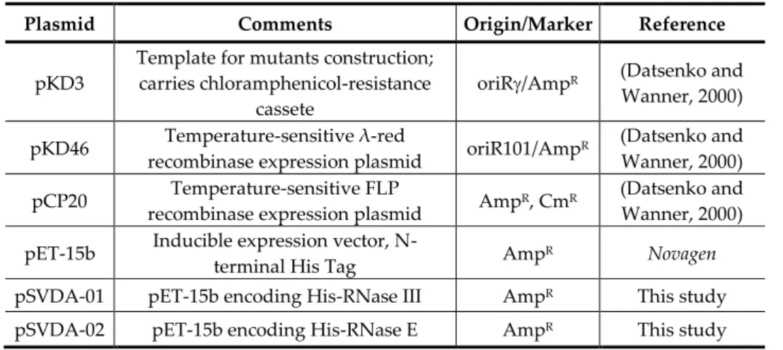
Amp ampicillin
A. aeolicus Aquifex aeolicus ATP adenosine triphosphate
bp base pair
B. subtillis Bacillus subtillis BSA bovine serum albumin
°C degree Celsius
C cytosine
cAMP-CRP cyclic adenosine
monophosphate-cAMP receptor protein
c-di-GMP cyclic diguanylic acid
cDNA complementary DNA
ChIP chromatin immunoprecipitation
Cm chloramphenicol
cpm counts per minute
CRISPR clustered regularly interspaced short
palindromic repeats
CS cold-shock
CSD cold-shock domain
deletion
DTT dithiothreitol
DNA deoxyribonucleic acid
DNase deoxyribonuclease
dsRBD double-stranded RNA binding domain
dsRNA double stranded RNA
E. coli Escherichia coli
EDTA ethylenediaminetetraacetic acid
G guanine
g relative centrifugal force
GMP deoxyguanosine monophosphate
h hour
Hfq-coIP-seq Hfq coimmunoprecipitation
coupled with RNA-seq
His histidine
IgG immunoglobulin G
IP immunoprecipitate
IPTG isoPropyl--D-thiogalactopyranoside kDa kilodalton
L. lactis Lactococcus lactis
L. monocytogenes Listeria monocytogenes LB luria-bertani broth
Log logarithm
LPS lipopolysaccharide
M molar/ molarity (mol/L)
M9 minimal medium
Mg magnesium
mg milligram
g microgram
l microliter
Mg magnesium
ml milliliter
min minute
mM milliMolar
MOPS 3-(N-morpholino)propanesulfonic acid
mRNA messenger RNA
ng nanogram
nM nanomolar
nt nucleotide
OD optical density
Oligo oligonucleotide
OMP outer membrane protein
32P phosphorus 32 radionucleotide
p p-value
PAA polyacrylamide
PAGE polyacrylamide gel electrophoresis
PAP I poly(A) polymerase I
PBS phosphate-buffered saline buffer
PCR polymerase chain reaction
PDB protein data bank
PMSF phenylmethylsulfonyl fluoride
PNPase polynucleotide phosphorylase
Poly(A) polyadenylate
PPIase peptidyl-prolyl cis/trans isomerase
psi pressure unit
RBS ribosome binding site
RNA ribonucleic acid
RNAi RNA interference
RNase ribonuclease
r.p.m. rotations per minute
rRNA ribosomal RNA
RT-PCR reverse transcriptase polymerase chain
reaction
SD Shine-Dalgarno
SD standard deviation
SDS sodium dodecyl sulphate
siRNA small interfering RNA
SOC super optimal broth with catabolite
repression medium
SPI Salmonella pathogenicity island
Sr streptomycin
sRNA small RNA
SSC sodium chloride/sodium citrate
S. aureus Staphylococcus aureus T thymine
TAE tris/acetic acid/EDTA
TBE tris/borate/EDTA
TCA tricarboxylic acid cycle
TCS two-component system
TF trigger factor
tmRNA transfer messenger RNA
Tris trishydroxymethylaminomethane
(2-Amino-2-(hydroxymethyl)propane-1,3-diol)
tRNA transfer RNA
T. thermophilus Thermus thermophilus U uracil
U units
UTP uracil triphosphate
UTR untranslated region
UV ultraviolet radiation
V volt
v/v volume/volume
wt wild-type
w/v weight/volume
Chapter 1
INTRODUCTION
Part of this chapter was based on:
Silva I.J., Saramago M., Dressaire C., Domingues S., Viegas S.C., Arraiano C.M. 2011.
Importance and key events of prokaryotic RNA decay: the ultimate fate of an RNA
Introduction ... 5
RNA decay: global approaches ... 6
Bacterial RNA degradation mechanisms ... 8
The original model and the initiator RNase E... 8 The case of the double-stranded specific RNase III... 12 An alternative mechanism involving different endonucleases ... 14
Role of RNA degradation in quality control ... 17
Structural determinants of RNA degrading enzymes ... 19
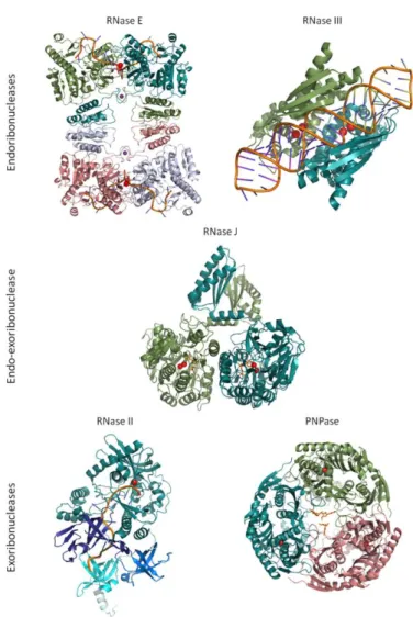
RNase E ... 19 RNase III ... 20 RNase J ... 21 RNase II ... 23 RNase R ... 24 PNPase... 25
Other players ... 28
Polyadenylation ... 28 Hfq ... 32 RNA regulators ... 34
Regulation of RNases ... 38
Auto-regulation and cross-regulation ... 38 Regulation by environmental conditions and other cellular modulations ... 39
Concluding remarks ... 42
Aim of this Dissertation ... 44
I
NTRODUCTION
In the Introduction chapter is presented the current knowledge on prokaryotic RNA degradation mechanisms. Several questions are discussed like: which are the main players involved? What is their mode of action? Are there any differences between bacterial species? After a brief overview of the genome-wide studies of RNA decay, prokaryotic RNA degradation mechanisms are described. The RNA features and the structural details of ribonucleases, the enzymes that process and degrade RNA, are also discussed. Finally, additional factors which have an impact on RNA stability are focused. Together, these key events determine the ultimate fate of an RNA molecule.
RNA
DECAY
:
GLOBAL APPROACHES
The development of transcriptome analysis has enabled the monitoring of intracellular RNA levels at the genomic scale. The rate of degradation of a given RNA can be estimated by determining its half-life in the cell. Combining inhibition of transcription with microarray technology has proven to be a powerful tool for assessing genome-wide mRNA decay. Most of these global studies in microorganisms have been carried out on the model yeast
aureus (Anderson et al., 2006). The short half-life of bacterial mRNAs is a key factor to allow the rapid adaptation to changing environmental conditions.
Altogether these global studies not only report average values in the same range of magnitude but also highlight a wide variation of half-lives among mRNAs. Furthermore, global stabilities have been observed to be dependent on growth conditions (Anderson et al., 2006; Redon et al., 2005). For example, the average half-life for mRNAs in L. lactis increases from 5.8 to 19.4 min when comparing exponential and carbon starvation phases, respectively (Redon et al., 2005). It has also been noted that genes which share related biological functions usually display similar messenger decay rates (Bernstein et al., 2002; Hambraeus
et al., 2003; Selinger et al., 2003). For instance, most house-keeping genes have long half-lives. Proteins which are central in E. coli protein–protein interaction network also tend to be encoded by stable transcripts (Janga and Babu, 2009).
B
ACTERIAL
RNA
DEGRADATION MECHANISMS
The original model and the initiator RNase E
Turnover of RNA molecules involves cleavage reactions that are carried out by RNases, a diverse collection of cellular enzymes, whose functions and properties have been elucidated through the study of mutants (Arraiano et al., 2010; Arraiano et al., 1988). Although E. coli possesses a plethora of RNases only a few are devoted to the RNA degradation. The conventional model for RNA decay in this bacterium usually begins with an endonucleolytic cleavage at one or more internal sites on the RNA molecule (Figure 1A). Two endonucleases have been associated with the initial cleavage event: RNase III and RNase E. However, RNase E is believed to be the main endonuclease involved in the RNA turnover in
E. coli (Arraiano et al., 2010). In fact, a recent report shows that in the absence of RNase E 60% of the annotated coding sequences were either increased or decreased in their steady-state levels (Stead et al., 2011). In contrast, only 12% of the coding sequences were affected by the absence of RNase III (Stead et al., 2011). RNase E is an essential single-stranded endonuclease that exhibits a preference for A/U-rich regions in close proximity to stem-loops (Mackie, 1992; McDowall et al., 1994). This characteristic is also shown by its paralogue, RNase G. This endonuclease, which has a strong resemblance with the amino-terminal portion of RNase E (McDowall et al., 1993), is also involved in the degradation and processing of RNA (Carpousis et al., 2009). Both enzymes display higher activity over substrates bearing a monophosphorylated than over substrates with
-phosphorylation status. This occurs in molecules with multiple single-stranded sites that allow the direct entry of RNase E through a different pathway, called
‘bypass’ or ‘internal entry’ (Baker and Mackie, 2003; Kime et al., 2010).
RppH is an RNA pyrophosphohydrolase that removes the
pyrophosphate from the 5’-termini and preferentially acts on single-stranded RNA. The discovery of this enzyme presented an alternative pathway in which the initial event is non-nucleolytic (Deana et al., 2008). Conversion of 5’
-triphosphate to 5’-monophosphate by RppH provides the ideal substrate for RNase E, and the preference of RppH for single-stranded RNA explains why 5’ -stem-loops are mediators of stability. Ribosome loading is also known to mediate RNA stability. A poor ribosome binding site, possibly by increasing the distance between the actively translating ribosomes, exposes putative internal cleavage sites and may increase message instability.
2005). This complex of enzymes assures the coordination of the endo- and exonucleolyticdegradation of an RNA molecule. After the initialendonucleolytic cleavage step the upstream fragment, lacking the 3’-terminal hairpin, can be readilydigested by 3’-exonucleases. The activity of theseenzymes is impaired by
a 3’-stem-loop, whichprotects the majority of the primary transcripts (Andrade et al., 2009). The downstream fragment generated after the initial endonucleolytic cleavage is usually more prone todegradation. It bears a monophosphorylated 5’ -end and therefore may be the ideal substrate foran additional cleavage by RNase E. The turnoverof malEF transcript illustrates how the endo- andexonucleolytic enzymes can act in a concerted way. PNPase degradation of malEF is only accomplished in the presence of RNase E and RhlB, indicating that the degradosome participates in its degradation (Stickney et al., 2005).
Three exonucleases are mainly involved in RNAdecay in E. coli: PNPase, RNase R and RNase II (Figure 1A). All of these enzymes degrade RNA processively and non-specifically from the 3’-end. While PNPase is a phosphorolytic exonuclease yielding nucleoside diphosphates as reaction products, both RNase R and RNase II catalyse the hydrolysis of the RNA substrates, producing nucleoside monophosphates.Among the three, only RNase R is able todigest structured RNA by itself (Andrade et al., 2009). The degrading activity of PNPase or RNase II is stalled by the presence of secondary structures (Spickler and Mackie, 2000). However, PNPase can also proceed through extensive folded RNA when acting in association with other proteins. Its associationwith the helicase RhlB or integration into the degradosome allows the unwinding of the RNA stem-loops (Liou et al., 2002). Surprisingly, the PNPase homologue of Thermus termophilus, whose optimal temperature is 65ºC, has been reported to completely degrade RNAs with stable intramolecular secondary structures without the aid of a helicase (Falaleeva et al., 2008). Nonetheless, both
in order to be able to bind and initiate digestion of an RNA molecule (Vincent and Deutscher, 2006). By providing a single-stranded platform for the initiation of the
exonucleolytic attack, the degradation of RNA molecules containing 3’ -stem-loops is stimulated by the addition of poly(A) tails to the 3’-end of the RNA molecules (for details see below the ‘Polyadenylation’ section). These poly(A) tails constitute the preferred substrate for PNPase and RNase II (Lisitsky and Schuster, 1999; Marujo et al., 2000).
None of the three 3’-exonucleases seems to be indispensable for E. coli
growth at optimal temperature. However the combined absence of both PNPase and RNase II or PNPase and RNase R is lethal for the cell, indicating some overlapping role between these exonucleases (Cheng and Deutscher, 2003). For instance, both RNase R and PNPase are involved in the degradation of rRNA fragments, whose accumulation was proposed to lead to cell death (Cheng and Deutscher, 2003). A transcriptome analysis revealed that, although RNase II accounts for 90% of exonuclease activity in the cell, PNPase probably plays a greater role in mRNA degradation than previously thought (Deutscher and Reuven, 1991; Mohanty and Kushner, 2003). RNase II is the major exonuclease involved in E. coli RNA decay and other enterobacteriaceae but, this enzyme is absent in several other bacterial species,such as B. subtilis, Legionella pneumophila
The degradative action of the ribonucleases described above releases RNA fragments of 2–5 nucleotides, whose accumulation may be deleterious to the cell (Ghosh and Deutscher, 1999). E. coli possesses another exonuclease, termed oligoribonuclease, which acts as a scavenger of these short oligoribonucleotides (Niyogi and Datta, 1975) (see Figure 1A). This essential enzyme processively hydrolyses RNA in the 3’–5’ direction. Overall, oligoribonuclease is a finishing enzyme in RNA metabolism, and the presence of proteins with analogous functions seems to be widespread. Two homologues, NrnA and NrnB, have been described in B. subtilis (Fang et al., 2009).
The case of the double-stranded specific RNase III
Even though RNase E has been considered the main enzyme in E. coli that catalyses the initial cleavage event, the RNase III family of enzymes has emerged as an important group of endonucleases in the control of RNA stability (Jaskiewicz and Filipowicz, 2008). RNase III deletion in E. coli causes a slow growth phenotype (Babitzke et al., 1993), while its homologue in B. subtilis is essential for viability (Herskovitz and Bechhofer, 2000). A second B. subtilis
RNase III-like enzyme (called Mini-III) has been described (Redko et al., 2008). Both enzymes seem to act mostly in bacteriophage mRNA and rRNA processing, since no endogenous mRNA targets are known (Bechhofer, 2009). Interestingly, a recent study performed using the human pathogen S. aureus revealed the existence of an overlapping sense/antisense RNA processing by the activity of RNase III (Lasa et al., 2012). This process also occurs in other Gram-positive bacteria such as B. subtilis, Listeria monocytogenes and Enterococcus faecalis.
FIGURE 1 - Mechanisms of RNA decay in the Gram-negative and Gram-positive bacterial models. (A) In E. coli the decay of the majority of transcripts starts with an endonucleolytic cleavage by RNase E. The enzyme has a preference for 5’ -monophosphorylated substrates. A possible pathway for RNase E cleavage involves a primary cleavage by the RNA pyrophosphohydrolase RppH, which converts the 5’ -triphosphorylated terminus of primary transcripts to monophosphate. However, some substrates are cleaved by RNase E regardless of the 5’-phosphorylation status, through an
alternative pathway called ‘bypass’ or ‘internal entry’, which involves the direct entry of
RNase E at single-stranded sites. RNase III is double-stranded specific and can also initiate the decay of structured RNAs. After endonucleolytic cleavage, breakdown products are ready for exonucleolytic digestion by any of the three main exonucleases in this bacterium. Unlike RNase R, both RNase II and PNPase are sensitive to secondary structures. Exonucleolytic activity is promoted by the 3’-polyadenylation of substrates. The activity of PAP I, the main polyadenylating enzyme in E. coli, is modulated by the RNA-chaperone Hfq. PNPase can synthesize heteropolymeric tails that also facilitate degradation. Cycles of polyadenylation and exonucleolytic degradation have been proposed as one way to overcome secondary structures. A minor alternative pathway in the cell is the direct exonucleolytic degradation of full length transcripts (represented by a dashed arrow). Exonucleolytic degradation releases short fragments which are subsequently degraded to mononucleotides by oligoribonuclease. (B) In B. subtilis, transcripts can be degraded from the 5’-end through the 5’–3’ exonuclease activity of RNase J1, or they can be first endonucleolytically cleaved. Since the 5’–3’exonuclease activity of RNase J1 is blocked by 5’-PPP, before this cleavage the recently discovered BsRppH removes the pyrophosphate. The endonucleolytic cleavage can be either performed by RNase J1/RNase J2 or RNase Y. The breakdown products can be then further degraded by the 3’–5’exonucleases, PNPase and RNase R (unprotected 3’-ends), or by the 5’–3’ exonuclease activity of RNase J1 (newly
generated monophosphorylated 5’-ends). RNase J1 is able to fully degrade its RNA substrates to mononucleotides. The final products released by RNase R and PNPase are further degraded by the oligoribonuclease homologues in B. subtilis. Here we represent ribonucleases acting independently. However some of these enzymes can act together in degradation complexes. For instance in E. coli the degradosome (RNase E, PNPase, RhlB and enolase) and in B. subtilis the putative complex formed by RNase J1/J2, RNase Y, PNPase, the RNA helicase CshA and two glycolytic enzymes.
An alternative mechanism involving different endonucleases
degradation. One answer came from the discovery of two different ribonucleases, the paralogous RNase J1 and J2, which are present in almost all bacteria lacking RNase E (Even et al., 2005). Curiously, both RNase E and RNase J1 orthologs have been found in Sinorhizobium meliloti (Madhugiri and Evguenieva-Hackenberg, 2009). Although there is no sequence homology, the RNase J enzymes share a similar architecture with RNase E (de la Sierra-Gallay et al., 2008) and exhibit equivalent endonucleolytic activity. RNase J1, which is essential for cell viability, is involved in RNA turnover (Mäder et al., 2008) (Figure 1B). Surprisingly, this
enzyme is also able to catalyse the exonucleolytic degradation of RNA in the 5’-3’ direction (Mathy et al., 2007). To date this is the only 5’-exonuclease known in prokaryotes and its discovery has had important implications in the RNA decay
model. The exonucleolytic decay from the 5’-end may explain the stabilizing
effect conferred by 5’-stem-loops, 5’-protein binding, 5’-ribosome stalling and the
presence of a 5’-triphosphate in B. subtilis (Bechhofer, 2009). It has been suggested that this dual-function enzyme (alone or in complex with RNase J2) catalyses not only the endonucleolytic cleavage of an RNA substrate but also continues the
degradation of the generated 5’-end by switching to the 5’-exonucleolytic mode (de la Sierra-Gallay et al., 2008). In fact, global analysis of RNase J-depleted B. subtilis strains showed an altered abundance for a large number of mRNA transcripts, indicating that this ribonuclease affects gene expression on a global scale (Durand et al., 2012; Mäder et al., 2008). Interestingly, only the exonucleolytic
activity of RNase J1 is dependent on the 5’-end phosphorylation status, as it is blocked by triphosphorylated RNA (de la Sierra-Gallay et al., 2008). The complex formed by RNase J1 and J2 changes their individual cleavage activities and specificities (Mathy et al., 2010).
(Figure 1B), that shares extensive functional homologies with RNase E. In fact,
several reports suggest that the predominant activity of RNase J1 is the 5’-3’ exonucleolytic activity (Condon, 2010; Durand et al., 2012; Lehnik-Habrink et al., 2012; Newman et al., 2011), while RNase Y has an effect on the global mRNA half-life in B. subtilis comparable to that of RNase E in E. coli. There are several studies reporting an increase of the bulk mRNA half-life in B. subtilis upon RNase Y deletion (Durand et al., 2012; Lehnik-Habrink et al., 2011b; Shahbabian et al., 2009). Like RNase E, RNase Y is sensitive to the phosphorylation state of the 5’-end, exhibiting a marked preference for monophosphorylated RNA. Hence, two essential enzymes in B. subtilis RNA decay are dependent on a
monophosphorylated 5’-end. Indeed, it was recently discovered the existence of a RNA pyrophosphohydrolase in B. subtilis (BsRppH) (Richards et al., 2011).
BsRppH was shown to remove the γ and β phosphates from the 5’-end of the RNA as orthophosphate (Richards et al., 2011), whereas the one from E. coli
releases them mainly as a pyrophosphate (Deana et al., 2008). Interestingly, the results obtained with rppH deletion mutant suggested the existence of other(s) RNA pyrophosphohydrolase(s) in B. subtilis (Richards et al., 2011).
Based on the amino acid sequence, RNase Y seems to comprise at least four domains: a short N-terminal trans-membrane domain (Hunt et al., 2006); a coiled-coil domain seemingly important for oligomerization (Lehnik-Habrink et al., 2011a); a KH domain required for RNA binding; and an HD domain that contains the catalytic site (Condon, 2003). The presence of an N-terminal trans -membrane domain in RNase Y suggests -membrane localization further extending the analogy to RNase E (Shahbabian et al., 2009) (Figure 1B). In fact, the proper membrane localization of RNase Y is essential for B. subtilis (Lehnik-Habrink et al., 2011a).
phosphofructokinase, has been reported (Commichau et al., 2009). This complex brings together some of the degrading activities necessary to achieve full degradation of an RNA molecule. The RNA fragments released by the RNase Y
endonucleolytic cleavage could be good substrates for the 3’-exonucleolytic
activity of PNPase and for the 5’-exonucleolytic degradation by RNase J1/J2 (Bechhofer, 2009). This putative degradosome-like complex indicates that the presence of such an RNA degradative machine may be a common feature in prokaryotes, even those that lack an RNase E homologue. Nonetheless, the existence of this complex in B. subtilis is still controversial. The presence of a degradosome-like complex in this organism has been challenged by the failure of isolating it as a complex in its native state and by the absence of detection of degradosome interactions in yeast two- and three-hybrid screens (Mathy et al., 2010). However, these screens failed to detect also the established self-interactions of the RNase J1 and J2 (Commichau et al., 2009; de la Sierra-Gallay et al., 2008; Newman et al., 2011). Accordingly, the current knowledge does not yet allow accurate conclusions about the presence or absence of an RNA degradosome in this bacterium.
R
OLE OF
RNA
DEGRADATION IN QUALITY CONTROL
One of the common errors is the presence of a premature stop codon in some messages, which produces a truncated protein. In E. coli the fast degradation of messages carrying a premature stop codon is thought to begin
with a 5’-independent RNase E cleavage at internal sites exposed by the premature release of ribosomes (Baker and Mackie, 2003). The resulting RNA molecules are further degraded through the pathways described above.
Messages without an in-frame stop codon may lead to ribosome stalling
at the 3’-end, significantly affecting translational efficiency. An elegant surveillance pathway, termed trans-translation, targets deficient proteins and mRNA for degradation while rescuing stalled ribosomes (see (Keiler, 2008; Richards et al., 2008) for a review). This process relies on the association of two molecules: a sRNA called transfer-messenger RNA (tmRNA), and a small RNA-binding protein (SmpB), whose homologues have been identified in every sequenced eubacterial genomes. Some situations that delay the progress of ribosomes during translation were also observed to elicit tagging by the trans -translation machinery in a process that involves RNase II (Garza-Sánchez et al., 2009; Richards et al., 2008).
Finally, errors in macromolecular processes like tRNA and rRNA synthesis also occur. Li and co-workers had shown that an aberrant precursor tRNA molecule is degraded by a mechanism that involves PAP I and PNPase (Li
et al., 2002), leading to the proposition that polyadenylation could serve as a signal to promote degradation of defective tRNAs. PNPase and RNase R have been implicated in the removal of defective rRNAs (Cheng and Deutscher, 2003).
S
TRUCTURAL DETERMINANTS OF
RNA
DEGRADING ENZYMES
The intrinsic degradative nature of ribonucleases and the fact that they share the cellular environment with a pool of different types of RNA molecules
raises some questions: How do these ‘molecular killers’ specifically select their
targets? What are the structural determinants that allow these enzymes to spare some molecules while dictating the destruction of others? As underlined above there are several features of the RNA molecules that are key factors in determining their fate. The interplay between the structural determinants of the enzymes and their specific degradation preferences, depending on the characteristics of each RNA molecule, seems to dictate the final decision.
RNase E
mechanism for RNA recognition and cleavage (Koslover et al., 2008). The
influence of 5’-phosphorylation is a consequence of the pocket formed between
the S1 and the RNase H subdomains, which binds 5’-monophosphorylated RNA and promotes downstream degradation. After binding, a conformational change induced by the movement of the RNA-binding domains clamps the substrate down and organizes the active site (Koslover et al., 2008). The catalytic site contains conserved residues of the DNase I domain and a single metal-binding site that coordinates an Mg2+ ion implicated in catalysis. The internal flexibility within the quaternary structure may be related to the deformation required to accommodate structured RNA for processing by internal entry. An amphipathic segment (called segment A) at the C-terminal region of RNase E directs the enzyme to the inner membrane (Khemici et al., 2008). It was recently published the existence of another RNase E-membrane binding interaction involving the catalytic domain of the enzyme (Murashko et al., 2012). The N-terminal RNase E membrane binding alters its enzymatic activity by increasing the substrate affinity, and affects the secondary structure of the catalytic domain stabilizing the folding state of the protein (Murashko et al., 2012). Membrane localization of this enzyme could be determinant for spatial discrimination of the RNA substrates.
RNase III
are found in the dimer interface, one at each end of the valley. Although dsRNA binding is governed by the combined dsRBD of the dimer, the NucD domains contribute to substrate specificity (Gan et al., 2008). Substrate selection consists of a combination of structural and sequence elements, such as the strength of base pairing, the occurrence of specific nucleotide pairs and the helix length (Pertzev and Nicholson, 2006). Several conserved residues in the catalytic centers were shown to be essential for catalysis, which involves amino acids from both subunits, and thus, dimerization of the NucD domains is necessary for RNase III function (see (Gan et al., 2006) and references therein). The crystal structure indicates that a single RNA cleavage event occurs on each strand of the dsRNA within each cleavage site, generating products with a two-base 3’-overhang. Mg2+ is required for the formation of a catalytical competent protein–RNA complex and two divalent cations are coordinated by each active site. When Mg2+ is absent the RNA is bound outside the catalytic valley (Gan et al., 2008). Indeed, RNase III is also known to be involved in the control of gene expression by binding a dsRNA molecule without cleaving. In this non-catalytic functional form the enzyme plays the role of a dsRNA-binding protein (Blaszczyk et al., 2004).
RNase J
RNase J is unique among bacterial RNases in that it possesses both endo-
and 5’–3’ exonucleolytic activity (de la Sierra-Gallay et al., 2008). The crystal structure of an RNase J homolog of T. thermophilus (sharing 43% and 39% sequence identity with B. subtilis RNase J1 and J2, respectively) was determined in the closed state, unable to accommodate the bulk of an RNA structure (de la Sierra-Gallay et al., 2008) (Figure 2). More recently, the structures of T. thermophilus RNase J in complex with short RNA oligomer and of B. subtilis
reported (Dorléans et al., 2011; Newman et al., 2011). In both structures, while there are small differences in the detail of RNA binding, the general mode of binding and the path taken by the RNA is the same.
The enzyme is active as a dimer in solution, and each monomer contains three distinct domains: a metallo-β-lactamase core, a β-CASP and a C-terminal domain. This last domain may be involved in substrate recognition and maintenance of the dimeric state. Two Zn2+ ions, essential for catalysis, are coordinated in an octahedral environment by residues located deep in the cleft
between the β-lactamase core and the β-CASP domain. Specific attack of 5’ -monophosphorylated transcripts is related to a binding pocket near the catalytic
center that precludes the accommodation of a 5’-triphosphorylated substrate. Only one catalytic center was obvious from the structures, suggesting that a single active site is responsible for the dual activity of RNase J. In fact, the model of B. subtilis RNase J1 bound to RNA contains a significant region of unoccupied
space at the free 5’-end of the RNA perhaps explaining how both endonuclease
and 5’-to-3’ exonuclease activities are incorporated in the same active site (Newman et al., 2011). Indeed, the 5’-monophosphate generated after the endonucleolytic cleavage may be directly placed in the binding pocket with a single translocation of the RNA molecule, allowing RNase J to start the exonucleolytic degradation. In both B. subtilis and T. thermophilus “open” state
RNase II
E. coli RNase II is the prototype of the RNase II family of enzymes, a widespread family that also includes RNase R and the catalytic subunit of the eukaryotic exosome, Rrp44/Dis3. The overall crystallographic structure of E. coli
RNase II reveals a monomeric protein with four domains: three RNA binding domains comprising CSD1 and CSD2 in the N-terminal region, and an S1 fold at the C-terminus; and one RNB catalytic domain in the central region, which is a hallmark of the RNase II family of proteins (Amblar et al., 2006; Frazão et al., 2006; Zuo et al., 2006) (Figure 2). The RNA binding domains are grouped together in one side of the structure, and play a role in the RNA substrate selection and binding (Frazão et al., 2006; Matos et al., 2009).
Interestingly, a truncated enzyme lacking all the RNA binding domains is still able to degrade RNA (Matos et al., 2009; Vincent and Deutscher, 2009b). The structure of the enzyme in complex with an RNA molecule reveals two main non-contiguous interaction points between the protein and the RNA fragment. A 10-nucleotide segment is the shortest RNA able to retain both contacts, explaining why RNase II becomes distributive on substrates shorter than 10 nt (Frazão et al., 2006). A single amino acid change in the catalytic region alters the final end-product from 4 to 10 nt, probably due to loosening of the RNA substrate at the catalytic site (Barbas et al., 2008). The access to the catalytic pocket is restricted to single-stranded RNAs by steric hindrance, explaining the inability of RNase II to degrade dsRNA. A specific interaction with ribose rings precludes DNA cleavage (Frazão et al., 2006). Several residues in the catalytic region are important for catalysis (Amblar and Arraiano, 2005; Barbas et al., 2008; Barbas et al., 2009; Frazão
exonucleolytic activity (120-times higher), consequently called ‘super-enzyme’ (Barbas et al., 2009).
RNase R
RNase R shares a similar domain organization with RNase II, and a three-dimensional model of this enzyme has been proposed based on the structure of RNase II (Barbas et al., 2008). Besides the domains identified in RNase II, RNase R also contains a helix-turn-helix in the N-terminus (Domingues et al., 2009), and a highly basic region after the S1 fold at the C-terminus (Vincent and Deutscher, 2009b). The RNB domain of RNase R alone is sufficient to bind and degrade an RNA duplex (Matos et al., 2009; Vincent and Deutscher, 2009b). Paradoxically, when the RNA-binding domains are present in the wild-type enzyme a short 3’ -overhang is necessary in order to initiate degradation. These domains are
PNPase
Symmons et al., 2000). Mg2+ is required for PNPase enzymatic activity, though Mn2+ can also support catalysis. The metabolite citrate has recently been shown to
directly modulate the enzyme’s activity, connecting RNA degradative pathways with the central metabolism (Nurmohamed et al., 2011). Moreover, PNPase was recently shown to be directly activated by the second messenger c-di-GMP (Tuckerman et al., 2011).
In this section, it were highlighted the structural features of the ribonucleases that are specifically involved in the recognition of a given RNA
FIGURE 2 - Structures of RNA degrading enzymes in complex with RNA substrates. On the top of the image are shown the crystal structures of endonucleases (catalytic domain of
E. coli RNase E, PDB ID 2C4R, on the left; A. aeolicus RNase III, PDB ID 2NUF, on the right). The crystal structures of exonucleases are on the bottom (E. coli RNase II, PBD ID 2IX1, on the left; E. coli PNPase, PDB ID 3GCM, on the right). The crystal structure of T. thermophilus
RNase J (PDB ID 3BK2), which has a dual function as endo- and exonuclease, is in the middle. RNA substrates in complex with the enzymes are coloured in orange and the metal ions that assist catalysis are shown as red spheres. Purple spheres denote the Zn2+
catalytic domain RDB is coloured in dark cyan). A model for RNase R has been proposed but the structure is not yet available. However functional and structural data available indicates that the structure will be quite similar to RNase II. Structures were drawn using PyMOL (http://pymol.sourceforge.net).
O
THER PLAYERS
Polyadenylation
Polyadenylation is a post-transcriptional event that involves the addition
of untemplated adenosine residues to the 3’-ends of RNA substrates. This widespread phenomenon occurs in bacteria, organelles, archaea, and in the nucleus and cytoplasm of eukaryotic cells. Eukaryotic poly(A) tails, present in the majority of mRNAs, are usually long, uniform in length and have traditionally been viewed as a stabilizing element. However, recent studies have also shown evidence of a polyadenylation-induced decay of non-functional RNAs in eukaryotes (Houseley and Tollervey, 2009). Prokaryotic polyadenylated transcripts are generally low in abundance and poly(A) tails are very short and unstable. This fact, together with a lack of evidence for a physiological role, made polyadenylation in bacteria almost overlooked for several years after its discovery. It has been estimated that less than 2% of total RNA in E. coli is polyadenylated. Despite this intriguingly low percentage, a transcriptome comparison between the wild-type and a strain defective in polyadenylation has indicated that the majority of transcripts (∼90%) undergo some degree of polyadenylation during exponential growth, either as full-length transcripts or decay intermediates (Mohanty and Kushner, 2006). The rapid turnover of polyadenylated mRNAs accounts in part for their low abundance.
Poly(A) tails provide a single-stranded extension region that works like a
activity. Paradoxically, the higher affinity of RNase II for these poly(A) stretches has been shown to protect mRNAs from degradation, as this enzyme degrades the tails that allow PNPase and RNase R to proceed through secondary structures (Marujo et al., 2000; Mohanty and Kushner, 2003).
Initially identified 50 years ago in E. coli (August et al., 1962), Poly(A) Polymerase I is responsible for approximately 90% of the polyadenylating activity in the cell (Mohanty and Kushner, 2011). The enzyme catalyzes the addition of homopolymeric poly(A) tails (15–30 nt long in average) to 3’- hydroxyl termini of RNA molecules using ATP as a substrate (Mohanty and Kushner, 2006; Mohanty
enzyme responsible for the addition of nucleotides to the 3’-end of RNAs in this organism (Campos-Guillén et al., 2005).
Regarding the nature of the poly(A) tail, several observations indicate that transcripts which terminate in a Rho-dependent fashion tend to contain only heteropolymeric tails generated by PNPase. On the other hand, Rho-independent transcription terminators serve as polyadenylation signals for PAP I (Mohanty and Kushner, 2006; Mohanty et al., 2004). Moreover, heteropolymeric tails are mainly added to breakdown products, whereas poly(A) tails are added to both breakdown and full-length transcripts (Mohanty and Kushner, 2011). In stationary phase, the addition of heteropolymeric tails is predominant over the homopolymeric tails commonly found in exponentially growing cells (Cao and Sarkar, 1997). The lack of energy resources in stationary phase could be a reason for this choice, since the generation of adenylated tails at the expense of several ATP molecules is more justifiable under exponential growth (Cao and Sarkar, 1997). Moreover, polyadenylation promotes high mRNA turnover rates which are characteristic of the exponential phase (Mohanty and Kushner, 2000).
In both bacteria and organelles, RNA breakdown products generated by endonucleolytic cleavages are considered the most favoured substrates for 3’ -tailing (Goodrich and Steege, 1999; Haugel-Nielsen et al., 1996; Lupold et al., 1999; Perrin et al., 2004). Single-stranded segments at either 5’- or 3’-end of RNA molecules and monophosphorylation at an unpaired 5’-terminus were reported to increase the RNA susceptibility to polyadenylation by PAP I (Feng and Cohen, 2000). This suggests that the endonucleolytically-generated RNA fragments containing single-stranded monophosphorylated 5’-termini would be preferential
substrates for 3’-end polyadenylation. However, this is not always required as
Kushner, 2006; Mohanty et al., 2004). PAP I over-synthesis is highly toxic, which may be the reason for such a low enzyme level (Cao and Sarkar, 1992; Mohanty and Kushner, 1999).
It has long been presumed that 3’-polyadenylation was only restricted to mRNAs, nevertheless PAP I and PNPase can polyadenylate almost any RNA species, including rRNAs, tRNAs, and sRNAs. Interestingly, while the tails found on rRNAs resemble the ones found on mRNAs, the tails on tRNAs and sRNAs tend to be very short (1–8 nt) (Argaman et al., 2001; Xu et al., 1993). A few sRNAs have been reported to be destabilized by polyadenylation (Argaman et al., 2001; Dam Mikkelsen and Gerdes, 1997; Reichenbach et al., 2008; Söderbom et al., 1997; Viegas et al., 2007). Polyadenylation was also implicated in the promotion of the exonucleolytic degradation of defective tRNAs (Li et al., 2002). A defective tRNATRP does not accumulate to the normal wild-type levels due to the rapid degradation of its precursor. Using PAP I and/or PNPase mutant strains, it has been proposed that polyadenylation of the defective precursor serves as a signal to promote its fast degradation by PNPase (Li et al., 2002).
PAP I has been reported to have physical interactions with Hfq, PNPase, RNase E, and the RNA helicase RhlB (Mohanty et al., 2004; Raynal and Carpousis, 1999), which suggests that the polymerase could act as part of a multiprotein complex (Mohanty et al., 2004). It has been demonstrated in E. coli that PAP I is localized either in the membrane or in cytosol, depending on the growth phase (Carabetta et al., 2009; Jasiecki and Wegrzyn, 2005). Such localization may, however, be indirect through a loose association with RNase E (Khemici et al., 2008; Liou et al., 2001). The release of PAP I from the membrane in the transition from the exponential to the stationary phase has been found to be dependent on the adaptor protein SprE (RssB), previously known for its role in governing the