A Novel
SERPINA1
Mutation Causing Serum Alpha
1
-Antitrypsin Deficiency
Darren N. Saunders1,2,3*, Elizabeth A. Tindall1¤a, Robert F. Shearer1,2, Jacquelyn Roberson4,
Amy Decker4, Jean Amos Wilson5¤b, Vanessa M. Hayes1¤a
1Cancer Research Program, Garvan Institute of Medical Research, Sydney, Australia,2Kinghorn Cancer Centre, Sydney, Australia,3St Vincent’s Clinical School, Faculty of Medicine, University of New South Wales, Sydney, Australia,4Medical Genetics, Henry Ford Hospital, Detroit, Michigan, United States of America,5Quest Diagnostics Nichols Institute, Valencia, California, United States of America
Abstract
Mutations in theSERPINA1gene can cause deficiency in the circulating serine protease inhibitora1-Antitrypsin (a1AT).a1AT
deficiency is the major contributor to pulmonary emphysema and liver disease in persons of European ancestry, with a prevalence of 1 in 2500 in the USA. We present the discovery and characterization of a novelSERPINA1mutant from an
asymptomatic Middle Eastern male with circulatinga1AT deficiency. This 49 base pair deletion mutation (T379D), originally
mistyped by IEF, causes a frame-shift replacement of the last sixteena1AT residues and adds an extra twenty-four residues.
Functional analysis showed that the mutant protein is not secreted and prone to intracellular aggregation.
Citation:Saunders DN, Tindall EA, Shearer RF, Roberson J, Decker A, et al. (2012) A NovelSERPINA1Mutation Causing Serum Alpha1-Antitrypsin Deficiency. PLoS
ONE 7(12): e51762. doi:10.1371/journal.pone.0051762
Editor:Rory Edward Morty, University of Giessen Lung Center, Germany
ReceivedJune 15, 2012;AcceptedNovember 6, 2012;PublishedDecember 12, 2012
Copyright:ß2012 Saunders et al. This is an open-access article distributed under the terms of the Creative Commons Attribution License, which permits unrestricted use, distribution, and reproduction in any medium, provided the original author and source are credited.
Funding:Supported by grants from the Cancer Institute New South Wales as a Career Development Fellowship (DNS and VMH) and a National Health and Medical Research Council of Australia training Fellowship (EAT). The funders had no role in study design, data collection and analysis, decision to publish, or preparation of the manuscript.
Competing Interests:The authors have declared that no competing interests exist. * E-mail: [email protected]
¤a Current address: J. Craig Venter Institute, San Diego, California, United States of America ¤b Current address: Berkeley HeartLab, Alameda, California, United States of America
Introduction
Mutations in the SERPINA1 (PI) gene can cause loss or deficiency in the circulating serine protease inhibitor, a1 -Anti-trypsin (a1AT).a1AT is primarily secreted by the liver and plays a key role in protecting the lower respiratory tract from proteolytic damage by inhibiting neutrophil elastase. Normal a1AT levels, resulting from two copies of the common SERPINA1 M allele, range between 1.5 and 3.5 g/l.a1AT deficiency is one of the most common hereditary disorders, with an estimated incidence rate of 1 case per 2500 individuals, yet the condition remains un-diagnosed in many patients [1,2]. Clinical conditions associated witha1AT deficiency primarily arise from either tissue damage due to uncontrolled elastase activity in the lungs, or from accumulation of misfolded or aggregated protein in the liver [3].
The most commona1AT deficient variants are known as the Z(E342K)and S(E264V)mutants, with the Z allele being the major contributor to pulmonary emphysema and liver disease in persons of European ancestry [4]. Protein assays based on isoelectric focusing (IEF) and differing migration patterns are the pre-dominant method for identifying SERPINA1 ‘deficiency’ muta-tions.
SERPINA1alleles are expressed codominantly, thus the type and combination of mutations will result in varying levels of circulating a1AT and associated clinical manifestation. Over 100SERPINA1
mutations have been identified to date, at least 30 of which have been implicated in disease pathogenesis [5]. a1AT deficiency is best managed with early and accurate diagnosis, which presents
challenges because of the polymorphic nature of this gene as well as limitations associated with IEF testing. In this study we describe a novel 49 base pair deletion of theSERPINA1gene in a patient presenting with deficiency of circulatinga1AT.
Materials and Methods
Mutation Detection and Variant Confirmation
A previously described denaturing gradient gel electrophoresis (DGGE) method was used for screening the entire coding region and splice junction regions of the SERPINA1 gene for DNA variants [6]. In brief, using optimal DGGE fragment selection and primer design [7], and improvements on DGGE conditions [8], all seven amplicons were screened within two gel lanes for a single individual, allowing for overnight analysis. Aberrant DGGE bands were excised from the 40% to 80% urea and formamide denaturing polyacrylamide gel, the amplified mutated fragment allowed to elute from the band overnight in distilled water before undergoing direct Sanger sequencing. Cleaned PCR products were sequenced using the non-GC-clamped primer and Big Dye Terminator chemistry on a 3100 Genetic analyzer (Applied Biosystems). This approach allows for both variant confirmation and nucleotide-specific classification.
Ethics
approval for this study. However, this study is exempt from requiring ethical approval under Australia’s National Health and Medical Research Council guidelines and National Statement on Ethical Conduct in Human Research (2007). Any patient in-formation has been sufficiently anonymised so that neither the patient nor anyone else could identify the patient with certainty.
Cloning
An ORF clone encoding wild-type SerpinA1 was obtained from the Human ORFeome library [9]. To generate the T379Dmutant ORF we employed gene synthesis (Geneart) to generate a short fragment containing the 39/C-terminal extension flanked by XbaI and BstXI sites and then subcloned this fragment into the wild-type clone by restriction digestion and ligation. Subcloning was verified by restriction digest and sequencing using the following primers (GGTGCCTATGATGAAGCGTT and CAGGAAA-CAGCTATGAC). Expression clones encoding for wild-type and mutant SerpinA1 with either N- or C-terminal EGFP fusions were generated by GatewayTM recombination cloning onto the pcDNA6.2-DEST-emGFP or pDEST47 backbones (Invitrogen) and fusion integrity was verified by sequencing with the following primers (CGCAAATGGGCGGTAGGCGTG and CCATC-TAATTCAACAAGAATTGGGACAAC).
Cell Culture
HEK293T cells (grown in DMEM with 10% FBS) were seeded into 6-well plates containing glass coverslips. Media was replaced with serum-free Optimem prior to transfection with 1mg plasmid DNA in 2ml Lipofectamine 2000 (Invitrogen), and cells were cultured back into complete medium 24 hours post-transfection. Coverslips, lysates, and conditioned media were harvested 48 hours post-transfection. Conditioned medium (1.5 ml) was con-centrated (to ,50ml) using Amicon Ultra-4 10 kDa centrifugal filters (Millipore). Cell lysates were prepared using RIPA buffer with CompleteTMprotease inhibitor cocktail (Roche).
GFP-trap Affinity Purification
Conditioned media (500mL) from transfected HEK293T cells was collected after 48 hrs and secreted GFP-a1AT fusion protein purified by immunoprecipitation using the GFP-Trap-A reagent (Chromotek) according to manufacturer’s standard protocol.
Western Blotting and Fluorescence Microscopy
SDS-PAGE followed by western blotting was performed on cell lysates, insoluble pellets, and concentrated conditioned media (15mg and 30mg total protein, respectively). Blots were blocked in
5% Skim milk powder in TBS/Tween and probed with 1:1000 anti-GFP (A11122, Invitrogen) or 1:1000 anti-a1AT (ab129354, Abcam) rabbit polyclonal antibody, followed by 1:5000 HRP-linked Donkey anti-Rabbit IgG (NA934V, GE Healthcare). Mouse anti-B-actin (A5441, Sigma Aldrich) was used as a loading control. Cells for fluorescence microscopy were grown on coverslips and prepared using Vectashield Mounting Medium containing DAPI (Vector Laboratories).
Results
Patient
A Middle Eastern male in his twenties presented as an asymptomatic carrier with seruma1AT levels in the low-carrier range of 0.58 g/l (11mM) as measured by nephelometry, and a Z/
M2 phenotype classification as measured by IEF. Attempted confirmation of a1AT allele status using the InvaderTM-based assay (Focus Diagnostics Inc., Cypress, CA) for Z and S allele detection, and targeted Sanger sequencing over the codon 342 region (extending 300 bases) suggested an incorrect IEF diagnosis.
Identification of SERPINA1 Mutation
Using our previously described SERPINA1 DGGE-based variant detection method [7], we confirmed the incorrect Z/M2 diagnosis and definitively identified the patient as heterozygous for two variants; including the M3 variant (E376D) on an M1 (V213)
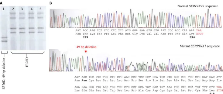
Figure 1. Identification of a novel SerpinA1 Mutant. A.DGGE banding patterns representing four controls (lanes 2–5) heterozygous for the M3 mutation (E376D), while our patient (lane 1), although also heterozygous for the M3 variant also presents with a shifted (faster migrating) banding depicting the novel deletion mutation (T379D).B.Sanger sequencing defines the deleted base pairs. The predicted amino acid sequence resulting from the novel 49 bp deletion (denoted by * on lower chromatogram) observed in our patient, results in the replacement of 16 amino acids and the addition of 24 amino acids through partial translation of the 39UTR.
doi:10.1371/journal.pone.0051762.g001
background, and a novel 49 base deletion mutation (g.12052_12100del #K02212 genomic sequence). This deletion results in a frame-shift at position T379 that replaces the last 16 amino acids of a1AT and adds an additional 24 amino acids through partial translation of the 39 UTR (Figure 1). This mutation has not previously been reported and joins the Z (E342K), S (S53F) and Mm (F52D) as pathogenic mutants causing profound plasma deficiency [10]. The additional amino poly-peptide sequence has very little homology to any known protein sequence and hence the likely structural implications of replacing the additional residues are not immediately apparent.
Functional Analysis: Mutant Protein Expression and Secretion
Consistent with the clinical observation of low circulatinga1AT levels in the patient, functional analysis showed clearly that a1ATT379Dis not secreted and is prone to intracellular aggrega-tion. We observed expression of both wild-type and T379D a1AT protein in HEK293T and HeLa cell lysates following transfection (Figure 2). The slightly slower migration of the mutant form reflects the larger protein resulting from the C-terminal extension. Notably, with high-level expression in HEK293 cells there is a striking accumulation of a1ATT379D
in the insoluble fraction following cell lysis (Figure 2D), likely indicating misfolding and/or aggregation of the mutant form. Immunofluorescence microscopy indicated the presence of intracellular aggregates ofa1ATT379D
in HEK293T cells (Figure 2F). Significantly, although wild-type a1AT is clearly detectable in conditioned media from transfected
HEK293 or HeLa cells, the mutant form is not detectable (Figure 2A, D).
Impaired secretion of a1ATT379D
was also confirmed by performing GFP-based affinity purification of conditioned media from transfected HEK293T cells, followed by immunoblot detection ofa1AT (Figure 2B). These experiments clearly showed secretion of wta1AT, while no secretion ofa1ATT379D
could be detected, even after GFP-trap enrichment. Cleavage of an N-terminal GFP tag from both wild-type and a1ATT379D
confirms normal processing of the secretion signal tag (Figure 2C) and suggests that intracellular aggregation/misfolding inhibits secre-tion ofa1ATT379D
.
Discussion
A link between circulating deficiency ofa1AT and misfolding or polymerisation of the protein has been known for over 20 years. However, despite some elegant and detailed structural analyses, the precise mechanism and exact nature of the pathogenic polymeric forms has been difficult to define. Understanding the structural and/or environmental factors drivinga1AT misfolding
are key to understanding a1AT deficiency and improving di-agnosis and therapy.
We describe here a novel SERPINA1 mutant from an asymptomatic patient with circulatinga1AT deficiency. A 49 base pair deletion results in a frame-shift at amino acid T379, replacing the last 16 amino acids of a1AT and adding an additional 24 amino acids through partial translation of the 39 UTR. In-tracellular accumulation and failed secretion of the a1ATT379D mutant in cultured cells is consistent with clinical observation of low circulatinga1AT in the patient and establishes the mutation, along with the Z, S and Mm variants, as abone fide pathogenic variant. Importantly, this represents the first pathogenic mutation identified in the C-terminal domain ofa1AT, which was recently implicated in the formation of pathogenica1AT polymers [11,12]. Normal circulating levels ofa1AT range from 104 to 276 g/L (20– 53 uM). Lung disease associated with diminished neutrophil elastase inhibitory capacity is typically observed in patients with decreased circulatinga1AT (0.36–0.57 g/L (5–11mM)) [13]. The
circulating a1AT level of 0.58 g/L (11mM) observed in this patient lies at threshold of this disease-associated range.
The T379Dmutation occurs in the C-terminal region ofa1AT, quite distinct from the Z(E342K) and S(E264V) mutants found commonly in European populations but relatively rarely in African populations [6,14]. It is noteworthy that the patient was of Middle Eastern descent, and it is highly likely that as yet unidentified deleteriousa1AT mutations exist in other population groups that have not been well studied. Critically, these novel mutants may be missed by commonly used phenotyping approaches, further emphasizing the importance of specific genotype-based assays for accurate classification of mutants and diagnosis ofa1AT deficiency [6,15]. This point is highlighted by the fact that the patient in this study was originally mistyped by IEF as having aZ/M2phenotype classification. This study further highlights the significance of rare mutations in clinically relevanta1AT deficiency.
Serpins are flexible molecules capable of extreme conforma-tional change, making them highly susceptible to polymerization. Polymer-causing mutations (such as thea1ATZmutant) influence the folding pathway by increasing the lifetime of a polymergenic folding intermediate. Serpin polymers are favored when secondary structural domain swaps occur at a faster rate than folding into the native state. The various pathological serpin mutants identified to date have been shown to accelerate this domain swapping [11,12]. Using a monoclonal antibody specific for hepatocellular inclusions of a1AT, Yakasaki etal [12] recently proposed a mechanism of pathological polymerization involving a C-terminal domain swap, distinct from the accepted model involving an s4As/5A swap. The implication of this observation is that the native state ofa1AT is achieved by rapid folding of the C-terminal domain [16]. However, the exact nature of the toxic form of a1AT polymers
Figure 2. Functional Characterisation ofa1ATD379Mutant.(A) Immunoblot (anti-GFP) detection ofa1AT-GFP fusion protein (C-terminal tag) in
whole-cell lysate and concentrated conditioned media (ie secreted) from HEK293T cells transfected with plasmids expressing either wild-type orD379 mutanta1AT-GFP. Red arrow denotes position of,75 kDaa1AT-GFP band, note the absence of this band in conditioned media from cells transfected withD379 mutant, indicating impaired secretion of mutant protein; (B) Immunoblot (anti-a1AT) detection ofa1AT-GFP fusion protein (C-terminal tag) in whole-cell lysate, or following immunprecipitation from conditioned media (i.e. secreted) from HEK293T cells transfected with plasmids expressing either wild-type orD379 mutanta1AT-GFP; (C) Transfection of either wild-type orD379 mutanta1AT with an N-terminal EGFP fusion into HEK293T cells clearly indicated normal proteolytic processing of the secretion signal peptide. Both,75 kDA and,27 kDA bands are visible, representing
full-length and processed (i.e. signal peptide cleaved)a1AT-GFP fusion protein respectively; (D) At higher expression levels, accumulation of insoluble D379 mutanta1AT was observed in HEK293T cells, clearly denoted by the presence of a darker band in the insoluble fraction from cells transfected with D379 mutant; (E) Detection of soluble (whole-cell lysate), insoluble and secreted (concentrated conditioned media) a1AT in HeLa cells transfected with either wild-type orD379 mutanta1AT-GFP. Red arrow denotes position of,75 kDaa1AT-GFP band. Note the absence of this band
in conditioned media from cells transfected withD379 mutant, indicating impaired secretion of mutant protein; (E) Fluorescent micrographs of HEK293T cells following transfection with either wild-type orD379 mutanta1AT-GFP expression plasmids. Increased intracellular aggregation of mutant protein is clearly visible. NB: Loading controls representa-tubulin immunoblot or PonceauS staining in lysate or secreted (conditioned media) samples, respectively.
doi:10.1371/journal.pone.0051762.g002
in the liver is yet to be determined and may involve heterogeneous populations of polymers [11]. Although the structural conse-quences of the T379Dmutation are not immediately obvious, it is highly significant that the mutation introduces an entirely new, extended C-terminal sequence into a1AT. This is likely to drastically modify the folding rate of the C-terminal domain of the T379Dmutant, possibly favoring polymerization.
Interventions modifying the folding pathway ofa1AT might be of therapeutic value in treating both loss and gain of function manifestations of a1AT deficiency [12]. Indeed, a number of strategies designed to attenuate polymerization are under in-vestigation as potential therapies for a1AT deficiency, including peptide analogues, chemical chaperones, and small molecule allosteric regulators [13,17,18]. Some of these strategies (particu-larly peptide analogues and allosteric regulators) target specific polymerigenic mutations (e.g. ZE342K) and so would not necessarily be effective against the T379Dvariant. This further highlights the need to better describe the range of pathogenica1AT mutations
and for detailed understanding of the exact mechanisms of polymer formation.
Conclusions
In summary, we describe a novel pathogenic SERPINA1 mutation causing circulating a1AT deficiency. This mutation provides novel insight into mechanisms ofa1AT misfolding in liver and lung disease, with important implications for molecular diagnosis and therapeutic development.
Author Contributions
Conceived and designed the experiments: DNS EAT RFS JR AD JAW VMH. Performed the experiments: EAT DNS RFS VMH. Analyzed the data: DNS EAT RFS JR AD JAW VMH. Contributed reagents/ materials/analysis tools: DNS EAT VMH JAW. Wrote the paper: DNS EAT RFS VMH.
References
1. Luisetti M, Seersholm N (2004) Alpha1-antitrypsin deficiency. 1: epidemiology of alpha1-antitrypsin deficiency. Thorax 59: 164–9.
2. de Serres FJ (2003) Alpha-1 antitrypsin deficiency is not a rare disease but a disease that is rarely diagnosed. Environ Health Perspect 111: 1851–4. 3. Silverman EK, Sandhaus RA (2009) Clinical practice. Alpha1-antitrypsin
deficiency. N Engl J Med 360: 2749–57.
4. Dickens JA, Lomas DA (2011) Why has it been so difficult to prove the efficacy of alpha-1-antitrypsin replacement therapy? Insights from the study of disease pathogenesis. Drug Des Devel Ther 5: 391–405.
5. Zaimidou S, van Baal S, Smith TD, Mitropoulos K, Ljujic M, et al. (2009) A1ATVar: a relational database of human SERPINA1 gene variants leading to alpha1-antitrypsin deficiency and application of the VariVis software. Hum Mutat 30: 308–13.
6. Hayes VM (2003) Genetic diversity of the alpha-1-antitrypsin gene in Africans identified using a novel genotyping assay. Hum Mutat 22: 59–66.
7. Wu Y, Hayes VM, Osinga J, Mulder IM, Looman MW, et al. (1998) Improvement of fragment and primer selection for mutation detection by denaturing gradient gel electrophoresis. Nucleic Acids Res 26(23): 5432–40. 8. Hayes VM, Wu Y, Osinga J, Mulder IM, van der Vlies P, et al. (1999)
Improvements in gel composition and electrophoretic conditions for broad-range mutation analysis by denaturing gradient gel electrophoresis. Nucleic Acids Res 27: e29.
9. Lamesch P, Li N, Milstein S, Fan C, Hao T, et al. (2007) hORFeome v3.1: a resource of human open reading frames representing over 10,000 human genes. Genomics 89: 307–15.
10. Lomas DA (2005) Molecular mousetraps, alpha1-antitrypsin deficiency and the serpinopathies. Clin Med 5: 249–57.
11. Yamasaki M, Sendall TJ, Pearce MC, Whisstock JC, Huntington JA (2011) Molecular basis of a1-antitrypsin deficiency revealed by the structure of
a domain-swapped trimer. EMBO Rep. 12(10): 1011–7.
12. Bottomley SP (2011) The structural diversity in a1-antitrypsin misfolding. EMBO Rep. 12(10): 983–4.
13. Greene CM, McElvaney NG (2010) Protein misfolding and obstructive lung disease. Proc Am Thorac Soc. 7(6): 346–55.
14. Blanco I, Bustillo EF, Rodriguez MC (2001) Distribution of alpha1-antitrypsin PI S and PI Z frequencies in countries outside Europe: a meta-analysis. Clin Genet 60(6): 431–41.
15. Rachelefsky G, Hogarth D (2008) Issues in the diagnosis of alpha 1-antitrypsin deficiency. J Allergy Clin Immunol. 121(4): 833–8.
16. Tsutsui Y, Dela Cruz R, Wintrode PL (2012) Folding mechanism of the metastable serpina1-antitrypsin. Proc Natl Acad Sci U S A. 109(12): 4467– 4472.
17. Ekeowa UI, Gooptu B, Belorgey D, Ha¨gglo¨f P, Karlsson-Li S, et al. (2009) Alpha1-Antitrypsin deficiency, chronic obstructive pulmonary disease and the serpinopathies. Clin Sci (Lond). 116(12): 837–50.