Direct Inhibition of the
Longevity-Promoting Factor SKN-1
by Insulin-like Signaling in
C. elegans
Jennifer M.A. Tullet,1,5Maren Hertweck,2,5Jae Hyung An,1,3,5Joseph Baker,1Ji Yun Hwang,3Shu Liu,2 Riva P. Oliveira,1Ralf Baumeister,2,4and T. Keith Blackwell1,*
1Section on Developmental and Stem Cell Biology, Joslin Diabetes Center; Department of Pathology, Harvard Medical School;
Harvard Stem Cell Institute, 1 Joslin Place, Boston, MA 02215, USA
2Center of Biochemistry and Molecular Cell Research (ZBMZ, Faculty of Medicine), Bioinformatics and Molecular Genetics
(Faculty of Biology), University of Freiburg, Scha¨nzlestr. 1, D-79104 Freiburg, Germany
3Protein Network Research Center, Yonsei University, 134 Shinchon-dong, Seodaemun-gu, Seoul, 120-749, Republic of Korea 4Center for Systems Biology (ZBSA), University of Freiburg, D-79104 Freiburg, Germany
5These authors contributed equally to this work.
*Correspondence:[email protected]
DOI 10.1016/j.cell.2008.01.030
SUMMARY
Insulin/IGF-1-like signaling (IIS) is central to growth
and metabolism and has a conserved role in aging.
In
C. elegans
, reductions in IIS increase stress
resis-tance and longevity, effects that require the
IIS-inhibited FOXO protein DAF-16. The
C. elegans
tran-scription factor SKN-1 also defends against oxidative
stress by mobilizing the conserved phase 2
detoxifica-tion response. Here we show that IIS not only opposes
DAF-16 but also directly inhibits SKN-1 in parallel. The
IIS kinases AKT-1, -2, and SGK-1 phosphorylate
SKN-1, and reduced IIS leads to constitutive SKN-1 nuclear
accumulation in the intestine and SKN-1 target gene
activation. SKN-1 contributes to the increased stress
tolerance and longevity resulting from reduced IIS
and delays aging when expressed transgenically.
Furthermore, SKN-1 that is constitutively active
in-creases life span independently of DAF-16. Our
find-ings indicate that the transcription network regulated
by SKN-1 promotes longevity and is an important
direct target of IIS.
INTRODUCTION
In many studies of aging, extended longevity correlates with en-hanced resistance against oxidative damage and other stresses (Finkel and Holbrook, 2000; Lithgow and Walker, 2002). In C. elegans, both life-span and stress resistance can be increased by the alteration of several processes, including insulin/IGF-1-like signaling (IIS) (Friedman and Johnson, 1988; Gottlieb and Ruvkun, 1994; Kenyon et al., 1993; Larsen, 1993), caloric intake (Houthoofd et al., 2002), mitochondrial respiration (Dillin et al., 2002; Lakowski and Hekimi, 1996; Lee et al., 2003), and germline function (Arantes-Oliveira et al., 2002). It is a formidable but
crit-ical challenge to understand how modulation of these processes might inhibit or defend against stresses that contribute to aging. Reductions in IIS have been associated with increased stress resistance and longevity in diverse species (Kenyon, 2005). Studies in worms and flies have established the paradigm that these effects derive from increased activity of the FOXO tran-scription factors, which are inhibited through IIS-induced phos-phorylation (Figure 1A) (Antebi, 2007; Kenyon, 2005). FOXO pro-teins are also important in many of the biological effects of insulin in mammals (Accili and Arden, 2004; van der Horst and Burger-ing, 2007). InC. elegans, signaling through the IIS receptor DAF-2 ultimately directs the related kinases AKT-1, -DAF-2, and SGK-1 to phosphorylate the FOXO protein DAF-16, thereby inhibiting its accumulation in nuclei (Henderson and Johnson, 2001; Hert-weck et al., 2004; Lin et al., 2001; Ogg et al., 1997). DAF-16 is required for phenotypes that are associated with decreased IIS, including increases in stress resistance, longevity, and the propensity to undergo diapause, the formation of long-lived dauer larvae that can survive adverse environmental conditions (Kenyon, 2005; Kenyon et al., 1993; Kimura et al., 1997). DAF-16 regulates genes that represent diverse processes, including resistance to oxidative and other stresses (Antebi, 2007; Kenyon, 2005).
SKN-1 is mediated by its expression in the intestine (digestive system) (Bishop and Guarente, 2007), where SKN-1 accumulates in nuclei and activates phase 2 gene expression inducibly in re-sponse to stress (An and Blackwell, 2003). In the intestine, phos-phorylation of SKN-1 by p38/mitogen-activated protein kinase (MAPK) signaling is required for its accumulation in nuclei, while negative regulation via glycogen synthase kinase-3 (GSK-3) phosphorylation is needed to prevent this from occurring consti-tutively (Figure 1A) (An et al., 2005; Inoue et al., 2005).
Asdaf-16is required for the increased stress resistance and longevity that are seen when IIS is reduced, it has seemed likely that these effects of decreased IIS could be accounted for by
increased DAF-16 activity. However, we reasoned that if it is ad-vantageous for IIS to inhibit stress response genes by acting on DAF-16, then IIS might also oppose SKN-1 (Figure 1A). Accord-ingly, here we show that reductions in IIS cause SKN-1 to accu-mulate constitutively in intestinal nuclei in the absence of stress and to activate phase 2 target genes. Importantly, these events do not require daf-16. AKT-1, -2, and SGK-1 phosphorylate SKN-1 at multiple sites, and mutation of an AKT phosphorylation site results in SKN-1 being present in intestinal nuclei constitu-tively.skn-1mutations significantly suppress the oxidative stress resistance and longevity phenotypes associated with reduced IIS. Aging is delayed when SKN-1 is expressed transgenically, Figure 1. Accumulation of SKN-1 in Intestinal Nuclei Is Inhibited by DAF-2
(A) A partial schematic ofC. elegansinsulin-like signaling and our working model.
(B)skn-1isoforms, alleles, and transgenes. The SKN-1A isoform is transcribed from an upstream operon promoter and differs from SKN-1C by the addition of 90 amino acids at its N terminus. SKN-1B and SKN-1C are each expressed from their own distinct upstream promoters (An and Blackwell, 2003; Bishop and Guarente, 2007). Theskn-1(zu67), (zu129),and(zu135)mutations each create a premature stop codon (B. Bowerman, personal communication). Green fluores-cent protein (GFP) is fused to the C terminus of SKN-1 in the transgenes indicated at the bottom. Strains that carry these and other transgenes are described in
Table S2. Theskn-1a/cRNAi construct includes only coding sequence and targets both SKN-1A and SKN-1C but does not alter the levels of SKN-1B in the ASI neurons (Bishop and Guarente, 2007).
(C) SKN-1::GFP accumulates in intestinal nuclei when DAF-2 activity is reduced. (D) Intestinal expression ofSKN-1B/C::GFPindaf-2(e1370)derives from SKN-1C.
and a mutant SKN-1 form that localizes constitutively to intesti-nal nuclei increases life span in the absence ofdaf-16. In sum-mary, our data indicate that IIS directly inhibits SKN-1 in parallel to DAF-16, that SKN-1 contributes to the stress and longevity phenotypes associated with reduced IIS, and that SKN-1 defines an important pro-longevity mechanism.
RESULTS
DAF-2 Signaling Inhibits SKN-1 Accumulation in Intestinal Nuclei
If IIS inhibits SKN-1 analogously to DAF-16 (Figure 1A), then re-duced DAF-2 signaling should allow SKN-1 to accumulate in intestinal nuclei. To test this idea, we first investigated how reduc-tions in DAF-2 activity affect the distribution of SKN-1 that is ex-pressed from the previously describedSKN-1B/C::GFP trans-gene (Figure 1B). This transgene rescues the maternal lethality, stress sensitivity, and DR-associated longevity defects ofskn-1
mutants (An and Blackwell, 2003; An et al., 2005; Bishop and Guarente, 2007).SKN-1B/C::GFPencodes two of three SKN-1 isoforms, SKN-1C and SKN-1B, which are expressed in the intestine and ASI neurons, respectively (Bishop and Guarente, 2007). In wild-type (WT) animals that carry extrachromosomal or integratedSKN-1B/C::GFP, SKN-1::GFP is generally unde-tectable in intestinal nuclei except under stress conditions or af-ter inhibition of its phosphorylation by GSK-3 (An and Blackwell, 2003; An et al., 2005). In contrast, SKN-1::GFP was constitutively present in intestinal nuclei of transgenicdaf-2(e1370)and daf-2(e1368)animals or in WT transgenics exposed todaf-2RNA interference (RNAi) at either 20C or low temperature (16C) ( Fig-ures 1C and 1E; see alsoFigures S1A–S1C available online). This nuclear SKN-1 appeared to correspond to SKN-1C because RNAi against this isoform prevented SKN-1::GFP from ac-cumulating in intestinal nuclei in response to daf-2 mutation (Figure 1D).
We also analyzed a transgene that encodes all three SKN-1 isoforms in the context of a large operon (SKN-1op::GFP; Fig-ure 1B). In the WT background this transgene was expressed in a similar pattern toSKN-1B/C::GFP(not shown), and in daf-2(e1370)SKN-1 that was expressed fromSKN-1op::GFPalso accumulated in intestinal nuclei constitutively (Figure 1E). In contrast to these effects in the intestine,daf-2mutation did not obviously alter the levels of nuclear SKN-1::GFP in the ASI neu-rons (Figures 1C and 1D; data not shown). We conclude that sig-naling through DAF-2 may be required to prevent SKN-1 from accumulating constitutively in intestinal nuclei.
DAF-2 Signaling Inhibits SKN-1 Directly, through Phosphorylation
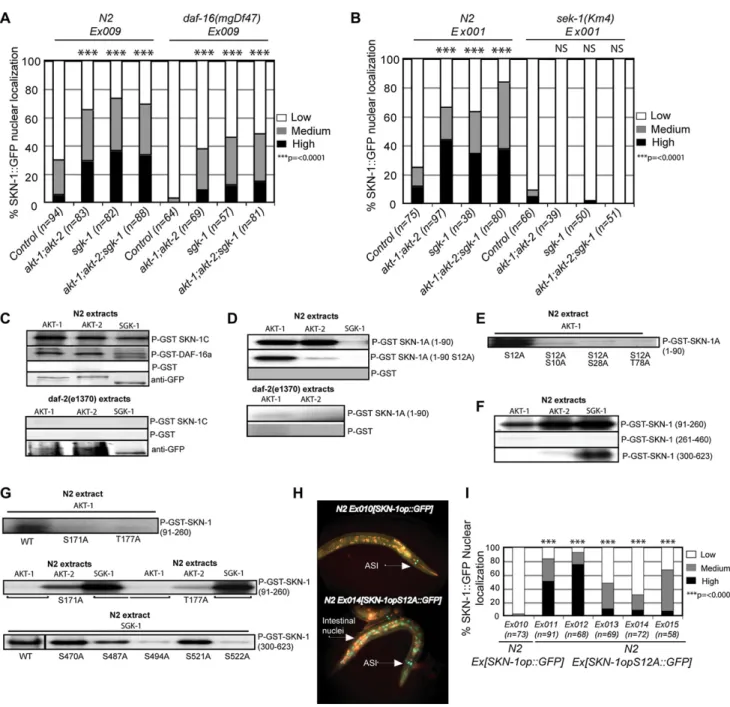
Signaling through DAF-2 ultimately activates the AKT-1, -2, and SGK-1 kinases, which phosphorylate and inhibit DAF-16 ( Fig-ure 1A) (Cahill et al., 2001; Hertweck et al., 2004; Lin et al., 2001). RNAi knockdown ofakt-1;akt-2orsgk-1dramatically in-creased the presence in intestinal nuclei of SKN-1::GFP that was expressed from eitherSKN-1B/C::GFPorSKN-1op::GFP
(Figures 2A and 2B), supporting the model that IIS inhibits SKN-1 directly. Importantly,daf-16was not required for SKN-1 expression, or for the nuclear accumulation of SKN-1 that
re-sulted from reductions in IIS. A constitutively nuclear SKN-1 sub-stitution mutant in which a critical inhibitory GSK-3 phosphoryla-tion site has been altered (SKN-1(S393A);An et al., 2005) was appropriately present in intestinal nuclei in nulldaf-16animals (Figure S2). In addition, in a nulldaf-16background, RNAi of the IIS kinases still increased the levels of intestinal nuclear SKN-1::GFP expressed fromSKN-1op::GFP(Figure 2A). Finally, indaf-2(e1370)animals, the nuclear accumulation of SKN-1 ex-pressed fromSKN-1B/C::GFPwas not reduced bydaf-16RNAi (Figure S1D). We conclude that the IIS pathway inhibits SKN-1 in the intestine, and that this regulation occurs independently of DAF-16.
We previously observed that p38 MAPK signaling is required for SKN-1 to accumulate in intestinal nuclei not only in response to stress but also when GSK-3 phosphorylation of SKN-1 is inhibited under normal conditions (An et al., 2005; Inoue et al., 2005). In the latter case, even though negative regulation of SKN-1 is impaired, the low-level background p38 signaling that is seen under normal conditions is still required. To test whether p38 signaling is also needed for SKN-1 to be present in intestinal nuclei when IIS is reduced, we performedakt-1;akt-2andsgk-1
RNAi in animals that lacked the p38 pathway MAPK kinase SEK-1 (Inoue et al., 2005). Insek-1(km4)mutants, SKN-1::GFP failed to accumulate in intestinal nuclei in response to IIS kinase RNAi (Figure 2B). Similarly, RNAi of the IIS kinases did not allow nuclear accumulation of a SKN-1 mutant in which two key p38 phosphorylation sites had been altered (Figure S3). We conclude that the SKN-1 nuclear accumulation that is allowed when IIS is reduced is appropriately dependent upon p38 MAPK signaling. We next investigated whether SKN-1 is phosphorylated by the downstream IIS kinases AKT-1, -2, and SGK-1. Analysis using SCANSITE identified seven potential AKT phosphorylation motifs within SKN-1C (Table S1). Four additional potential AKT phosphorylation motifs were detected within the unique N-termi-nal 90 amino acids of SKN-1A (SKN-1A(1–90);Table S1), which is predicted by expressed sequence tag analysis to be the most abundant SKN-1 isoform (http://www.wormbase.org). AKT-1::GFP, AKT-2::GFP, and SGK-1::GFP that were purified from WT C. elegans(Hertweck et al., 2004) each phosphorylated full-length SKN-1C in vitro at least as robustly as they phosphor-ylated DAF-16 (Figure 2C). In contrast, SKN-1A(1–90) was phos-phorylated by AKT-1 and AKT-2 but not SGK-1 (Figure 2D), con-sistent with the previous finding that AKT-1, -2, and SGK-1 differ with respect to their phosphorylation sites within the mammalian DAF-16 homolog FOXO3a (Brunet et al., 2001). Significantly, SKN-1 was not phosphorylated robustly by these same kinases if they were purified fromdaf-2(e1370)animals (Figures 2C and 2D). We conclude that SKN-1 is a direct substrate of the down-stream IIS kinases, and that their phosphorylation of SKN-1 depends upon signaling through DAF-2.
Figure 2. DAF-2 Signaling Inhibits Nuclear Accumulation of SKN-1 Directly, and Independently of DAF-16
(A)akt-1;akt-2orsgk-1RNAi allowsdaf-16-independent accumulation of SKN-1 in intestinal nuclei. Expression ofSKN-1op::GFPwas monitored.akt-1orakt-2
RNAi had a similar effect to combinedakt-1;akt-2RNAi (data not shown).
(B) RNAi depletion of the downstream IIS kinases causessek-1-dependent accumulation of SKN-1 in intestinal nuclei. Expression ofSKN-1B/C::GFPwas monitored.
(C) SKN-1C is phosphorylated by AKT-1, -2, and SGK-1 comparably to DAF-16, dependent upondaf-2. Here and in (D)–(G), AKT-1::GFP, AKT-2::GFP, and SGK-1::GFP were immunoprecipitated from WT ordaf-2(e1370) C. eleganslysates with anti-GFP antibody, quantitated by western blotting with anti-GFP, and used to phosphorylate purified GST-fused SKN-1 or DAF-16 proteins with [gP32]ATP. GST controls are shown inFigure S4B. These kinases appropriately required
activation inC. elegans, as they did not phosphorylate SKN-1 after they had been expressed inE. coli(Figure S4A).
(D) The unique 90 aa region at the SKN-A N terminus (SKN-1A(1–90)) is phosphorylated by the AKT kinases but not SGK-1, dependent upondaf-2. (E) Partial mapping of AKT-1 sites within SKN-1A(1–90).
(F) AKT and SGK phosphorylate particular regions of SKN-1C. Here and in (G), residues are numbered as in SKN-1A, so that the first residue of SKN-1C is numbered as 91.
(G) Partial mapping of AKT-1, -2, and SGK-1 sites within SKN-1C fragments.
different regions or individual sites within the remainder of SKN-1, which corresponds to the SKN-1C isoform (Figures 2F and 2G; Table S1). Together, our data show that AKT-1, -2, and SGK-1 phosphorylate SKN-1 at multiple overlapping and distinct sites. To investigate whether SKN-1 is regulated directly by IIS ki-nase phosphorylation in vivo, we substituted Ala at its most strongly predicted AKT site (Ser12 of SKN-1A) (Table S1), within theSKN-1op::GFPtransgene. In each transgenic line examined, this substitution resulted in dramatic constitutive intestinal nu-clear accumulation of SKN-1A (Figures 2H and 2I). This finding shows that SKN-1A is expressed in the intestine, and that the AKT phosphorylation site Ser12 is essential for its appropriate regulation. This strongly supports the model that signaling through the DAF-2 pathway directly inhibits SKN-1 in vivo.
Inhibition of SKN-1 Target Genes by IIS
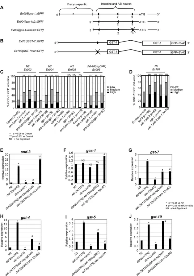
Given that IIS directly inhibits the SKN-1 protein in the intestine, it follows that IIS should also reduce expression of SKN-1 target genes. To test this idea, we first asked how reductions in IIS af-fect expression of two SKN-1 target reporter transgenes (Figures 3A and 3B). In the intestine, SKN-1 directly induces expression ofgcs-1 (g-glutamyl cysteine synthetase), which encodes an
enzyme rate-limiting for glutathione biosynthesis (Figure 3A) (An and Blackwell, 2003). In the intestine agcs-1::GFP transcrip-tional reporter is generally expressed at very low levels in the ab-sence of stress but was upregulated byakt-1;akt-2orsgk-1RNAi (Figure 3C). This induction was abolished by mutation of an important SKN-1 binding site(gcs-1D2mut3;Figure 3A) but not by absence of daf-16 (Figure 3C), indicating that SKN-1 but not DAF-16 was required for IIS kinase RNAi to activate the
gcs-1promoter. Interestingly,gcs-1was not induced bydaf-2
RNAi, implying that its regulation by AKT and SGK-1 might in-volve an additional input (data not shown). In contrast, a transla-tional fusion reporter for the SKN-1 target genegst-7(glutathione S-transferase;Figure S5) was comparably induced in the intes-tine by eitherdaf-2or IIS kinase RNAi (Figures 3B and 3D).
We next used quantitative (Q)-PCR to analyze howdaf-2mutation affected the levels of mRNA expressed from a set of known or pre-dicted SKN-1 target phase 2 genes (An and Blackwell, 2003; Kell et al., 2007). As would be expected, SKN-1 was not required for
daf-2mutation to induce expression of a negative control for our ex-periments, the DAF-16 targetsod-3(Figure 3E) (Antebi, 2007). The levels ofgcs-1mRNA were largely unaffected in thisdaf-2mutant (Figure 3F), in agreement with our reporter data and the high levels ofskn-1-independentgcs-1expression seen in the pharynx (An and Blackwell, 2003). In contrast, four differentgstgenes were strongly upregulated bydaf-2mutation in a manner that was substantially or completely dependent uponskn-1. (Figures 3G–3J). DAF-16 con-tributed to induction of some of these genes, but the SKN-1 target
gst-10was upregulated bydaf-2mutation independently ofdaf-16
(Figures 3G–3J). Together, our results indicate that when DAF-2 pathway signaling is reduced, SKN-1 induces a gene expression program that overlaps only partially with that of DAF-16.
skn-1Contributes to the Increased Longevity
and Stress Resistance Associated with Reduced IIS Having established that DAF-2 and IIS inhibit SKN-1 directly, and thereby suppress SKN-1-dependent gene expression, we next
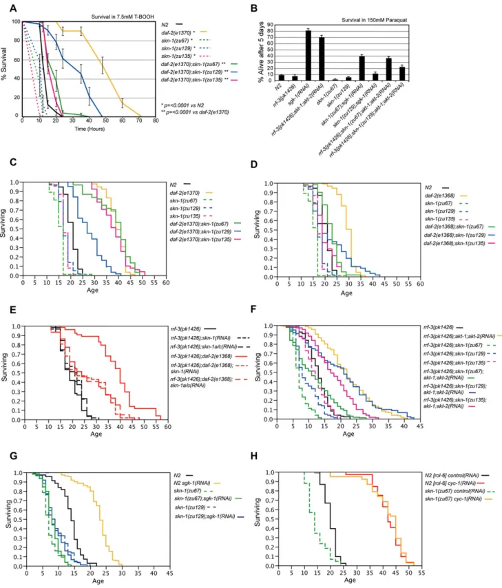
asked whether SKN-1 contributes to phenotypes that are asso-ciated with decreased IIS. We engineereddaf-2;skn-1 double mutants that each carried one of threeskn-1mutations, along with one of twodaf-2alleles. Eachskn-1mutation (Figure 1B) results in reduced stress resistance and longevity (Figure 4; Ta-ble 1) (An and Blackwell, 2003), failure of DR to extend life span (Bishop and Guarente, 2007), and maternally derived embryonic lethality (Bowerman et al., 1992). The class 2daf-2mutante1370
is stress resistant and lives much longer than WT at 20C ( Fig-ures 4A and 4C), but this increased life span is accompanied by various dauer-like abnormalities (Gems et al., 1998; Kimura et al., 1997). The class 1 daf-2mutatione1368increases life span to a lesser extent (Figure 4D), but these animals appear es-sentially normal. We also tested howskn-1mutations affect sim-ilar phenotypes caused by RNAi of the IIS downstream kinases. At 25C,sgk-1RNAi increases both oxidative stress resistance and life span, dependent upondaf-16(Hertweck et al., 2004). Previously, life span was not extended whenakt-1RNAi was per-formed in theakt-2(ok393)mutant (Hertweck et al., 2004), but we have now observed thatakt-1;akt-2RNAi significantly increases stress resistance and life span in an RNAi hypersensitive strain (Figures 4B and 4F;Table 1). This suggests that AKT-1, -2, and SGK-1 may contribute comparably to the effects of DAF-2 signaling on stress resistance and longevity.
Initially, we tested whether the IIS-associated phenotypes of dauer formation and elevated stress resistance requireskn-1.
skn-1RNAi did not prevent the constitutive dauer entry associ-ated with thedaf-2(e1370)mutation, orakt-1;akt-2RNAi ( Sup-plemental Data). In contrast, and consistent with its role in phase 2 detoxification, mutations inskn-1dramatically suppressed the increases in oxidative stress resistance seen in each of these contexts of reduced IIS, and also withsgk-1RNAi (Figures 4A and 4B).
We then investigated howskn-1contributes to the increased lon-gevity deriving from decreaseddaf-2function. Onlyskn-1(zu129)
suppressed the increased longevity of the class 2 daf-2 allele
e1370(Figure 4C;Table 1). In contrast, all threeskn-1mutations significantly reduced the life span of the class 1daf-2 mutant
e1368(Figure 4D;Table 1). Theseskn-1mutations also altered the shape of thedaf-2(e1368)survival plot, so that the longest-living double mutant animals appeared to be less dependent uponskn-1
than the remaining population (Figure 4D). Accordingly, when we excluded the 20% most long-lived animals from statistical analysis, two of threeskn-1mutations affected death rate more severely in
daf-2(e1368)animals than in WT (Figure S7;Tables S3 and S4). Importantly, an RNAi-sensitive genetic background skn-1 RNAi decreased the life span ofdaf-2(e1368)but not control animals (Figure 4E;Tables 1andS4), indicating that thisdaf-2mutant is more susceptible than WT to a reduction in SKN-1 levels. daf-2(e1368)life span was similarly decreased by eitherskn-1or skn-1a/cRNAi (Figure 4E;Tables 1andS4), indicating that the SKN-1 A and/or C isoforms are required for the longevity associated withdaf-2(e1368). daf-2(e1368)life span was also decreased by feeding ofskn-1RNAi from the beginning of adulthood (Figure S8), indicating an effect on aging, not development.
Mutations inskn-1also suppressed the life-span extensions associated with reductions in IIS kinase levels. Each of twoskn-1
Figure 3. SKN-1 Target Genes downstream of IIS
(A and B) Schematics of thegcs-1promoter (An and Blackwell, 2003) and theGST-7::GFPtranslational fusion. Triangles mark SKN-1 binding sites.gcs-1D2::GFP
the dramatic increase in life span deriving from sgk-1 RNAi (Figure 4G;Tables 1andS4). In addition, two of threeskn-1alleles significantly reduced the life spans ofakt-1;akt-2RNAi animals (Figure 4F;Table 1). We conclude that SKN-1 makes important contributions to the increases in stress resistance and longevity that are associated with reductions in IIS activity.
Next, we investigated whetherskn-1affects the long life spans of animals with reduced mitochondrial electron transport chain activity to find out whetherskn-1mutations might cause a gen-eral sickness that limits life span (Dillin et al., 2002; Lee et al., 2003; Rea et al., 2007). In agreement with a recent report (Rea et al., 2007), askn-1mutation did not reduce the large increases in life span associated with RNAi against either cytochromec re-ductase(cyc-1)or cytochromecoxidase(cco-1)(Figures 4H and S9;Table 1). This indicates that whileskn-1is important for life span in long-lived animals in which IIS is reduced, lack of
skn-1does not limit longevity nonspecifically.
SKN-1 Promotes Longevity
The involvement ofskn-1in the longevity effects of IIS led us to investigate whether SKN-1 has an independent pro-longevity function. Previously, life span was modestly increased by trans-genic overexpression of either WT or constitutively nuclear forms of DAF-16 (Henderson and Johnson, 2001; Lin et al., 2001). To investigate whether modulation of SKN-1 expression can extend life span, we first analyzed otherwise WT strains in which either
the SKN-1B/C::GFP or SKN-1B/C S393A::GFP transgenes
were present. TheSKN-1B/C S393A::GFPtransgene, which ex-presses constitutively nuclear SKN-1 in the intestine, increased mean life span variably but consistently (by 5%–21%;Figure 5A; Table 2). A modest increase was also seen withSKN-1B/C::GFP
(Ex001;Figure 5B;Table 2).
We had previously observed thatskn-1transgenes not only res-cue the oxidative stress sensitivity ofskn-1(zu67)mutants but also increase their stress resistance to well beyond that of WT (An et al., 2005). Similarly, strains in whichskn-1(zu67)was rescued with these transgenes also lived longer than wild-type animals that carried the same transgene arrays (arrays Ex001 and
Ex008,Figures 5A and 5B;Table 2). This difference was corrobo-rated in independent strains that were genecorrobo-rated by breeding the
Ex001[SKN-1B/C::GFP]array from the WT intoskn-1(zu67)and
skn-1(zu129)(FiguresS10A, S10B, andS11), and in an additional
skn-1(zu67)strain carryingSKN-1B/C S393A::GFP(Ex020, Fig-ure 5B;Table 2). Perhaps an optimal window of SKN-1 expression is important for longevity because we found that expression of SKN-1 from high-copy arrays can be toxic (data not shown). Sig-nificantly, rescuedskn-1(zu67) Ex001[SKN-1B/C::GFP]animals maintained the ability to move longer than WT (Figure S10C), indi-cating that transgenicskn-1expression delayed the aging
pro-cess. RNAi that was targeted to transgenic SKN-1C (skn-1a/c
RNAi, Figure 1B) eliminated expression of intestinal nuclear SKN-1 fromSKN-1B/C S393A::GFP(Figure S12) and suppressed the extended life span of a transgenically rescuedskn-1(zu67)
strain (Figure 5C). Taken together, our results indicate that SKN-1 promotes longevity, an effect that appears to require the SKN-1C isoform.
We next investigated whetherdaf-16is required for transgenic SKN-1 to increase life span because of the general importance of
daf-16forC. eleganslongevity and because SKN-1 and DAF-16 overlap in regulating some target genes (Figures 3E–3J). When we bred a SKN-1 S393A transgenic array (Ex008) into the null mutantdaf-16(mgDf47), life span was modestly but significantly increased in two of three experiments (Figure 5D;Tables 2and S6). Similarly, after daf-16RNAi the life spans of skn-1(zu67)
animals were reduced, but presence of an array that expresses constitutively nuclear SKN-1(S393A) (Ex008 or 020) still in-creasedskn-1(zu67)life span to beyond that of WT (Figure 5E; Table 2). Interestingly,daf-16RNAi completely suppressed the life-span extension associated with transgenic WT SKN-1 ( Fig-ure S13;Table 2), suggesting that reductions in DAF-16 levels not only reduce life span but also might free up negative regula-tory mechanisms that act on SKN-1. Taken together, our data suggest that SKN-1 can promote longevity independently of DAF-16, provided that SKN-1 is released from negative regulation in the intestine.
DISCUSSION
It is well established that FOXO proteins are inhibited directly by the IIS pathway, and that the FOXO protein DAF-16 is required for the increased longevity and stress resistance that result from decreased IIS in C. elegans (Kenyon, 2005). This has made it difficult to imagine that another transcription factor might be regulated similarly by IIS in parallel to FOXO and could play a major role in IIS-regulated processes. However, here we have identified SKN-1 as the second transcription factor that is known to be inhibited directly by IIS. We have also shown that IIS inhibits SKN-1 target genes and that SKN-1 contributes to the stress re-sistance and longevity phenotypes of reduced IIS and functions as a pro-longevity factor.
Direct Regulation of SKN-1 by IIS
Multiple lines of evidence indicate that IIS inhibits SKN-1 in the intestine directly (Figure 1A). Reductions in IIS activity resulted in SKN-1A and SKN-1C being present in intestinal nuclei in the absence of either stress or DAF-16 and in induction of SKN-1 target genes (Figures1E,2A, 2B, and3). SKN-1 isoforms were phosphorylated by AKT-1, -2, and SGK-1 at multiple sites, and
(C)gcs-1::GFPis induced in the intestine independently ofdaf-16in response toakt-1;akt-2andsgk-1RNAi. Here and in (D), results of multiple experiments performed on L4 larvae have been combined and scored as described in theSupplemental Experimental ProceduresandAn and Blackwell (2003)andAn et al. (2005). p values were derived from a chi2test.akt-1andakt-2RNAi were also performed separately and shown to have a similar effect to combined akt-1;akt-2RNAi (data not shown).
(D)GST-7::GFPis induced in response todaf-2, akt-1;akt-2,orsgk-1RNAi.
Figure 4. skn-1Mutations Suppress the Longevity and Stress Resistance Phenotypes Conferred by Reduced IIS
(A)skn-1contributes to the oxidative stress resistance phenotype ofdaf-2(e1370)mutants. A representative experiment is shown in which animals were exposed to 7.5 mMt-butyl hydrogen peroxide. Here and in (B), error bars represent the SEM.
(B)skn-1contributes to the stress resistance phenotypes conferred bysgk-1orakt-1;akt-2RNAi. Animals were scored after 5 days exposure to 150 mM paraquat. Data were combined from at least three experiments with n > 90 for each group.
mutation of an AKT site (Ser12) led to SKN-1A being present in intestinal nuclei constitutively (Figures 2H and 2I). The AKT and SGK kinases differed in their preferences for sites within SKN-1, consistent with previous analyses of their phosphorylation of FOXO (Brunet et al., 2001). The complexity of this regulation could potentially allow SKN-1 to integrate inputs from multiple signals or molecular interactions.
We also observed that SKN-1 contributes significantly to some IIS-associated phenotypes.skn-1mutations dramatically sup-pressed the increases in stress resistance deriving fromsgk-1
RNAi,akt-1;akt-2RNAi, and thedaf-2(e1370)mutation (Figures 4A and 4B). Lack ofskn-1completely suppressed the increased life span deriving fromsgk-1RNAi, reduced the extended lon-gevity ofakt-1;akt-2RNAi animals (Figures 4F and 4G), and sup-pressed the increased life span of the class 1daf-2allelee1368
(Figure 4D). Importantly, skn-1RNAi reduced the life span of
daf-2(e1368) but not WT animals (Figure 4E), indicating that
daf-2(e1368) was more severely affected than WT by partial loss ofskn-1function. In contrast,skn-1mutation did not sup-press the long life associated with RNAi of mitochondrial respira-tory chain components (Figures 4H andS9), indicating that lack ofskn-1does not reduce life span nonspecifically.skn-1 muta-tions also did not consistently suppress the life-span extension ofdaf-2(e1370)mutants (Figure 4C). However, dauer-like pheno-types are associated with this class 2 allele (Gems et al., 1998), suggesting that this difference might reflect the apparent dispensability of SKN-1 for dauer formation (Supplemental Data). The simplest model to explain the contributions of SKN-1 to IIS-associated longevity is that they derive from phase 2 detoxifi-cation gene expression in the intestine. Our finding thatskn-1a/c
RNAi suppressesdaf-2(e1368)longevity (Figure 4E) is consistent with this idea, although we cannot exclude that other tissues or SKN-1 functions are involved. We conclude thatdaf-16is neces-sary but not sufficient to confer the increases in stress resistance and longevity that are associated with reduced IIS, and that these increases also involveskn-1.
In contrast to the regulation of SKN-1 by IIS in the intestine,
daf-2mutation did not obviously affect SKN-1 levels in the ASI neurons (Figure 1C; not shown), in which SKN-1B is required for DR to extend life span (Bishop and Guarente, 2007). How-ever, we cannot exclude that IIS might subtly influence the levels or activity of endogenous SKN-1 in ASI. This possibility could help resolve the apparent paradox that inC. elegansmost DR protocols extend life span independently ofdaf-16, and there-fore seemingly without input from IIS (Antebi, 2007; Bishop and Guarente, 2007; Greer et al., 2007; Houthoofd et al., 2003; Kenyon, 2005; Panowski et al., 2007).
SKN-1 is the sequence and functional counterpart to the mammalian NF-E2-related factor (Nrf) proteins (An and
Blackwell, 2003), which orchestrate the phase 2 detoxification response (Hayes and McMahon, 2001). Our results predict that the conserved IIS pathway might also regulate mammalian Nrf proteins, which include potential AKT sites (Table S1). Why would it be advantageous for IIS to decrease stress resistance? Many signaling cascades are influenced by redox status (Tonks, 2005). In particular, IIS is attenuated by protein tyrosine phos-phatase (PTP)1B, which is inhibited by reactive oxygen species (ROS) via the oxidation of conserved cysteine residues ( Gold-stein et al., 2005; Tonks, 2005). Thus, by inhibiting DAF-16/ FOXO and SKN-1/Nrf, IIS could suppress antioxidant defenses that would lead to a reducing environment, and that in turn might decrease IIS effectiveness. This is consistent with evidence that insulin induces starved cells to produce a ‘‘burst’’ of ROS, and that IIS functions more effectively under oxidative conditions (Goldstein et al., 2005).
Pro-longevity Functions of SKN-1
Our finding that transgenic SKN-1 expression extends life span (Figures 5A and 5B) demonstrates thatskn-1has an independent pro-longevity function. This activity appears to require the action of the intestinal SKN-1C isoform because it is suppressed by
skn-1a/cRNAi (Figure 5C). This pro-longevity activity of SKN-1 thus appears to be distinct from its ASI-mediated role in DR-induced longevity (Bishop and Guarente, 2007). The observation that transgenic SKN-1 increased life span more robustly inskn-1
mutants than in the WT (Figures 5A and 5B) suggests that the systemic effects of SKN-1 expression may be complex. How-ever, taken together, our data demonstrate that SKN-1 levels can be adjusted to extend life span and delay aging. We propose that the ancestral function of SKN-1 in phase 2 detoxification can be modulated by either reductions in IIS or adjustment of SKN-1 expression to extend life span, raising the exciting possibility that this detoxification system might be of general and conserved im-portance for longevity.
In the context of reduced IIS, SKN-1 and DAF-16 appeared to have both distinct and overlapping functions (Figure 3).daf-16
contributed to expression of someskn-1-dependent phase 2 de-toxification genes (Figures 3E–3I), but other SKN-1 targets were
daf-16independent (Figure 3J), consistent with our finding that transgenic expression of SKN-1 S393A increased longevity inde-pendently ofdaf-16(Figures 5D and 5E). Our data indicate that SKN-1 has important activities that are largely independent of DAF-16, making it of intense interest to dissect the extent to which these two transcription factors cooperate or act independently under normal, stress, and reduced IIS conditions. It is interesting that transgenic overexpression of neither DAF-16 or SKN-1 reca-pitulates the dramatic increases in longevity seen with reductions in DAF-2 function (Lin et al., 2001;Figure 5). One possibility is that
(D)skn-1suppresses the longevity ofdaf-2(e1368)mutants.
(E) In the RNAi-sensitiverrf-3background (Simmer et al., 2002), the life-span increase seen indaf-2(e1368)is comparably suppressed by eitherskn-1orskn-1a/c
RNAi. RNAi bacteria were provided from the L1 stage.
(F and G)skn-1mutations suppress the life-span extension conferred byakt-1;akt-2orsgk-1RNAi. Survival assays were carried out at 25C, are from adulthood, and represent the combined data from at least two experiments.
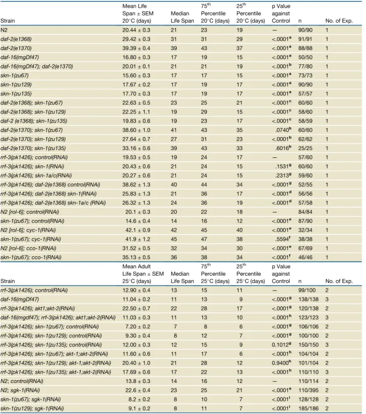
Table 1.skn-1Contributes to the Life-Span Extension Resulting from Reduced IIS
Strain
Mean Life Span±SEM 20C (days)
Median Life Span
75th Percentile 20C (days)
25th Percentile 20C (days)
p Value against
Control n No. of Exp.
N2 20.44±0.3 21 23 19 — 90/90 1
daf-2(e1368) 29.42±0.3 31 31 29 <.0001a 91/91 1
daf-2(e1370) 39.39±0.4 39 43 37 <.0001a 88/88 1
daf-16(mgDf47) 16.80±0.3 17 19 15 <.0001a 50/50 1
daf-16(mgDf47); daf-2(e1370) 20.01±0.1 21 21 19 <.0001b 77/80 1
skn-1(zu67) 15.60±0.3 17 17 15 <.0001a 73/73 1
skn-1(zu129) 17.67±0.2 17 19 17 <.0001a 90/90 1
skn-1(zu135) 17.70±0.3 17 19 17 <.0001a 57/57 1
daf-2(e1368); skn-1(zu67) 22.63±0.5 23 25 21 <.0001c 60/60 1
daf-2(e1368); skn-1(zu129) 22.25±1.1 19 29 15 <.0001c 58/60 1
daf-2 (e1368); skn-1(zu135) 19.83±0.6 19 23 17 <.0001c 58/59 1
daf-2(e1370); skn-1(zu67) 38.60±1.0 41 43 35 .0740b 60/60 1
daf-2(e1370); skn-1(zu129) 27.64±0.7 27 31 23 <.0001b 62/62 1
daf-2(e1370); skn-1(zu135) 33.16±0.6 39 43 33 .6016b 25/25 1
rrf-3(pk1426); control(RNAi) 19.53±0.5 19 24 17 — 57/60 1
rrf-3(pk1426); skn-1(RNAi) 20.43±0.6 21 24 15 .1531g 60/60 1
rrf-3(pk1426); skn-1a/c(RNAi) 20.27±0.6 21 24 15 .2313g 59/60 1
rrf-3(pk1426); daf-2(e1368) control(RNAi) 38.62±1.3 40 44 34 <.0001g 52/55 1
rrf-3(pk1426); daf-2(e1368) skn-1(RNAi) 25.83±1.3 21 36 17 <.0001d 56/56 1
rrf-3(pk1426); daf-2(e1368) skn-1a/c (RNAi) 26.32±1.3 24 36 19 <.0001d 57/58 1
N2 [rol-6]; control(RNAi) 20.1±0.3 20 22 18 — 84/84 1
skn-1(zu67); control(RNAi) 14.6±0.4 14 16 12 <.0001e 87/90 1
N2 [rol-6]; cyc-1(RNAi) 42.1±0.9 42 45 40 <.0001e 32/34 1
skn-1(zu67); cyc-1(RNAi) 41.9±1.2 45 47 38 .5594f 38/38 1
N2 [rol-6]; cco-1(RNAi) 31.52±0.5 32 34 30 <.0001e 67/69 1
skn-1(zu67); cco-1(RNAi) 35.13±0.5 36 38 34 <.0001f 46/46 1
Strain
Mean Adult Life Span±SEM 25C (days)
Median Life Span
75th
Percentile 25C (days)
25th
Percentile 25C (days)
p Value against
Control n No. of Exp.
rrf-3(pk1426); control(RNAi) 12.90±0.4 13 15 11 — 99/100 2
daf-16(mgDf47) 11.04±0.2 11 13 9 <.0001g 138/138 3
rrf-3(pk1426); akt1;akt-2(RNAi) 22.50±0.7 22 28 17 <.0001g 120/138 2
daf-16(mgdf47); rrf-3(pk1426); akt1;akt-2(RNAi) 11.03±0.3 11 13 10 <.0001h 123/123 3
rrf-3(pk1426); skn-1(zu67); control(RNAi) 7.20±0.2 7 8 6 <.0001g 106/106 2
rrf-3(pk1426); skn-1(zu129); control(RNAi) 9.30±0.4 8 12 7 <.0001g 100/100 2
rrf-3(pk1426); skn-1(zu135); control(RNAi) 12.00±0.3 12 15 9 0.1012g 150/150 3
rrf-3(pk1426); skn-1(zu67); akt-1;akt-2(RNAi) 11.60±0.6 11 17 6 <.0001h 104/104 2
rrf-3(pk1426); skn-1(zu129); akt-1;akt-2(RNAi) 20.40±1.0 21 28 12 0.9400h 101/104 2
rrf-3(pk1426); skn-1(zu135); akt-1;akt-2(RNAi) 17.69±0.6 17 22 13 <.0001h 110/110 3
N2;control(RNAi) 13.8±0.3 14 16 12 — 110/114 2
N2; sgk-1(RNAi) 22.6±0.4 23 25 21 <.0001e 110/395 2
skn-1(zu67); sgk-1(RNAi) 8.2±0.2 8 10 7 <.0001i 128/128 2
skn-1(zu129); sgk-1(RNAi) 9.1±0.2 8 11 7 <.0001i 185/186 2
Corresponds to the data inFigures 4C–4H. SEM: standard error of the mean. 75thand 25thpercentiles refer to the day at which 75% or 25% of the
population is dead. n represents total number of animals dying of old age versus those in total experiment (combined between trials where appropri-ate). In the case ofsgk-1(RNAi)the majority of exclusions are due toEgl/bag of worms phenotypes. p values were calculated as follows:aN2,b
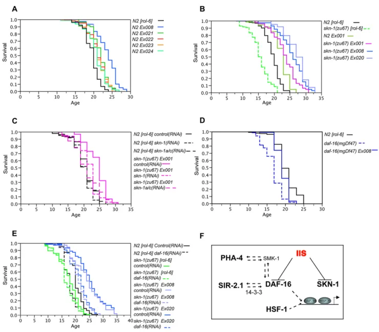
Figure 5. Transgenic Expression of SKN-1 Extends Life Span
(A) Life-span extension in otherwise WT animals carrying theSKN-1B/C S393A::GFPtransgene, which expresses constitutively nuclear SKN-1C in the intestine (see text). These independent extrachromosomal arrays were created by injecting 2.5 ng/ml transgene DNA.
(B) Life-span extension inskn-1(zu67)animals carryingskn-1transgenes. TheEx001[SKN-1B/C::GFP],Ex008[SKN-1B/C S393A::GFP], andEx020[SKN-1B/C S393A::GFP]extrachromosomal arrays were originally created in WT animals by injection of 2.5, 2.5, and 10 ng/ml transgene DNA, respectively, and then
bred intoskn-1(zu67). The presence of the transgene did not dramatically affect the developmental timing, brood size, or age-specific fecundity of either the WT orskn-1mutants (Figure S14). The life spans of WT animals carryingEx001[SKN-1B/C::GFP]is shown for comparison.
(C) SKN-1C contributes to the life span ofskn-1(zu67) Ex001[SKN-1B/C::GFP]animals. Life span is reduced comparably byskn-1andskn-1a/cRNAi (Figure 1B). (D) Presence ofEx008[SKN-1B/C S393A::GFP]increases the life span ofdaf-16(mgDf47)mutants.
(E) Transgene arrays that express constitutively nuclear SKN-1 (Ex008orEx020)increaseskn-1(zu67)life span beyond WT whendaf-16is depleted by RNAi. Life-span analyses were scored from hatching and performed at 20C unless otherwise stated and are summarized inTable 2. The experiments in (A) and (B) were carried out on OP50, and the others on HT115. (C) and (D) are representative experiments, but otherwise data were combined from at least two experiments, within which all sample sets were analyzed in parallel.
simultaneous overexpression of both transcription factors is nec-essary, although modifications of these proteins or other signals associated with decreased IIS might also be required. Our obser-vation thatdaf-16RNAi suppressed life-span extension by the WT
SKN-1B/C::GFPtransgene (Figure S13) suggests that DAF-16 may influence processes that regulate SKN-1. This could involve DAF-16 regulating genes that affect SKN-1, but a simpler model is that SKN-1 and DAF-16 compete for binding to negative regula-tors that include AKT-1, -2, and SGK-1 (Figure 1A). Thus, decreas-ing DAF-16 levels would increase the ratio of negative regulators available to sequester SKN-1 in the cytoplasm.
Our results add SKN-1 to the small group ofC. elegans tran-scription regulators that extend longevity when expressed
trans-genically (Figure 5F). SIR-2.1, PHA-4, DAF-16, and SKN-1 or their orthologs have all been shown to influence aspects of metabolism and thereby presumably may affect the rates of damage that lead to aging (Accili and Arden, 2004; Bishop and Guarente, 2007; Kenyon, 2005; Panowski et al., 2007; van der Horst and Burgering, 2007). Importantly, SKN-1, HSF-1, and each of these other proteins have also been implicated in resis-tance to free radical or other stresses (Figure 5F; see text), supporting the idea that stress defenses and detoxification are fundamentally important for longevity. A remarkable web of func-tional interactions among these factors has been uncovered, including our demonstration here that DAF-16 and SKN-1 are regulated in parallel by IIS (Figure 5F). These interactions could
Table 2. Transgenic Expression of SKN-1 Extends Life Span
Strain
Mean Life Span±SEM 20C (days)
75th Percentile 20C (days)
p value against control n
No. of Exp.
N2 Ex[rol-6] 19.57±0.2 21 — 129/130 2
N2 Ex008[SKN-1 S393A::GFP] 2.5 ng/ml 23.62±0.3 26 <.0001a 78/78 2
N2 Ex021[SKN-1 S393A::GFP] 2.5 ng/ml 21.80±0.2 23 <.0001a 117/122 2
N2 Ex022[SKN-1 S393A::GFP] 2.5 ng/ml 21.10±0.3 23 <.0001a 62/62 2
N2 Ex023[SKN-1 S393A::GFP] 2.5 ng/ml 20.89±0.3 23 <.0001a 96/96 2
N2 Ex024[SKN-1 S393A::GFP] 2.5 ng/ml 20.66±0.4 23 <.0001a 63/63 2
N2 Ex[rol-6] 19.52±0.2 21 — 236/243 3
skn-1(zu67) Ex[rol-6] 15.51±0.3 18 <0.0001a 96/96 2
N2 Ex001[SKN 1B/C::GFP] 2.5 ng/ml 22.14±0.3 25 <.0001b,c 146/148 2
skn-1(zu67) Ex001[SKN-1B/C::GFP] 2.5 ng/ml 23.53±0.6 26 <.0001b,c 48/51 2
skn-1(zu67) Ex008[SKN-1B/C S393A::GFP] 2.5 ng/ml 25.74±0.5 29 <.0001b,c 67/70 2
skn-1(zu67) Ex020[SKN-1B/C S393A::GFP] 10 ng/ml 27.16±0.6 29 <.0001b,c 30/40 1
N2 Ex[rol-6]; control(RNAi) 20.60±0.5 23 — 50/50 1
N2 Ex[rol-6]; skn-1(RNAi) 19.91±0.4 21 0.0699f 60/60 1
N2 Ex[rol-6]; skn-1a/c(RNAi) 20.37±0.4 23 0.7880f 59/60 1
skn-1(zu67) Ex001[SKN-1B/C::GFP] 2.5 ng/ml; control(RNAi) 24.20±0.4 27 <.0001f 62/64 1
skn-1(zu67) Ex001[SKN-1B/C::GFP] 2.5 ng/ml ; skn-1(RNAi) 21.58±0.4 23 <.0001d 84/84 1
skn-1(zu67) Ex001[SKN-1B/C::GFP] 2.5 ng/ml; skn-1a/c(RNAi) 21.06±0.5 25 0.0005d 69/69 1
N2 Ex[rol-6] 20.60±0.5 23 — 50/50 1
daf-16(mgDf47) 15.87±0.3 17 <.0001f 8/61 1
daf-16(mgDf47) Ex008[SKN-1 S393A::GFP] 2.5 ng/ml 18.88±0.3 21 <.0001e 48/48 1
N2 Ex[rol-6]; control(RNAi) 20.02±0.2 22 — 256/259 4
N2 Ex[rol-6]; daf-16(RNAi) 17.77±0.1 19 <.0001f 266/266 4
skn-1(zu67) Ex[rol-6]; control(RNAi) 17.38±0.3 20 <.0001f 145/146 3
skn-1(zu67) Ex[rol-6]; daf-16(RNAi) 16.59±0.3 19 0.0240g 142/145 3
skn-1(zu67)Ex001[SKN-1b/c::GFP] 2.5 ng/ml; control(RNAi) 22.95±0.5 26 <.0001g 62/62 2
skn-1(zu67) Ex001[SKN-1b/c::GFP] 2.5 ng/ml; daf-16(RNAi) 16.53±0.3 18 0.1655h 59/59 2
skn-1(zu67) Ex008[SKN-1 S393A::GFP] 2.5 ng/ml; control(RNAi) 24.28±0.6 28 <.0001g 64/76 3
skn-1(zu67) Ex008[SKN-1 S393A::GFP] 2.5 ng/ml; daf-16(RNAi) 21.12±0.4 23 <.0001h,i 76/87 3
skn-1(zu67) Ex020[SKN-1 S393A::GFP] 10 ng/ml; control(RNAi) 25.10±0.6 28 <.0001g 121/132 3
skn-1(zu67) Ex020[SKN-1 S393A::GFP] 10 ng/ml; daf-16(RNAi) 21.39±0.4 24 <.0001h,i 124/140 3 Corresponds to the data inFigures 5A–5E. p values were calculated as follows:aN2 Ex[rol-6],bskn-1(zu67)[rol-6],csignificantly enhanced compared to
N2 [rol-6](p = < 0.0001 in each case),dskn-1(zu67) Ex001 control(RNAi),edaf-16(mgDf47) control(RNAi),fN2 Ex[rol-6] control(RNAi),gskn-1(zu67)[rol-6] control(RNAi),hskn-1(zu67)[rol-6] daf-16(RNAi),icompared toN2 Ex[rol-6] control(RNAi)(p = 0.0021 forskn-1(zu67) Ex008and p = 0.0001 for
be important for coordinating how these proteins respond in different tissues to various scenarios of nutrient availability and stress. As these regulators are also evolutionarily conserved, it seems likely that a deeper understanding of interactions among them will be broadly relevant to elucidating how these proteins influence metabolism, stress resistance, and possibly longevity across species.
EXPERIMENTAL PROCEDURES
AllSupplemental Dataand experimental procedures, including details of clon-ing, strains, RNAi, GFP scoring system, and assays for life span, movement, and reproduction are available online.
In Vitro Kinase Assay
Kinases were isolated from sonicated worm lysates ofN2 Ex[AKT-1::GFP], N2 Ex[AKT-2::GFP], andN2 Ex[AKT-1::GFP](Hertweck et al., 2004), using a GFP antibody bound to protein A/G agarose beads. These purified kinases were used to phosphorylate 6 mg of bacterially expressed GST-SKN-1 fusion
proteins in the presence of 6mCi [gP32]ATP. After phosphorylation, samples
were washed in 50 mM Tris/HCl (pH 8.0), 100 mM NaCl, 10% glycerol, and 1% Triton X-100 and analyzed by SDS-PAGE and autoradiography.
RNAi
Feeding RNAi was carried out as previously described, with empty pL4440 as the control (Supplemental Experimental Procedures). To avoid maternal lethal-ity in experiments usingskn-1orskn-1a/cRNAi, animals were hatched on HT115 bacteria expressing control RNAi and were transferred toskn-1RNAi at the L1 stage or on the first day of adulthood. For life-span experiments that required feeding of other RNAi, animals were exposed to bacteria expressing the appropriate construct continually starting from hatching.
Life-Span Analysis
Prior to experiments, all animals were maintained at the permissive tempera-ture and grown for at least two generations in the presence of food to assure health. Life-span assays were performed essentially as described (Hsin and Kenyon, 1999; Supplemental Experimental Procedures). Survival plots, p values (Log-Rank), and proportional hazards were determined using JMP software, version 5.1.
Oxidative Stress Resistance Assay
To assess stress resistance, young adults were transferred to plates that contained 7.5 mMt-butyl hydrogen peroxide (Sigma) or 150 mM paraquat (Sigma) in nematode growth media. These plates had been seeded with OP50 or HT115 expressing the appropriate dsRNAs. Animals were incubated on these plates at 20C and periodically scored for survival.
RNA Isolation and Quantitative PCR
Animals were picked to clean plates to minimize contamination, then total RNA was extracted from approximately 200 animals suspended in 50ml M9. RNA
was extracted using Trizol (Sigma) and cDNA was synthesized using the Invi-trogen Superscript Reverse Transcriptase Kit. SYBR Green Real Time PCR was carried out using the ABI 7700 and normalized toact-1. Primer sequences are available on request.
SUPPLEMENTAL DATA
Supplemental Data include Experimental Procedures, six tables, and fourteen figures and can be found with this article online athttp://www.cell.com/cgi/ content/full/132/6/1025/DC1/.
ACKNOWLEDGMENTS
We thank Siu Sylvia Lee, Gary Ruvkun, and Laura Mitic for generously provid-ing advice and strains; Stacey Robida, Anne Oelmann, Jeeyong Lee, and
Michael Lucke for invaluable technical support; and members of the Blackwell lab, Javier Apfeld, and Rohit Kulkarni for critically reading this manuscript. Work was supported by funding from the NIH (grant number 2 R01 GM062891) and Iacocca Foundation (T.K.B.), the Fonds der Chemischen In-dustrie, BMBF FRISYS, Qualitaetsoffensive BW, and Deutsche Forschungsge-meinschaft CRC746 (R.B.), and EC 6thFramework Network of Excellence
LIFE-SPAN (LSHG-CT-2007-036894) (R.B. and M.H.), as well as an NRSA (number 5 F32 GM070088-02) (R.P.O.), an NIH training grant (number T32 DK07260) (J.B.), and KRF (2006-353-C00035) and MOST/KOSEF(R112000078010010) (J.H.A.).
Received: June 29, 2007 Revised: December 18, 2007 Accepted: January 18, 2008 Published: March 20, 2008
REFERENCES
Accili, D., and Arden, K.C. (2004). FoxOs at the crossroads of cellular metabolism, differentiation, and transformation. Cell117, 421–426.
An, J.H., and Blackwell, T.K. (2003). SKN-1 links C. elegans mesendodermal specification to a conserved oxidative stress response. Genes Dev. 17, 1882–1893.
An, J.H., Vranas, K., Lucke, M., Inoue, H., Hisamoto, N., Matsumoto, K., and Blackwell, T.K. (2005). Regulation of the Caenorhabditis elegans oxidative stress defense protein SKN-1 by glycogen synthase kinase-3. Proc. Natl. Acad. Sci. USA102, 16275–16280.
Antebi, A. (2007). Genetics of aging in Caenorhabditis elegans. PLoS Genet.3, 1565–1571. 10.1371/journal.pgen.0030129.
Arantes-Oliveira, N., Apfeld, J., Dillin, A., and Kenyon, C. (2002). Regulation of life-span by germ-line stem cells in Caenorhabditis elegans. Science295, 502–505.
Berdichevsky, A., Viswanathan, M., Horvitz, H.R., and Guarente, L. (2006).C. elegansSIR-2.1 interacts with 14–3-3 proteins to activate DAF-16 and extend life span. Cell125, 1165–1177.
Bishop, N.A., and Guarente, L. (2007). Two neurons mediate diet-restriction-induced longevity in C. elegans. Nature447, 545–549.
Bowerman, B., Eaton, B.A., and Priess, J.R. (1992). skn-1, a maternally expressed gene required to specify the fate of ventral blastomeres in the early
C. elegansembryo. Cell68, 1061–1075.
Brunet, A., Park, J., Tran, H., Hu, L.S., Hemmings, B.A., and Greenberg, M.E. (2001). Protein kinase SGK mediates survival signals by phosphorylating the forkhead transcription factor FKHRL1 (FOXO3a). Mol. Cell. Biol.21, 952–965.
Cahill, C.M., Tzivion, G., Nasrin, N., Ogg, S., Dore, J., Ruvkun, G., and Alexander-Bridges, M. (2001). Phosphatidylinositol 3-kinase signaling inhibits DAF-16 DNA binding and function via 14–3-3-dependent and 14–3-3-independent pathways. J. Biol. Chem.276, 13402–13410.
Dillin, A., Hsu, A.L., Arantes-Oliveira, N., Lehrer-Graiwer, J., Hsin, H., Fraser, A.G., Kamath, R.S., Ahringer, J., and Kenyon, C. (2002). Rates of behavior and aging specified by mitochondrial function during development. Science
298, 2398–2401.
Finkel, T., and Holbrook, N.J. (2000). Oxidants, oxidative stress and the biology of ageing. Nature408, 239–247.
Friedman, D.B., and Johnson, T.E. (1988). A mutation in the age-1 gene in Caenorhabditis elegans lengthens life and reduces hermaphrodite fertility. Genetics118, 75–86.
Gems, D., Sutton, A.J., Sundermeyer, M.L., Albert, P.S., King, K.V., Edgley, M.L., Larsen, P.L., and Riddle, D.L. (1998). Two pleiotropic classes of daf-2 mutation affect larval arrest, adult behavior, reproduction and longevity in Caenorhabditis elegans. Genetics150, 129–155.
Gottlieb, S., and Ruvkun, G. (1994). daf-2, daf-16 and daf-23: genetically inter-acting genes controlling Dauer formation in Caenorhabditis elegans. Genetics
137, 107–120.
Greer, E.L., Dowlatshahi, D., Banko, M.R., Villen, J., Hoang, K., Blanchard, D., Gygi, S.P., and Brunet, A. (2007). An AMPK-FOXO pathway mediates longevity induced by a novel method of dietary restriction inC. elegans. Curr. Biol.17, 1646–1656.
Hayes, J.D., and McMahon, M. (2001). Molecular basis for the contribution of the antioxidant responsive element to cancer chemoprevention. Cancer Lett.
174, 103–113.
Henderson, S.T., and Johnson, T.E. (2001). daf-16 integrates developmental and environmental inputs to mediate aging in the nematodeCaenorhabditis elegans. Curr. Biol.11, 1975–1980.
Hertweck, M., Gobel, C., and Baumeister, R. (2004).C. elegansSGK-1 is the critical component in the Akt/PKB kinase complex to control stress response and life span. Dev. Cell6, 577–588.
Houthoofd, K., Braeckman, B.P., Lenaerts, I., Brys, K., De Vreese, A., Van Eygen, S., and Vanfleteren, J.R. (2002). Axenic growth up-regulates mass-specific metabolic rate, stress resistance, and extends life span in Caenorhabditis elegans. Exp. Gerontol.37, 1371–1378.
Houthoofd, K., Braeckman, B.P., Johnson, T.E., and Vanfleteren, J.R. (2003). Life extension via dietary restriction is independent of the Ins/IGF-1 signalling pathway in Caenorhabditis elegans. Exp. Gerontol.38, 947–954.
Hsin, H., and Kenyon, C. (1999). Signals from the reproductive system regulate the lifespan of C. elegans. Nature399, 362–366.
Hsu, A.L., Murphy, C.T., and Kenyon, C. (2003). Regulation of aging and age-related disease by DAF-16 and heat-shock factor. Science300, 1142–1145.
Inoue, H., Hisamoto, N., An, J.H., Oliveira, R.P., Nishida, E., Blackwell, T.K., and Matsumoto, K. (2005). The C. elegans p38 MAPK pathway regulates nuclear localization of the transcription factor SKN-1 in oxidative stress response. Genes Dev.19, 2278–2283.
Kell, A., Ventura, N., Kahn, N., and Johnson, T.E. (2007). Activation of SKN-1 by novel kinases in Caenorhabditis elegans. Free Radic. Biol. Med. 43, 1560–1566.
Kenyon, C. (2005). The plasticity of aging: insights from long-lived mutants. Cell120, 449–460.
Kenyon, C., Chang, J., Gensch, E., Rudner, A., and Tabtiang, R. (1993). A C. elegans mutant that lives twice as long as wild type. Nature366, 461–464.
Kimura, K.D., Tissenbaum, H.A., Liu, Y., and Ruvkun, G. (1997). daf-2, an insu-lin receptor-like gene that regulates longevity and diapause in Caenorhabditis elegans. Science277, 942–946.
Lakowski, B., and Hekimi, S. (1996). Determination of life-span in Caenorhab-ditis elegans by four clock genes. Science272, 1010–1013.
Larsen, P.L. (1993). Aging and resistance to oxidative damage in Caenorhab-ditis elegans. Proc. Natl. Acad. Sci. USA90, 8905–8909.
Lee, S.S., Lee, R.Y., Fraser, A.G., Kamath, R.S., Ahringer, J., and Ruvkun, G. (2003). A systematic RNAi screen identifies a critical role for mitochondria in C. elegans longevity. Nat. Genet.33, 40–48.
Lin, K., Hsin, H., Libina, N., and Kenyon, C. (2001). Regulation of the Caeno-rhabditis elegans longevity protein DAF-16 by insulin/IGF-1 and germline signaling. Nat. Genet.28, 139–145.
Lithgow, G.J., and Walker, G.A. (2002). Stress resistance as a determinate of C. elegans lifespan. Mech. Ageing Dev.123, 765–771.
McMahon, M., Itoh, K., Yamamoto, M., Chanas, S.A., Henderson, C.J., McLellan, L.I., Wolf, C.R., Cavin, C., and Hayes, J.D. (2001). The Cap’n’Collar basic leucine zipper transcription factor Nrf2 (NF-E2 p45-related factor 2) controls both constitutive and inducible expression of intestinal detoxification and glutathione biosynthetic enzymes. Cancer Res.61, 3299–3307. Ogg, S., Paradis, S., Gottlieb, S., Patterson, G.I., Lee, L., Tissenbaum, H.A., and Ruvkun, G. (1997). The Fork head transcription factor DAF-16 transduces insulin-like metabolic and longevity signals in C. elegans. Nature389, 994–999. Panowski, S.H., Wolff, S., Aguilaniu, H., Durieux, J., and Dillin, A. (2007). PHA-4/Foxa mediates diet-restriction-induced longevity of C. elegans. Nature447, 550–555.
Rea, S.L., Ventura, N., and Johnson, T.E. (2007). Relationship between mitochondrial electron transport chain dysfunction, development, and life extension in Caenorhabditis elegans. PLoS Biol.5, e259. 10.1371/journal. pbio.0050259.
Simmer, F., Tijsterman, M., Parrish, S., Koushika, S.P., Nonet, M.L., Fire, A., Ahringer, J., and Plasterk, R.H. (2002). Loss of the putative RNA-directed RNA polymerase RRF-3 makes C. eleganshypersensitive to RNAi. Curr. Biol.12, 1317–1319.
Tissenbaum, H.A., and Guarente, L. (2001). Increased dosage of a sir-2 gene extends lifespan in Caenorhabditis elegans. Nature410, 227–230.
Tonks, N.K. (2005). Redox redux: revisiting PTPs and the control of cell signal-ing. Cell121, 667–670.