online | memorias.ioc.fiocruz.br
Identification of the alpha-enolase P46 in the extracellular
membrane vesicles of
Bacteroides fragilis
Thais Gonçalves Ferreira1, Camilla Nunes dos Reis Trindade1, Petra Bell6, André Teixeira-Ferreira2,3, Jonas E Perales2,3, Rossiane C Vommaro4, Regina Maria Cavalcanti Pilotto Domingues1/+, Eliane de Oliveira Ferreira1,5
1Universidade Federal do Rio de Janeiro, Instituto de Microbiologia Paulo de Góes, Departamento de Microbiologia Médica, Laboratório
de Biologia de Anaeróbios, Rio de Janeiro, RJ, Brasil
2Fundação Oswaldo Cruz-Fiocruz, Instituto Oswaldo Cruz, Laboratório de Toxinologia, Rio de Janeiro, RJ, Brasil 3Rede Proteômica do Rio de Janeiro, Rio de Janeiro, RJ, Brasil
4Universidade Federal do Rio de Janeiro, Instituto de Biofísica Carlos Chagas Filho, Laboratório de Ultraestrutura Celular Hertha Meyer,
Rio de Janeiro, RJ, Brasil
5Universidade Federal do Rio de Janeiro, Duque de Caxias, RJ, Brasil
6University of Leeds, Faculty of Biological Sciences, School of Biology, Leeds, UK
BACKGROUND Members of the Bacteroides fragilis group are the most important components of the normal human gut microbiome, but are also major opportunistic pathogens that are responsible for significant mortality, especially in the case of bacteraemia and other severe infections, such as intra-abdominal abscesses. Up to now, several virulence factors have been described that might explain the involvement of B. fragilis in these infections. The secretion of extracellular membrane vesicles (EMVs) has been proposed to play a role in pathogenesis and symbiosis in gram-negative bacteria, by releasing soluble proteins and other molecules. In B. fragilis, these vesicles are known to have haemagglutination and sialidosis activities, and also contain a capsular polysaccharide (PSA), although their involvement in virulence is still not clear.
OBJECTIVE The aim of this study was to identify proteins in the EMV of the 638R B. fragilis strain by mass spectrometry, and also to assess for the presence of Bfp60, a surface plasminogen (Plg) activator, previously shown in B. fragilis to be responsible for the conversion of inactive Plg to active plasmin, which can also bind to laminin-1.
METHODS B. fragilis was cultured in a minimum defined media and EMVs were obtained by differential centrifugation, ultracentrifugation, and filtration. The purified EMVs were observed by both transmission electron microscopy (TEM) and immunoelectron microscopy (IM). To identify EMV constituent proteins, EMVs were separated by 1D SDS-PAGE and proteomic analysis of proteins sized 35 kDa to approximately 65 kDa was performed using mass spectrometry (MALDI-TOF MS).
FINDINGS TEM micrographs proved the presence of spherical vesicles and IM confirmed the presence of Bfp60 protein on their surface. Mass spectrometry identified 23 proteins with high confidence. One of the proteins from the B. fragilis EMVs was identified as an enolase P46 with a possible lyase activity.
MAIN CONCLUSIONS Although the Bfp60 protein was not detected by proteomics, α-enolase P46 was found to be present in the EMVs of B. fragilis. The P46 protein has been previously described to be present in the outer membrane of B. fragilis as an iron-regulated protein.
Key words: proteomics - Bacteroides fragilis - extracellular membrane vesicles - α-enolase
doi: 10.1590/0074-02760170340 Financial support: FAPERJ, CNPq
+ Corresponding author: [email protected] Received 31 August 2017
Accepted 17 November 2017
Extracellular membrane vesicles (EMVs) are formed by many microorganisms, spanning both prokaryotes (gram-positive and negative bacteria) and eukaryotes (Chernov et al. 2014). Protein secretion into the extracellular environ-ment is a fundaenviron-mental process in the bacterial communi-cation. There are several secretion pathways described in gram-negative bacteria that can promote the delivery of toxins and other specific proteins to host cells. Among them, are the secretion type III (TSS3) system, for exam-ple, which after contact with the cell surface and assembly, can inject effector proteins into host cells. The release of
vesicles is involved in the response to environmental stress, virulence factors, the secretion of components destined for the cell surface, antigens, and in the case of pathogens, for host interaction (Deatherage & Cookson 2012).
These vesicles, ranging from 20 to 250 nm in diam-eter, contain components of the cell wall and outer mem-brane (OM), flagellin, lipopolysaccharide, cytosine, and pathogen associated molecular patterns (PAMPS) that can activate significantly inflammatory immune responses (Kuehn 2012). In fact, they have provided an alternative for producing acellular vaccines (Avila-Calderón et al. 2012) and this has proven to be effective in the specific case of serogroup B of Neisseria meningitidis (Holst et al. 2009).
flo-ra (Qin et al. 2012), Bacteroides fflo-ragilis contributes to T helper cell development in particular. However, as a pathogen, it also causes severe infections (abscesses and peritonitis), and is the most frequently isolated gram-negative anaerobic bacterium found in clinical infec-tions (Patrick & Blakely 2012). Several virulence factors have been described in B. fragilis to explain this duality within its host. Among them are proteases (Patrick et al. 1996), enterotoxin (Wu et al. 1998), and lipopolysac-charide (Pumbwe et al. 2006), but their roles in pathoge-nicity have not been elucidated. A capsular polysaccha-ride (PSA) in the large capsule polysacchapolysaccha-ride complex (CPC), produced by B. fragilis, has also been shown to have an immunomodulatory effect and to prevent colitis (Shen et al. 2012). PSA is a large molecule, so it has been proposed by Shen et al. (2012) that PSA is released in EMVs and helps to induce immunomodulatory effects and prevent colitis in an animal model. The mechanism by which PSA is delivered to the immune system is still unclear. PSA is however not the only molecule present in EMVs; Patrick et al. (1996) have described the presence of a hemagglutination and enzymatic activity in EMVs, and Domingues et al. (1997) have described a sialidase activity. In 2014, the first antimicrobial molecule, re-ferred to as BSAP-1 (Bacteroidales Secreted Antimicro-bial Protein-1), was identified that promotes interference among Bacteroidales strains. BSAP-1 is released in B. fragilis EMVs, and contains a membrane attack com-plex/perforin (MACPF) domain, demonstrating that se-creted molecules can promote competitive interference among human gut bacteria (Chatzidaki-Livanis et al. 2014). More recently, Wilson et al. (2015) have identi-fied several putative secretion mechanisms in the outer membrane (OM) of B. fragilis, including a family of au-totransporters, multiple potential type I secretion system proteins, and possible type VI secretion system proteins.
Bacteria have evolved several virulence strategies to interact with host factors, such as plasminogen (Sun 2006). The fibrinolytic system comprises, among other proteins, plasminogen (Plg), an abundant component of blood that is the zymogenic form of the serine protease plasmin. More than 40 different proteins have been im-plicated as Plg receptors in pathogenic and commensal bacteria (Sanderson-Smith et al. 2012). In B. fragilis, a Plg cell-surface binding protein p60, referred to as Bfp60, was characterized (Ferreira et al. 2013) and shown to be responsible for the conversion of Plg into plasmin and also for laminin-1 recognition. Therefore, the aim of this study was to identify components of the EMVs in B. fra-gilis, using a proteomics approach, and to assess whether Bfp60 was present in these subcellular structures.
MATERIALS AND METHODS
Bacterial culture - The clinical isolate 638R from the rifamycin-resistant B. fragilis strain (Privitera et al. 1979) was used in this study. The strain was routinely grown anaerobically in a chamber containing H2 (10%), N2 (80%), and CO2 (10%), using brain heart infusion broth supplemented (BHIS) with hemin (5 mg/mL) and L-cysteine (0.5 g/L) (Jousiemies-Somier et al. 2002). To isolated EMVs, an inoculum was made in a minimum
defined medium (MDM) broth containing per litre, 2 g (NH4)2SO4; 0.5 g sodium citrate; 5 mg vitamin B12; 7 g KH2PO4; 8 g K2HPO4; 10 mg MnCl2.4H2O; 20 mg MgCl2.2H2O; 0.3 mg FeCl3.6H2O; 30 mg CaCl2.2H2O; 4 g NaHCO3; 0.5 g cysteine HCl; 10 g glucose; 5 mg hemin; and 1 mg resazurin (Patrick et al. 1996).
Purification of EMVs - In order to obtain a culture en-riched in EMVs (Patrick et al. 1996), bacteria were inocu-lated in MDM broth (500 mL), and the culture was main-tained until the late exponential-phase was reached. The culture was then centrifuged at 7245 × g using a Beckman GS-6R centrifuge to pellet the bacterial cells. The superna-tant fraction was then passed through a 0.45 µm pore-mem-brane (Millipore) to remove any residual cells, followed by a 0.22 µm polyvinylidene difluoride (PVDF) filter (Mil-lipore). The filtered supernatant was then centrifuged at 30790 × g for 4 h using a Sorvall Ultra Pro 80 ultracentri-fuge. The supernatant fraction removed and the pellet re-suspended in 1 mL of phosphate buffered saline (0.01 M), pH 7.4 (PBS). The presence of EMVs and the absence of bacterial cells were confirmed by electron microscopy.
Transmission electron microscopy (TEM) analysis - Electron microscopy of the EMVs was performed ac-cording to the method of Théry et al. (2006). Briefly, 10 µL of the purified vesicles from B. fragilis was al-lowed to settle onto formvar-carbon coated copper grids (300 mesh) for 25 min. After that, the grids were washed twice with phosphate buffered saline (PBS) and fixed with glutaraldehyde (Type I; Sigma-Aldrich Co.) pre-pared in 0.1 M sodium cacodylate buffer. The grids were negatively stained with 4% uranyl acetate and 2% meth-ylcellulose (1:9) and observed using a Zeiss 900 TEM.
For immunoelectron microscopy, the grids were fixed with 2% paraformaldehyde in PBS for 30 min in a fume hood at room temperature (RT). Following this, the grids were washed four times with PBS containing 100 mM gly-cine and blocked with 3% bovine serum albumin (BSA, Sigma-Aldrich Co), for 10 min at RT. Before labelling with the antibodies, a solution of 1% BSA and 0.1% saponin in PBS was used to permeabilise the cells for 20 min at RT. The grids were placed in a humidity chamber and incu-bated with the primary antibody (polyclonal rabbit anti-Bfp60) in PBS containing 1% BSA and 0.1% saponin for 30 min at RT. Grids were washed six times with PBS con-taining 0.2% BSA and 0.1% saponin, and incubated with a secondary antibody conjugated to 5 nm gold particles (BB International) in PBS containing 0.2% BSA and 0.1% sapo-nin for 1 h. After washing eight times with PBS, the grids were fixed in 1% glutaraldehyde for 5 min and washed eight times with water. Grids were negatively stained with 4% uranyl acetate and 2% methylcellulose (1:9) at 4ºC (Théry et al. 2006) and observed with a Zeiss 900 TEM.
bis-acrylamide gel (4% stacking; 12% separating) in a Tris-glycine running buffer (3 g/L Tris, 72 g/L glycine,
and 5 g/L SDS) (Laemmli 1970). Protein samples (10 μg)
were mixed with sample buffer (Tris-HCl, pH 6.8, 10% SDS, 10% glycerol, 5% 2-b-mercaptoethanol and 0.05% bromophenol blue; Sigma), boiled (100ºC) for 5 min, and applied to the SDS-PAGE gel. Protein standards (Spec-tra Multicolor Broad Range Protein Ladder; Invitrogen) were used as molecular weight markers. The gel was sub-sequently stained with colloidal Coomassie Blue G-250. Proteins corresponding to a molecular mass range of 35 kDa to approximately 65 kDa were excised from the gel and processed for proteomic analyses (Section 2.5).
For western blotting (Ferreira et al. 2009), proteins were separated by SDS-PAGE and transferred to a nitro-cellulose membrane overnight at 4ºC (30 V; 40 mA). The PVDF membrane containing the blotted proteins was washed three times with PBS (0.01 M * , 150 mM NaCl) and blocked with PBST (0.03% Tween 20) containing 5% skimmed milk overnight at 4ºC. After washing three times with PBST (0.3% Tween 20), the membrane was incubated with the primary antibody anti-Bfp60 (1:100 for 2 h at RT. The membrane was washed three times with PBST (0.3% Tween 20) and incubated with the sec-ondary antibody anti-rabbit IgG conjugated with peroxi-dase (1:5000; Sigma) in PBST (0.3% Tween 20) for 1 h at RT. The membrane was then washed three times with PBS and immunoreactive bands were detected by the ad-dition of 50 mg DAB (3,3’diaminobenzidine) dissolved in 30% H2O2 in 0.01 M PBS. To stop the reaction, the membrane was washed with distilled water.
In-gel- digestion: mass spectrometry analysis - After proteins were separated by one-dimensional (1D) SDS-PAGE, bands were excised from the gel and destained in 50% acetonitrile/25 mM ammonium bicarbonate pH 8.0, until no blue colour remained. The gel bands were dehy-drated with two incubations of 5 min each in 100% ace-tonitrile, and air-dried for 10 min, followed by a reduc-tion in 65 mM DTT (30 min at 56ºC) and alkylareduc-tion in 200 mM iodoacetamide (30 min in the dark). Following this, the bands were hydrated with 100 mM ammonium bicarbonate (10 min) and dehydrated twice with 100% acetonitrile (5 min each time), and dried using a Speed-vac (Thermo Scientific™). Digestion was carried out at 37ºC overnight in 40 mM ammonium bicarbonate
so-lution containing trypsin (20 ng/μL; Sequencing grade;
PromegaÒ). Peptides were extracted in 5% (v/v) formic
acid (TFA), 50% (v/v) acetonitrile (30 μL), with the help
of ultrasound (10 min). Samples were concentrated to 5
μL and purified with a ZipTip (C18 Millipore®) for di -rect spotting onto MALDI plates for MALDI-TOF/TOF analysis (Gharahdaghi et al. 1999).
Protein identification by mass spectrometry MAL-DI-TOF-TOF analysis was performed using a 5800
Proteomics Analyzer (ABSciex). Briefly, 0.5 μL of the micro-column eluate was mixed with 0.5 μL of alpha-cy -ano-4-hydroxycinnamic acid matrix (20 mg/mL in 50% ACN/0.1% TFA). Samples were spotted onto the ABI 192-targed MALDI plate by co-crystallisation, and mass spectrometry data were acquired in positive reflectron
mode, with a mass range 800-3.500 Da. Typically, analy-ses were conducted using 2.000 shots of MS and 4.000 shots of MS/MS to the ten most abundant ions. External calibration was performed using a mixture of four pep-tides: des-Arg1-bradykinin (m/z = 904.47), angiotensin I (m/z = 1296.69), Glu1-fibrin peptide B (m/z = 1570.68), and adrenocorticotropic hormone (18-39) (m/z = 2465.20) (mass standards kit for the 4800 Proteomics Analyzer).
Bioinformatics - Peptide sequences (protein identity) were determined by matching protein databases with the acquired fragmentation pattern using the software pro-gram MASCOT (Matrix Science), and all MS/MS spectra were searched against the entire NCBInr protein database or a modified NCBInr database created to search “fragi-lis” recognised proteins, assuming use of trypsin as the digestion enzyme, and allowing up two missed cleavages. MASCOT was searched with a product ion mass tolerance of 0.30 Da and a precursor ion tolerance of 0.60 Da. Only significant hits, as defined through MASCOT probability analysis (p < 0.05), and peptides identified with individ-ual ion scores greater than 30 were considered. Scaffold and the Blast2Go (https://www.blast2go.com/b2ghome) were used for functional annotation and analysis with the proteins identified. PsortB v. 3.0 was used to predict the subcellular localisation (www.psort.org/psortb/) and Sig-nalP to predict the presence and location of signal peptide cleavage. Kalign software in the Clustal 2.1 program was used to align the enolase protein sequences.
RESULTS
Isolation of EMVs and electron microscopy - EMVs were purified by differential centrifugation from a cell-free culture in a minimum defined medium (MDM), and subsequently pelleted by ultracentrifugation. In order to confirm purification of the EMVs from the B. fragilis 638R strain, electron micrographs of EMVs were per-formed using negative staining. TEM analysis of the pu-rified EMVs revealed them to be attached to the outer surface of B. fragilis (Fig. 1A) and consisted of spheri-cal vesicles with a pleomorphic bilayer (Fig. 1B-C). In general, the EMVs released from B. fragilis had a cir-cular shape, and were of differing sizes. We then used a polyclonal rabbit anti-Bfp60 antibody (Ferreira et al. 2009) to confirm the presence of the Bfp60 protein on the EMV membrane surface. The immuno-gold labelled TEM micrographs scattered the EMV surface mem-brane sporadically and also the cell debris (Fig. 1D). The antibody labelling was not as strong as expected, which could indicate lower quantities of Bfp60 in the EMVs or cross-reaction with another surface protein.
SDS-page and western blotting - The presence of
vesicular proteins was analysed by separating 10 μg
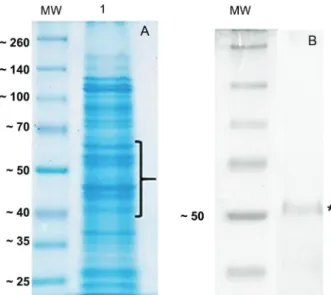
of total EMV protein by SDS-PAGE and staining with Coomassie blue colloidal stain, which showed the pres-ence of several different proteins (Fig. 2A) consistent with EMV proteins isolated from other gram-negative bacteria (Horstman & Kuehn 2000) as well as B. fragilis.
enolase, however we did not detect the Bfp60 protein in our mass spectrometry analysis. Interestingly, two proteins in the molecular function pie diagram were categorized as having lyase activity. We identified a protein [enolase (Bacteroides); gi number 492241000] that, according to the gene ontology (GO) annotation (Supplementary data, Tables I-II ), had enolase activ-ity. A protein blast search with the putative sequence against the NCBI sequence database (http://www.ncbi. nlm.nih.gov/BLAST/) revealed a 100% identity with the
P46 α-enolase previously identified in B. fragilis. In the B. fragilis EMVs this enolase had a cytoplasmic cellular location and molecular weight of approximately 46 kDa (Supplementary data, Tables I-II), the same character-istics found in the inner membrane protein fraction ob-tained from iron-depleted B. fragilis cells.
Proteomics analysis - Duplicate samples of B. fragilis 638R EMV proteins were separated using 1D SDS-PAGE and bands ranging from 40 kDa to approximately 65 kDa were excised and digested with trypsin. The extracted peptides were analysed by MALDI TOF/TOF and the data obtained were used to search a database contain-ing the B. fragilis genome. The analysis identified 23 proteins, which are listed in Supplementary data, Tables
Fig. 1: transmission electron microscopy (TEM) of the 638R Bacte-roides fragilis strain and the extracellular membrane vesicles (EMV) after negative staining with 4% uranyl acetate and 2% methylcellulose. (A) B. fragilis with EMVs attached to the cells. The asterisks show the
EMVs attached to the surface of the bacteria. Bar = 2 μm. (B-C) TEM
of the purified EMVs from the 638R strain. (B) Different sized EMVs
(arrow heads) Bar = 2 μm; (C) EMVs magnified with the plasma mem -brane well-defined (arrows). Magnification = 30,000 ×, Bar = 600 nm; (D) immunogold labelling of a preparation of purified EMVs stained with a rabbit anti-Bfp60 antibody and an anti-rabbit colloidal gold conjugate (5 nm). The arrows indicate staining for the surface protein Bfp60 in the EMVs and cell debris. Magnification = 50,000 ×, Bar = 600 nm.
Fig. 2: analysis of the protein profile derived from extracellular mem-brane vesicles (EMVs) isolated from the Bacteroides fragilis 638R strain; (A) ten micrograms of purified EMV protein were separated on a 12% SDS-PAGE gel followed by Coomassie colloidal staining (G-250). Lane 1 shows the EMV proteins profile; the brace indicates the selected bands excised from the gel and digested with trypsin. Peptides were enriched using Ziptip C18 columns and analysed by mass spectrometry (Maldi- TOF/TOF). MW indicates the molecular weight standards in kDa. (B) Western-blotting of the EMV protein extract showing the recognition of a protein (asterisk) of approximately 50 kDa by the rabbit anti-Bfp60 an-tibody. MW indicates the molecular weight standards in kDa.
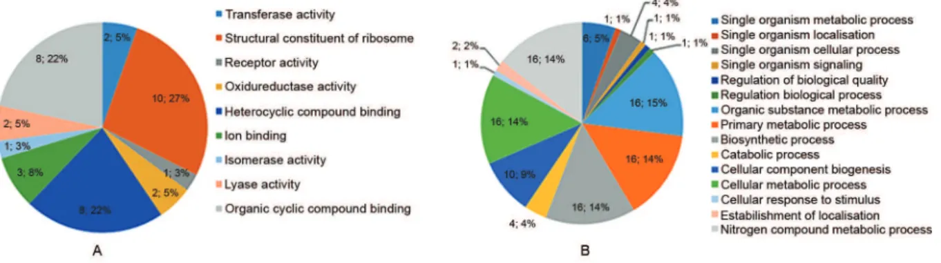
I-II. The distribution of these 23 proteins based on their biological process and molecular function is presented in Fig. 3. Based on the biological process bar chart graphic (Fig. 3B), most of the proteins are involved in binding, act as structural molecules, or possess a catalytic activity. With respect to molecular function (Fig. 3A), the majori-ty of the proteins were structural constituents of the ribo-some, which was consistent with the mass spectrometry identification (Supplementary data, Tables I-II).
All the EMV proteins identified were subjected to computer analysis using PsortB to identify their predict-ed protein localisation. The prpredict-edictpredict-ed subcellular locali-sation is shown in Fig. 4. Approximately 78.26% (18) of the proteins were classified as being cytoplasmic with 13.04% (3) being not categorised (unknown). Cytoplas-mic membrane (4.04%; 1) and outer membrane (4.04%; 1) proteins were also identified. No periplasmic proteins were identified. Of these 23 proteins, 13.04% were pre-dicted to be secreted (Supplementary data, Tables I-II). Of the proteins containing a signal peptide, three had an uncertain cellular localisation (membrane protein and hypothetic protein) and one was assigned to the outer membrane (putative exported protein).
species, including the P46 and P60 proteins sequences (Fig. 5), suggest that the anti-bfp60 antibody could
recog-nise the P46 α-enolase. In Fig. 5, the dark shaded and light
grey residues indicate identity and similarity among the amino acids in the protein sequences, respectively.
DISCUSSION
Gram-positive and negative bacteria use different types of secretion systems to transport important virulence fac-tors to the cell envelope and the extracellular milieu. One of the mechanisms employed by the bacterial cells to release proteins and mediators is through the shedding and accu-mulation of membrane vesicles (Théry et al. 2002), which seems to be important process in the intracellular crosstalk between living organisms, including bacteria.
Although several studies examining B. fragilis viru-lence factors have been conducted, there are not many examining EMVs in this species. A preliminary study conducted by Patrick et al. (1996) compared enzymatic activities, including esterase, lipase, alkaline
phospha-tase, glucosaminidase, and acid phosphatase activities in whole cells and their EMVs in two B. fragilis strains, NCTC 9343 and BE3 containing large capsule (LC) and electron-dense layer (EDL) populations. The purified EMVs exhibited both haemagglutination activity and an enzymatic activity suggesting a potential role for the EMV. Domingues et al. (1997) have shown that a sialidase activity was associated with these sub-cellular structures in all the strains analysed, suggesting that these surface components have a function in the commensal stages of B. fragilis. More recently (Shen et al. 2012), it was dis-covered that B. fragilis release PSA in EMVs, which induces immunomodulatory effects and prevent colitis in animal models. The authors imply that the EMV-me-diated delivery of a commensal molecule prevents dis-ease and provides a mechanism of inter-kingdom com-munication between the microbiota and mammals. In 2014, Elhenawy et al. (2014) performed a comparative proteomics analysis of the outer membrane vesicles of B. fragilis and B. thetaiotaomicron where they identified 40 proteins exclusively in B. fragilis EMVs. The authors found a high prevalence of glycosidases and proteases, most of the acidic proteins, and active in in vitro assays. According to their results the EMVs contained a number of hydrolytic enzymes that were found in both species. Due to the fact that B. fragilis and B. thetaiotaomicron are members of the human gut microbiota, they can de-grade a variety of glycans that are not substrates for hu-man glycosidases. So, the production of short-chain fatty acids generated by the degradation of these glycans can be beneficial to other members of the microbiota. We did not identify any glycosidases in B. fragilis EMVs, possi-bly because, Elhenawy et al. (2014) used a different broth media from ours, enriched with 0.5% glucose or fucose, and employed a different mass spectrometry protocol.
Fig. 3: distribution of the Bacteroides fragilis extracellular membrane vesicle proteins identified based on gene ontology annotations (GO). (A) Molecular function bar chart graphic and (B) Biological process pie diagram. The Blast2Go software was used to classify the proteins.
Fig. 4: distribution of subcellular locations of proteins identified in
Bacteroides fragilis extracellular membrane vesicles as determined by PSORTb.
Proteomics approaches have been used previously to determine the components of vesicles in attempts to pro-vide clues to the mechanisms of vesicle production and cargo loading (Lee et al. 2008). In this study, conventional SDS-PAGE coupled to mass spectrometry (MALDI-TOF/ TOF) was used to identify the composition of B. fragilis EMVs, which extends the list of previously known EMVs protein that are associated with B. fragilis pathogenicity.
Although most of the studies (Elmi et al. 2012, Altin-dis et al. 2014) refer to vesicles being secreted from the bacterial surface, we only observed vesicles attached to the outer membrane of bacterial cells. Spherical vesicles of different sizes were also observed in the micrographs, but no measure of size distribution was made. A mem-brane lipid bilayer was also observed in the purified EMVs, as has been observed in the EMVs derived from other bacterial species. To check whether the Bfp60 pro-tein was present in the EMVs a polyclonal rabbit anti-Bfp60 enolase antibody was used. The antibody was pro-duced by immunising rabbits with the recombinant Bfp60 enolase protein (Ferreira et al. 2013). Using this antibody, images showed gold labelling on the EMV surface, with gold particles labelling the bacteria EMV, as well as cell debris. Western blotting of total EMV proteins showed a strong recognition of a protein with a mass slightly great-er than the 50 kDa molecular weight markgreat-er.
Using mass spectrometry we have identified 23 EMV proteins, covering approximately a 35 kDa to 70 kDa mo-lecular weight range. The EMV proteins identified (GI number) and their putative subcellular localisation are listed in Supplementary data, Tables I-II. These proteins were predicted to be involved in transport activity, nutri-tion, and metabolism, which is consistent with most of the EMVs functions previously identified in bacteria (Elmi et al. 2012). Their predicted subcellular localisations were quite diverse, with proteins predicted to be in the cyto-plasmic membrane, the cytocyto-plasmic fraction and the outer membrane. In our analysis, we also obtained evidence for the presence of ribosomal proteins which appeared to be a contamination of the vesicles. We speculate that their presence is due to our protocol of extraction which did not use a density gradient during the purification.
Although numerous studies have described the pres-ence of periplasmic proteins in bacterial EMVs, we did not identify any in this study. This result might be related to the fact that only a limited range of proteins were used in our study, or that there are no periplasmic proteins in B. fragilis EMVs. In support of this latter conclusion, a comparative proteomic study of EMVs in B. fragilis and B. thetaiotaomicron (Elhenawy et al. 2014) did not identify any periplasmic proteins, and instead most of the putative proteins identified by MS/MS in both
or-ganisms were putative hydrolases, such as β-glucosidase and β-galactosidase. On the other hand, as previously
mentioned, no putative hydrolases were identified in our study. Elhenawy et al. (2014) used a basal medium sup-plemented with either glucose or fucose. The authors did not give any explanation as to why a supplemented media was used, and there was no reference given to support its specific use, but we assume that the presence of glucose or fucose in the broth might have influenced the produc-tion of hydrolases by B. fragilis and B. thetaiotaomicron.
EMVs are known to contain a full complement of surface antigens, secretory proteins, and toxins (Death-erage & Cookson 2012). The present study tried to iden-tify the presence of an outer membrane protein, Bfp60 (p60) which has been characterized as an enolase, in B. fragilis. The proteomics analysis that we performed iden-tified several proteins in the B. fragilis EMVs; among these was the P46 enolase. Enolase is an ancient and ubiquitous metalloenzyme, which catalyses the revers-ible dehydration of 2 phospho D-glycerate into phospho-enolpyruvate during glycolysis. Eubacteria and archae-bacteria both contain a single enolase gene and therefore multiple molecular forms (isoforms) are not observed. Overall, enolase sequences are highly conserved, show-ing a roughly 40% amino acid identity across eukary-otes, eubacteria, and archaebacteria, although a large number of insertions and deletions occur across phyla. When enolases from different species (Homo sapiens, Mus musculus, Escherichia coli, and B. fragilis) were aligned, P46 showed a good degree of amino acid simi-larity with the human, mice, and E. coli sequences. In contrast, the Bfp60 amino acid sequence did not show the same similarity, although some conserved regions (homology) could explain why the polyclonal antibody recognised a protein by western blotting. Enolases are
dimeric hydrolases encoded by three genes, alpha (α), beta (β), and gamma (γ) (Brown & Doolittle 1997). These
enzymes were initially studied only in neuronal, cancer-ous, and hematopoietic cells (Nakagima et al. 1994).
How α-enolase is transported through the cell membrane
and sorted to the cell surface without the presence of a signal sequence remains intriguing (Muesch et al. 1990).
The presence of α-enolase at the cell surface, where it
acts as a receptor and not an enzyme is not surprising because there is an ever-growing list of proteins which are targeted to more than one location in both prokary-otes and eukaryprokary-otes with variable biological functions.
In conclusion, we have identified 23 B. fragilis EMVs proteins, to our knowledge, the most comprehensive pro-teomic analysis related to this structure. Although we did not identify the Bfp60 protein, we believe that with fur-ther studies, B. fragilis virulence factors secreted within these EMVs will help us to comprehend the participation of those EMVs contents during pathogenesis, modula-tion of the host cell response to B. fragilis infecmodula-tion, and the establishment and maintenance of the species in the human microbiota. In addition, we have provided new insights in a biological field that is poorly characterised, namely that of commensal bacteria which are a major component of the human gut microbiome.
ACKNOWLEDGEMENTS
To the Rede de Plataformas Tecnológicas do Programa de Desenvolvimento Tecnológico em Insumos para Saúde (PDTIS). We thank Joaquim Santos and Semiramis Castro for their technical support.
AUTHORS’ CONTRIBUTION
manu-script; CNRJ carried out the experimental procedures; RCV performed the TEM methodology and analysed microscopy images; ATF and JEP provided the mass spectrometry proce-dures; RMCP conceived the study and revised the manuscript. All authors have read and approved the final manuscript.
REFERENCES
Altindis E, Fu Y, Mekalanos JJ. Proteomic analysis of vibrio choler-ae outer membrane vesicles. Proc Natl Acad Sci. 2014; 111(15): E1548-56.
Avila-Calderón ED, Lopez-Merino A, Jain N, Peralta H, López-Ville-gas EO, Sriranganathan N, et al. Characterization of outer mem-brane vesicles from Brucella melitensis and protection induced in mice. Clin Dev Immunol. 2012; 2012: 13 pp.
Brown JR, Doolittle WF. Archaea and the prokaryote-to-eukaryote transition. Microbiol Mol Biol Rev. 1997; 61(4): 456-502.
Chatzidaki-Livanis M, Coyne MJ, Comstock LE. An antimicrobial pro-tein of the gut symbiont Bacteroides fragilis with a MACPF domain of host immune proteins. Mol Microbiol. 2014; 94(6): 1361-74.
Chernov VM, Mouzykantov AA, Baranova NB, Medvedeva ES, Grygorieva TY, Trushin MV, et al. Extracellular membrane ves-icles secreted by mycoplasma Acholeplasma laidlawii PG8 are enriched in virulence proteins. J Proteomics. 2014; 110C: 117-28.
Deatherage BL, Cookson BT. Membrane vesicle release in bacteria, eukaryotes, and archaea: a conserved yet underappreciated as-pect of microbial life. Infect Immun. 2012; 80: 1948-57.
Domingues RM, de Souza WG, Moraes S, Avelar KE, Hirata Jr R. Fonseca ME, et al. Surface vesicles: a possible function in com-mensal relations of Bacteroides fragilis. Zentralbl Bakteriol. 1997; 285(4): 509-17.
Elhenawy W, Debelyy MO, Feldman MF. Preferential packing of acidid glycosidases and proteases into Bacteroides outer mem-brane vesicles. MBio. 2014; 5(2): e00909-14.
Elmi A, Watson E, Sandu P, Gundogdu O, Mills DC, Inglis NF, et al.
Campylobacter jejuni outer membrane vesicles play an impor-tant role in bacterial interactions with human intestinal epithelial cells. Infect Immun. 2012; 80(12): 4089-98.
Ferreira EO, Carvalho JB, Peixoto RJM, Lobo LA, Zingali RB, Smith CJ, et al. The interaction of Bacteroides fragilis with components of the human fibrinolytic system. FEMS Immunol Med Micro-biol. 2009; 56(1): 48-55.
Ferreira EO, Teixeira FL, Cordeiro F, Lobo AL, Rocha ER, Smith JC, et al. The Bfp60 surface adhesin is an extracellular matrix and plasminogen protein interacting in Bacteroides fragilis. Int J Med Microbiol. 2013; 303(8): 492-7.
Gharahdaghi F, Weinberg CR, Meagher DA, Imai BS, Mische SM. Mass spectrometric identification of proteins from silver-stained polyacrylamide gel: a method for the removal of silver ions to enhance sensitivity. Electrophoresis. 1999; 20(3): 601-5.
Holst J, Martin D, Arnold R, Huergo CC, Oster P, O’Hallahan J, et al. Properties and clinical performance of vaccines containing outer membrane vesicles from Neisseria meningitidis.Vaccine. 2009; 27(Suppl. 2): B3-12.
Horstman AL, Kuehn MJ. Enterotoxigenic Escherichia coli secretes active heat-labile enterotoxin via outer membrane vesicles. J Biol Chem. 2000; 275(17): 12489-96.
Jousiemies-Somier HR, Summanen P, Citron DM, Baron EJ, Wexler HM, Finegold FM. Wadsworth anaerobes bacteriology manual. 6th ed. Belmont: Star Publishing Company; 2002. 152 pp.
Kuehn MJ. Secreted bacterial vesicles as good Samaritans. Cell Host Microbe. 2012; 12(4): 392-3.
Laemmli UK. Cleavage of structural proteins during the assembly of the head of bacteriophage T4. Nature. 1970; 227(5259): 680-5.
Lee EY, Choi DS, Kim KP, Gho YS. Proteomics in Gram-negative bacterial outer membrane vesicles. Mass Spectrometry Reviews. 2008; 27(6): 535-55.
Muesch A, Hartmann E, Rohde K, Rubartelli A. A novel pathway for secretory proteins? Trends Biochem Sci. 1990; 15(3): 86-8.
Nakajima K, Hamanoue M, Takemoto N, Hattori T, Kato K, Kohsaka S. Plasminogen binds specifically to alpha-enolase on rat neu-ronal plasma membrane. J Neurochem. 1994; 63(6): 2048-57.
Patrick S, Blakely GW. Crossing the eukaryote-prokaryote divide: a ubiquitin homolog in the human commensal bacterium Bacte-roides fragilis. Mob Genet Elements. 2012; 2(3): 149-51.
Patrick S, McKenna JP, O’Hagan S, Dermott E. A comparison of the haemagglutinating and enzymic activities of Bacteroides fragilis
whole cells and outer membrane vesicles. Microb Pathog. 1996: 20(4): 191-202.
Privitera G, Sebald M, Fayolle F. Common regulatory mechanism of expression and conjugative ability of a tetracycline resistance plasmid in Bacteroides fragilis. Nature. 1979; 278(5705): 657-9.
Pumbwe L, Skilbeck CA, Wexler HM. The Bacteroides fragilis cell envelope: quarterback, linebacker, coach-or all three? Anaerobe. 2006; 12(5-6): 211-20.
Qin J, Li Y, Cai Z, Li S, Zhu J, Zhang F, et al. A metagenome-wide association study of gut microbiota in type 2 diabetes. Nature. 2012; 490(7418): 55-60.
Sanderson-Smith ML, de Oliveira DM, Ranson M, McArthur JD. Bacterial plasminogen receptors: mediators of a multifaceted re-lationship. J Biomed Biotechnol. 2012; 2012: 272148.
Shen Y, Torchia MLG, Lawson GW, Karp CL, Ashwell JD, Mazmani-an SK. Outer membrMazmani-ane vesicles of a humMazmani-an commensal mediate immune regulation and disease protection. Cell Host Microbe. 2012; 12(4): 509-20.
Sun H. The interaction between pathogens and the host coagulation system. Physiology (Bethesda). 2006; 21: 281-8.
Théry C, Amigorena S, Raposo G, Clayton A. Isolation and charac-terization of exosomes from cell culture supernatants and bio-logical fluids. Curr Protoc Cell Biol. 2006; Chapter 3: Unit 3.22.
Théry C, Zitvogel L, Amigorena S. Exosomes: composition, biogene-sis and function. Nat Rev Immunol. 2002; 2(8): 569-79.
Wilson MM, Anderson DE, Bernstein HD. Analysis of the outer membrane proteome and secretome of Bacteroides fragilis re-veals a multiplicity of secretion mechanisms. PLoS ONE. 2015; 10(2): e0117732.