Occurrence of xyloglucan containing protuberances in the storage cell walls of
cotyledons of
Hymenaea courbaril
L.
MARCO AURÉLIO SILVA TINÉ
1, ANGELO CORTELAZZO
2and MARCOS S. BUCKERIDGE
1,3(received: Abril 12, 2000; accepted: August 2, 2000)
ABSTRACT- (Occurrence of xyloglucan containing protuberances in the storage cell walls of cotyledons ofHymenaea courbarilL.). Despite the suggestions of its pectic composition, no clear evidence for this has been presented. Here we show the occurrence of such a structure in walls of cells from cotyledons ofHymenaea courbariIL.These cells are known to accumulate large amounts of storage xyloglucan in the wall and, in this case, the protuberances seem to contain this storage polysaccharide rather than pectin. A hypothetical sequence of events leading from wall strands to protuberances was assembled based on scanning electron microscopy observations. On this basis, a tentative model for how polysaccharides are distributed into the wall, near the regions where protuberances are found, is proposed to explain the presence of storage xyloglucan in their composition.
RESUMO- (Ocorrência de protuberâncias contendo xiloglucano nas paredes celulares da reserva deHymenaea courbarilL.). Apesar das sugestões da sua composição péctica, não foram apresentadas evidências diretas da sua composição. Neste trabalho é mostrada a ocorrência desta estrutura em paredes de células parenquimáticas de cotilédones deHymenaea courbarilL. Sabe-se que estas células acumulam grandes quantidades de xiloglucano nas suas paredes e, nesse caso, as protuberâncias parecem conter este polissacarídeo na sua constituição. Uma seqüência de eventos que leva ao aparecimento destas protuberâncias é sugerida e um modelo de parede celular é proposto para explicar a presença de xiloglucano de reserva na composição das protuberâncias.
Key words - Xyloglucan, pectin, cell wall, protuberances,Hymenaea courbaril
Introduction
The plant cell wall is a composite formed by
three different matrices (cellulose/hemicellulose,
pectin and protein) that coexist independently. In the
plant tissues, cells are cemented together by pectic
polysaccharides forming a structure named middle
lamella (Roland et al. 1992, Carpita & Gibeaut
1993). Whereas the cellulose/hemicellulose matrix
is assumed to be responsible for the mechanical
strength of the wall, the structural protein matrix is
usually associated with wall loosening. In some
seeds, the deposition of one of these matrices is
proportionally increased, generating a great deposit
of carbohydrate which functions as a storage
com-pound.
During the formation of certain tissues, the cell
wall develops structures known as Intercellular
Pec-tic Protuberances (IPP) (Potgieter & van Wyk 1992).
According to these authors, IPPs are thought to be
associated with the formation of intercellular spaces
during tissue expansion. IPPs are widespread
throughout the plant kingdom from ferns to
angios-perms (Carr et al. 1980b, Potgieter & van Wyk
1992). Largely ignored in anatomy books, their
func-tion is not clear yet. Among the proposed funcfunc-tions,
cell adhesion, cell wall hydration and apoplastic
transport have been proposed. They were first
re-ported in the 19
thcentury (see Potgieter & van Wyk
1992 for a review), but their composition has not
been defined exactly yet. Although a pectic
compo-sition has been proposed by some authors (Carr &
Carr 1975, Carr et al. 1980a and b, Machado & Sajo
1996), a complex composition has been proposed by
others (Butterfield et al. 1981). Being part of the
plant cell wall, it is reasonable to suppose that
differ-ent cell wall polysaccharides might be presdiffer-ent.
Here, we report the occurrence of protuberances
in the storage cell walls of cotyledons of
Hymenaea
courbaril
. These walls are known to accumulate
xyloglucan, which is also named “amyloid” due to
its characteristic blue colour when impregnated with
iodine. In the protuberances of walls of
H. courbaril,
1. Seção de Fisiologia e Bioquímica de Plantas, Instituto de Botânica, Caixa Postal 4005, CEP 01061-970 São Paulo, SP, Brasil.
2. Depto. de Biologia Celular e Morfologia, Instituto de Bi-ologia, UNICAMP, Campinas, SP, Brasil.
xyloglucan has been detected with iodine whereas no
pectin was detected with toluidine blue.
Material and methods
Seeds ofHymenaea courbarilL. were provided by the Seed Department of the Institute of Botany at São Paulo (Brazil). The seeds were scarified individually, soaked in tap water overnight and planted in vermiculite. The pots were kept at 25 °C under a photoperiod of 12 h for 40 days and cotyledons were collected for microscopic analyses.
Mesophyll tissues from cotyledons were dissected in order to produce cuboid blocks measuring approximately 5 mm length at all sides. The blocks were fixed withp-formaldehyde (4%) /glutaraldehyde (2.5%) in phosphate buffer 0.1 M pH 7.4 for 24 h at 5 °C, washed in the same buffer, dehydrated in a series of ethanol (70, 80, 95, 100%, successively) at room temperature, included in Paraplast and 8µm sections were obtained (Kierman 1981). For iodine impregnation, the sections were covered with a solution of I2/KI (0.5%/1%) and photographed immediately. For neutral carbohydrate staining, periodic acid-Schiff (PAS) method was used according to Cortelazzo (1992). For total anionic radi-cals, toluidine blue was used according to Vidal (1977).
For Scanning Electron Microscopy (SEM), pieces of coty-ledons were mounted on stubs, freeze-dried, coated with gold (Baltec SCD 050 coater), examined, and photographed in a Philips Scanning Electron Microscope XL20 at an acceleration voltage of 10 kV.
Results and Discussion
Cell wall protuberances were observed in the
intercellular spaces of the storage parenchyma from
seeds of
Hymenaea courbaril
(figures 1-5). The
analysis by scanning electron microscopy showed
that when tissue was broken, cells broke
preferen-tially at the pit fields (asterisk in figure 1). It is clear
in this picture that protuberances are present in the
intercellular spaces (white arrows in figure 1) and not
related with the pit field structure.
Cytochemical analysis using light microscopy,
showed that the protuberances stain in red with PAS,
indicating that it contains neutral sugars. On the
other hand, they also stained in blue with iodine
(figures 3, 5), suggesting the presence of storage
xyloglucan in their composition. The lack of colour
when stained with toluidine blue (a dye that interacts
with anionic radicals), indicates that little or no acid
polysaccharides are present in their composition
(figure 4).
Altogether, these results suggest that the
protu-berances visualised in cotyledon cells of
Hymenaea
courbaril
are composed mostly of xyloglucan and
either lack or contain very little pectin.
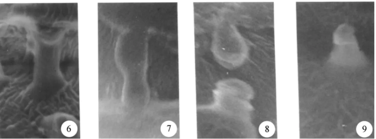
A close examination using SEM revealed that
besides protuberances, certain regions present
strand-like structures (open arrow in figures 1, 6 and
7), these regions being the ones in which intercellular
contact is still present. A careful search under the
SEM allowed the visualisation of different stages of
the process of strand breaking into protuberances.
These were assembled in figure 2.
The fact that strands are present where storage
walls are in close contact with each other is
consis-tent with the proposed origin of these protuberances
(Potgieter & van Wyk 1992). Some parts of walls of
contiguous cells would remain linked to each other
after the separation of the cells following imbibition.
These regions would form strands of wall material
between the cells that would eventually break and
form the protuberances observed (figures 8 and 9).
Based on the current models of the primary cell
walls (Carpita and Gibeaut 1993) and of storage cell
walls (Buckeridge et al. 2000) a possible explanation
for the existence of such strands of storage
xyloglu-can could be thought as hemicellulosic “bridges”
which would have to cross the primary cell wall. One
possibility is pictured in figure 10. In this picture, the
middle lamella is drawn as discontinuous and
stor-age material is crossing from one storstor-age wall to the
other. No transmission electron microscopy has been
reported for
H. courbaril
yet, but in another species
that accumulate xyloglucan (
Tropaeolum majus
) the
middle lamella presented cross reaction with a
xylo-glucan-specific probe (gold particles covered with
endo-
β
-glucanase nowadays named xyloglucan
endo-transglycosilase - see figure 8 in Vian et al.
1991).
Figures 1-5. Scanning electron microscopy and light microscopy of transversal sections of storage parenchyma of cotyledons of
these xyloglucan “bridges” might also be important
in the transport of solutes or water between
contigu-ous cells.
Acknowledgements - The authors are indebted with Mary Anne Heidi Dolder and Henrique Pessoa dos Santos for the help with the SEM. M.A.S. Tiné and M.S. Buckeridge thank help by CNPq
for fellowship funding. This work has been funded by BIOTA-FAPESP (98/05124-8).
References
BUCKERIDGE, M.S., SANTOS, H.P. & TINÉ, M.A.S. 2000. Mobilization of storage cell wall polysaccharides in seeds. Plant Physiology and Biochemistry 38:141-156.
BUTTERFIELD, B.G., MEYLAN, B.A. & EXLEY, R.R. 1981. Intercellular protuberances in the ground tissue ofCocos nuciferaL. Protoplasma 107:69-78.
CARPITA, N.C. & GIBEAUT, D.M. 1993. Structural models of primary cell walls in flowering plants: consistency of molecular structure with the physical properties of the cell wall during growth. The Plant Journal 3:1-30.
CARR, S.G.M. & CARR, D.J. 1975. Intercellular pectic strands in parenchyma: studies of plant cell walls by scanning electron microscopy. Australian Journal of Botany 23:95-105.
CARR, D.J., CARR, S.G.M. & JAHNE, R. 1980a. Intercellular strands associated with stomata: stomatal pectic strands. Protoplasma 102:177-182.
CARR, D.J., OATES, K. & CARR, S.G.M. 1980b. Studies on intercellular pectic strands of leaf palisade parenchyma. Annals of Botany 45:403-413.
CORTELAZZO, A.L. 1992. Detecção e quantificação do amido em cotilédones de Canavalia ensiformis e C. gladiata
durante o desenvolvimento inicial da planta. Revista Brasileira de Botânica 15:157-162.
KIERMAN, J.A. 1981. Histological and histochemical methods. Theory and practice. Pergamon Press, London.
MACHADO, S.R. & SAJO, M.G. 1996. Intercellular pectic protuberances in leaves of someXyrisspecies (Xyridaceae). Canadian Journal of Botany 74:1539-1541.
Figures 6-9. Proposed mechanism for the formation of protuberances on the surface of the storage cell walls of cotyledons from
Hymenaea courbarilassembled from different micrographies. During expansion caused by water imbibition, cells are pulled apart so that xyloglucan-containing strands (6) become under tension (7) and eventually break (8) into protuberances (9). 1cm corresponds to approximately 1µm.
Figure 10. Tentative model for the cell wall of storage cells of
POTGIETER, M.J. & VAN WYK, A.E. 1992. Intercellular pectic protuberances in plants: their structure and taxonomic significance. Botanical Bulletin of Academia Sinica 33:295-316.
ROLAND, J.C., REIS, D. & VIAN, B. 1992. Liquid crystal order and turbulence in the planar twist of the growing plant cell walls. Tissue and Cell 24:335-345.
VIAN, B., NAIRN, J. & REID, J.S.G. 1991. Enzyme-gold c y to c h e m is t r y o f s e e d x ylo glu c an s us in g tw o xyloglucan-specific hydrolases. Importance of prior heat-deactivation of the enzymes. Histochemical Journal 23:116-124.