ANTIMYCOBACTERIAL ACTIVITY OF A BREVIBACILLUS LATEROSPORUS STRAIN ISOLATED FROM A MOROCCAN SOIL
Mohammed Hassi 1; Souraya El Guendouzi 1; Abdelleatif Haggoud 1; Susana David 2; Saad Ibnsouda 1; Abdellah Houari 1; Mohammed Iraqui 1*
1
Laboratoire de Biotechnologie Microbienne, Faculté des Sciences et Techniques de Fès, B.P: 2202, Fès, Maroc; Instituto
Nacional de Saúde Dr. Ricardo Jorge (INSA,IP), 1649-016, Lisbon, Portugal.
2
Submitted: February 14, 2011; Approved: June 07, 2012.
ABSTRACT
The treatment of tuberculosis has become more difficult with the worldwide spread of multidrug-resistant
(MDR) and extensively drug-resistant (XDR) strains of Mycobacterium tuberculosis. Moreover, the
prevalence of human disease caused by atypical mycobacteria has also increased in the past two decades and
has further complicated the problem of the treatment of mycobacterial infections. It is therefore urgent to
develop new highly active molecules against these bacteria. The present study reports the isolation from a Moroccan soil of a Bacillus strain that exhibits an important antimycobacterial activity. The strain was
identified as Brevibacillus laterosporus using DNA sequencing of the 16S ribosomal RNA gene. The
antimycobacterial activity was assigned to a substance with a protein nature. This nature was revealed using
a liquid-liquid extraction with organic solvents, precipitation with ammonium sulfate and treatment with a
protease. This study suggested the identification and the characterization of this active metabolite enabling
therapeutic investigations further.
Key words: Mycobacteria; Antimycobacterial; Antibiotic; Brevibacillus.
INTRODUCTION
According to the latest report published by the World
Health Organization (WHO) in 2009 (27), the incidence of
tuberculosis (TB) worldwide is estimated at 9.27 million cases
with a mortality of 1.7 million, including 0.2 million
HIV-positive cases. Developing countries are the most affected, with
approximately 95% of the cases and 99% of the deaths. Indeed,
the estimated incidence is the highest in sub-Saharan Africa, followed by Southeastern Asia (18). In industrialized countries,
tuberculosis has again become a major threat after the emergence of drug-resistant forms in the early 1990s.
However, the incidence remains low and does not exceed an
average of 17 cases per 100000 inhabitants (18).
The causes of failure in the control of this disease are
multi-factorial and are primarily related to the advent of
acquired immunodeficiency syndrome (AIDS), the relative
effectiveness of the vaccine Bacille Calmette–Guérin (BCC)
(2) and the expansion of multidrug-resistant and extensively drug-resistant strains of Mycobacterium tuberculosis (6). This
resistance has been favored by the length of the currently
available therapeutic schemes and by the inappropriate use of antibiotics.
A multidrug-resistant strain (MDR-TB) is defined as a
strain resistant to at least the two major first-line TB drugs,
isoniazid (INH) and rifampicin (RIF). If a MDR-TB strain is
also resistant to at least one of the three second-line injectable
TB drugs (capreomycin, kanamycin or amikacin) in addition to
any one of the fluoroquinolones, the strain is considered to be
extensively drug-resistant (XDR-TB) (16). It is urgent to develop new highly active molecules against M. tuberculosis,
including MDR and XDR strains. These molecules must act
rapidly, have a long half-life for intermittent administration and
be capable of sterilizing the sites where the bacteria remain
persistent (10).
Telluric microorganisms are the primary source of
antibiotics (11). Bacillus is a bacterial genus that is abundant in
soil and contains several species that produce a large number of antibiotics with different chemical structures (29). These
antibiotics are mainly active against Gram-positive bacteria,
but compounds such as polymyxin, colistin and circulin inhibit
the growth of Gram-negative bacteria (3), while bacillomycin,
mycobacillin and fungistatin are active against yeast and fungi
(12). The antibiotics produced by Bacillus mainly have a
peptidic nature, and the majority of them are produced by
strains of Bacillus subtilis and Bacillus brevis (13).
The aim of this study was the isolation and identification
of a strain of Brevibacillus laterosporus that exhibits an
important antimycobacterial activity. Furthermore, This study
aimed to determine the nature of the substance responsible for
this activity. To our knowledge, this is the first report on a
strain of Brevibacillus laterosporus showing an
antimycobacterial activity.
MATERIALS AND METHODS
Samples and microbial strains
Soil samples were collected from ten Moroccan
ecosystems. Using a large sterile spatula, the first five
centimeters of the surface layer of the soil were removed. Then, with a small sterile spatula, 100 to 150 g of soil were
taken from the subjacent layer at 5 to 15 cm of depth and
deposited on a sterile aluminum sheet. The large debris, such as
stones and roots, were eliminated, and approximately 50 g of
the remaining material was placed in a sterile flask and
transported as quickly as possible to the laboratory.
The microbial strains used in this study were:
- Mycobacterium aurum A+: a non-pathogenic Mybacterium
species witha generation time of approximately 6 h. This strain
was usedas a model to evaluate the effect of active substances
onthe growth of M. tuberculosis (4),
- Mycobacterium smegmatis mc(2)155: a non-pathogenic
atypical mycobacterial strain with a generation time of
approximately 3 h,
- Mycobacterium kansasii ATCC 12478: an atypical
mycobacterial strain causing opportunistic infections,
- Mycobacterium bovis BCG IP: the vaccine strain,
- Mycobacterium vaccae ATCC 1548314 and
- Escherichia coli DH5α.
The mycobacteria were cultivated at 37 °C on Lowenstein
Jensen medium or Sauton medium (1, 21).
The Escherichia coli strain was cultivated on Luria
Bertani (LB) medium containing the following: peptone
(Biokarps Diagnostics, Beauvais, France), 10 g/l; yeast extract (Biokarps Diagnostics, Beauvais, France), 5 g/l; and sodium
chloride (Riedel- de Haën, Seelze, Germany), 10 g/l.
Screening for bacterial strains with antimycobacterial
activity
To isolate bacterial strains with antimycobacterial
activities, 4 g of each soil sample were dissolved in 36 ml of
sterile saline (NaCl, 9 g/l) and shaken for 2 h at ambient temperature. The supernatant was then recovered, and various
dilutions were prepared in LB medium (10-1 to 10-7). A volume
of 100 μl of each dilution was plated onto LB agar previously
smegmatis cultures. After incubation at 37°C for 48 h, the
bacterial clones surrounded by inhibition zones were recovered. One of these clones showed strong activity and was
maintained for this work.
Antimycobacterial activity assay
The isolated clone was cultivated under agitation on LB
medium at 37°C for 24 h. A volume of 10 ml of the bacterial
culture was centrifuged for 10 min at 6000 g. The supernatant
was then recovered and sterilized by filtration. The antimycobacterial activity was evaluated using an agar well
diffusion assay on plates pre-seeded with the indicator strains,
M. aurum A+ or M. smegmatis (23).
The agar wells were filled with 100 µl of the prepared
filtrates (20). The zones of inhibition were analyzed after 72 h
of incubation at 37°C.Each test was repeated 3 times. For the
negative control, an E. coli culture filtrate was used.
The antimycobacterial activity was also studied after a liquid-liquid extraction using ethyl acetate and ether solvents.
The isolated clones were cultivated under agitation on LB
medium at 37°C for 48 h. A volume of 100 ml of the bacterial
culture was centrifuged for 10 min at 6000 g. Then, the
supernatant was recovered, sterilized by filtration and added to
100 ml of organic solvent, either ethyl acetate or ether. After
agitation for one hour at room temperature, the organic extract
obtained was evaporated under vacuum at 37°C. The dry residue was taken up in 1 ml of sterile distilled water and
filtered. The antimycobacterial activity was measured using the
agar well diffusion assay on plates pre-seeded with the
indicator strains, M. aurum A+ and M. smegmatis, as described
above. For the negative control, an E. coli culture filtrate was
used.
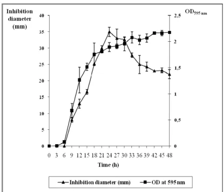
Kinetics of the active substance production
The bacterial growth was assessed by monitoring the
optical density at 595 nm (O.D595 nm). To determine the kinetics
of the production of the active substance, a colony of the tested
bacterial strain was inoculated in a volume of 100 ml of LB
medium and incubated at 37°C for 12 h. This culture was
diluted 104 fold in a volume of 2 l of fresh LB medium and incubated at 37°C under moderate agitation. At regular time
intervals, 100 ml of this culture was recovered and a
liquid-liquid extraction using ether was performed as previously
described. A volume of 100 μl of the organic extract was then
deposited in wells on Sauton agar medium already pre-seeded
with M. aurum A+ (O.D595 nm: 0.2). The diameters of inhibition
were measured after 72 h of incubation at 37°C.
Identification of the antimycobacterial-producing strain To identify the antimycobacterial-producing strain,
amplification and sequencing of the 16S rRNA gene was
performed. The use of 16S rRNA is a powerful tool that has
been used to identify bacteria from various sources, such as
environmental or clinical specimens, and to establish
phylogenetic relationships between bacteria (5, 19, 25, 28). The
DNA extraction from bacteria was carried out according to a standard method (17). For the PCR amplification, the universal
primers fD1 (5’- AGAGTTTGATCCTGGCTCAG-3’) and rP2
(5’-TACGGCTACCTTGTTACGACTT- 3’) were used to
amplify a 1.5-kb fragment of the 16S rDNA (9, 26). The PCR
was performed under the following conditions: 94°C for 5 min,
35 cycles of 94°C for 45 s, 55°C for 1 min and 72°C for 2 min
and finally 72°C for 10 min.
For sequencing, the PCR products were purified using a PCR product purification kit according to the manufacturer
(JETquick, Genomed). The sequencing reaction mixture
contained 2.0 µL of BigDye V 1.1, 2.7 µL of the PCR product
and 0.5 µM of the primer, either fD1 or rP2. A final reaction
volume of 10 µL was used. The amplification conditions
included an initial denaturation at 96ºC (3 min) followed by 35
cycles of denaturation at 96ºC (20 s), annealing at 50ºC (5 s)
and extension at 60ºC (4 min). The sequencing of the PCR products was performed on an ABI PRISM sequencing
apparatus (ABI Prism 310 Genetic Analyzer, Applied
Biosystems), and the data analysis was completed using
Characterization of the protein nature of the
antimycobacterial metabolite
The bacterial strain was grown in 100 ml of LB medium
at 37°C until the early stationary phase. The cells were
removed by centrifugation for 10 min at 6000 g and 4°C, and
the supernatant was recovered and filtered using a 0.22-μm
filter (Millipore, Molsheim, France).
A precipitation with 80% saturated ammonium sulfate
was carried out overnight at 4°C under agitation (29). The
resulting precipitate was harvested by centrifugation at 10000 g for 20 min at 4°C, resuspended in 1 ml of potassium phosphate
buffer (50 mM; pH 6) and dialyzed against the same buffer at
4°C for 24 h. The buffer was renewed several times to facilitate
a better dialysis. The antimycobacterial activity of the resulting
precipitate was measured using the agar well diffusion assay on
plates pre-seeded with the indicator strain M. aurum A+
(O.D595nm: 0.2).
The diameters of inhibition were determined after 72 h of incubation at 37°C. This experiment was repeated twice. The
control that was used corresponded to a dialyzed product
obtained from an E. coli culture prepared in the same
conditions as the test strain. The proteins in the resulting
precipitate were quantified using the Lowry method (15) and
analyzed using sodium dodecyl sulfate polyacrylamide gel
electrophoresis (SDS-PAGE).
To establish the protein nature of the antimycobacterial metabolite, the metabolite was also tested for its sensitivity to
proteinase K (29). Two controls were used: (1) the ammonium
sulfate extract untreated by proteinase K and (2) proteinase K
without the extract. The samples were incubated in the
presence of proteinase K (1 mg/ml) at 37°C for 3 h. The
antimycobacterial effect of these preparations was examined
using the agar well diffusion assay against M. aurum A+ as
previously described.
RESULTS AND DISCUSSION
Isolation of a bacterial strain with antimycobacterial
activity
Telluric microorganisms were the principal source of
antibiotics discovered in the second half of the 20th century
(13). In this work, we chose particular biotopes to isolate
bacteria that express antimycobacterial activity. A clone surrounded by an inhibition zone was obtained on LB-agar
medium pre-seeded with M. smegmatis. Furthermore, the
filtrate prepared from the isolated clone was able to inhibit the
growth of M. smegmatis and M. aurum A+ (Table 1),
indicating that the active substance was secreted by the clone.
The filtrate was also active against M. vaccae, M. bovis BCG
and M. kansasii (data not shown). In addition, the ethyl acetate and ether extracts showed a more accentuated
antimycobacterial activity than that of the filtrate (Table 1),
indicating that the solvents were capable of extracting and
concentrating the active substance responsible for the observed
biological activity.
The production kinetics of the active substance were also
studied (Fig. 1). The antimycobacterial activity was detected
after six hours and increased progressively during the exponential phase of growth. The maximum activity was
reached after 24 h of growth and then decreased rapidly. These
results (Table 1, Fig. 1) indicate that the active substance,
responsible for the observed antimycobacterial effect, is
synthesized during the exponential phase and early stationary
phase.
Table 1. Antimycobacterial activity of the bacterial isolate (measured in mm).
Microorganism Filtrate Ethyl acetate extract Ether extract
M. smegmatis M. aurum A+
10 ± 0.5 11.5 ± 0.7
32 ± 0.0 34.5 ± 2.12
Figure 1. Time course of the antimycobacterial metabolite production by the bacterial isolate.
Molecular identification of the antimycobacterial-producing
isolate
The lengths of the 16S rDNA sequences analyzed were 713
bp and 648 bp for the primers fD1 and rP2, respectively. Their
analysis in comparison with the sequences available in Gen Bank,
EMBL, DDJB and PDB databases showed that the
antimycobacterial-producing strain was closely related to
Brevibacillus laterosporus species with 100% similarity
(accession number: DQ371289.2).
Characterization of the protein nature of the
antimycobacterial metabolite
The protein extract of the Brevibacillus laterosporus strain
was prepared and shown to be capable of inhibiting M. aurum A+
growth (Table 2). Moreover, this activity was totally lost in the
presence of proteinase K (Table 2). The same result was also
obtained after treatment of the organic extracts, obtained with
ethyl acetate and ether, with proteinase K (Table 2). This result
indicates that the bioactive metabolite secreted by the
Brevibacillus strain is of a protein nature.
The concentration of the protein extract was determined to be
0.0104 mg/ml. An analysis of the extract using polyacrylamide gel
electrophoresis revealed the presence of nine bands (Fig. 2) with
molecular weights comprised between 40 and 240 kDa. These
bands were absent in the control, an E. coli protein extract. These
results (Table 2, Fig. 2) indicate that the antimycobacterial activity
of the bacterial isolate could be due to one protein or the
synergistic action between two or more of these proteins.
Table 2. Characterization of the protein nature of the antimycobacterial metabolite.
Inhibition diameters against M. aurum A+ (mm) Without treatment After treatment Controls with proteinase K with proteinase K T1 T2 T3
ASP 33 ± 0.7 0 0 0 0
EAE 34.5 ± 1.5 0 0 0 0
EE 24.5 ± 0.0 0 0 0 0
Bacillus species produce approximately 167 biological
compounds (12). Brevibacillus laterosporus comb. nov. (24), previously classified as Bacillus laterosporus (Laubach 1916),
has demonstrated a very wide spectrum of biologicalactivity
(7).Several antibiotics produced by Brevibacillus laterosporus
have been described, such as laterosporamine, laterosporin
(12), tauramamide and loloatin A.
Tauramamide is a lipopeptide antibiotic active against
pathogenic Enterococcus sp (8), while loloatin A was
identified as a cyclic decapeptide with cyanolytic activity (14). Moreover, an antimicrobial substance produced by a
Brevibacillus laterosporus strain isolated from a marine habitat
was recently purified and characterized. The antimicrobial
substance had high activities against Gram-positive bacteria,
such as Streptococcus, Staphylococcus aureus and
Clostridium; Gram-negative bacteria, such as Escherichia coli
and Pseudomonas putrefaciens; and against Candida albicans
(22).
As previously described, the Brevibacillus laterosporus
strain isolated in this work is able to secrete an active substance
of a protein nature that inhibits the growth of various
mycobacteria. To our knowledge, this study is the first report
describing such a substance produced by a strain of B.
laterosporus. Based on this study, our goal is to test the
antimycobacterial metabolite on M. tuberculosis and on
mycobacteria hosted by macrophages. Moreover, the identification and the characterization of this compound would
facilitate further therapeutic investigations.
REFERENCES
1. Allen, B.W. (1998). Mycobacteria: general culture methodology and safety considerations. Methods Mol. Biol. 101, 15-30.
2. Billy, C.; Lévy-Bruhl, D. (2007). Vaccin BCG et place de l’intradermoréaction en 2006. La Revue de médecine interne. 28 (3), 151-160.
3. Bottone, E.J.; Peluso, R.W. (2003). Production by Bacillus pumilus
(MSH) of an antifungal compound that is active against Mucoraceae and
Aspergillus species: preliminary report. J. Med. Microbiol. 52 (Pt 1), 69-74.
4. Chung, G.A.; Aktar, Z.; Jackson, S.; Duncan, K. (1995). High throughput screen for detecting antimycobacterial agents. Antimicrob. Agents. Chemother. 39 (10), 2235-2238.
5. Clarridge, J.E. (2004). Impact of 16S rRNA gene sequence analysis for identification of bacteria on clinical microbiology and infectious diseases. Clin. Microbiol. Rev. 17(4), 840-862.
6. Danilchanka, O.; Mailaender, C.; Niederweis, M. (2008). Identification of a novel multidrug efflux pump of Mycobacterium tuberculosis.
Antimicrob. Agents. Chemother. 52(7), 2503-2511.
7. de Oliveira, E.J.; Rabinovitch, L; Monnerat, R.G.; Passos, L.K.J.; Zahner, V. (2004). Molecular characterization of Brevibacillus laterosporus and its potential use in biological control. Appl. Environ. Microbiol. 70 (11), 6657-6664.
8. Desjardine, K.; Pereira, A.; Wright, H.; Matainaho, T.; Kelly, M.; Andersen, R.J. (2007). Tauramamide, a lipopeptide antibiotic produced in culture by Brevibacilluslaterosporus isolated from a marine habitat: structure elucidation and synthesis. J. Nat. Prod. 70 (12), 1850-1853. 9. Drancourt, M.; Bollet, C.; Carlioz, A.; Martelin, R.; Gayral, J.P.; Raoult,
D. (2000). 16S ribosomal DNA sequence analysis of a large collection of environmental and clinical unidentifiable bacterial isolates. J. Clin. Microbiol. 38 (10), 3623-3630.
10. Dutta, N.K.; Dastidar, S.G.; Kumar, A.; Mazumdar, K.; Ray, R.; Chakrabarty, A.N. (2004): Antimycobacterial activity of the antiinflammatory agent diclofenac sodium, and its synergism with streptomycin. Braz. J. Microbiol. 35, 316-323.
11. Gillespie, D.E.; Brady, S.F.; Bettermann, A.D.; Cianciotto, N.P.; Liles, M.R.; Rondon, M.R.; Clardy, J.; Goodman, R.M.; Handelsman, J. (2002). Isolation of antibiotics turbomycin A and B from a metagenomic library of soil microbial DNA. App. Environ. Microbiol. 68 (9), 4301-4306.
12. Katz, E.; Demain, A.L. (1977). The peptide antibiotics of Bacillus: chemistry, biogenesis and possible functions. Bacteriol. Rev. 41 (2), 449-474.
13. Kleinkauf, H.; von Döhren, H. (1990). Nonribosomal biosynthesis of peptide antibiotics. Euro. J. Biochem. 192 (1), 1-15.
14. Krachkovskiĭ, S.A.; Sobol', A.G.; Ovchinnikova, T.V.; Tagaev, A.A.; Iakimenko, Z.A.; Azizbekian, R.R.; Kuznetsova, N.I.; Shamshina, T.N.; Arsen'ev, A.S. (2002). Isolation, biological properties, and spatial structure of an antibiotic loloatin A. Bioorg. Khim. 28 (4), 298-302. 15. Lowry, O.H.; Rosebrough, N.J; Farr A. L.; Randall, R.J. (1951). Protein
measurement with the Folin phenol reagent. J. Biol. Chem. 193 (1), 265-275.
16. Marigot-Outtandy, D.; Perronne, C. (2009). Les nouveaux antituberculeux. Réanimation. 18 (4), 334-342.
17. Marmur, J. (1961). A procedure for the isolation of deoxyribonucleic acid from microorganisms. J. Mol. Biol. 3 (2), 208-218.
tuberculose et des autres mycobactérioses. Arch. Pediatr. 12, 96-101. 19. Mignard, S.; Flandrois. J.P. (2006). 16S rRNA sequencing in routine
bacterial identification: a 30-month experiment. J. Microbiol. Methods. 67 (3), 574-581.
20. Motta, A.S.; Cladera-Olivera, F.; Brandelli, A. (2004): Screening for antimicrobial activity among bacteria isolated from the Amazon Basin.
Braz. J. Microbiol. 35, 307-310.
21. Papa, F.; Rivière, M.; Fournié, J.J.; Puzo, G.; David, H. (1987). Specificity of a Mycobacterium kansasii phenolic glycolipid (mycoside A) immunoserum. J. Clin. Microbiol. 25 (12), 2270-2273.
22. Ren, Z.Z.; Zheng, Y.; Sun, M.; Liu, J.Z.; Wang, Y.J. (2007). Purification and properties of an antimicrobial substance from marine
Brevibacillus laterosporus Lh-1. Wei. Sheng. Wu. Xue. Bao. 47 (6), 997-1001.
23. Sánchez, J. G. B.; Kouznetsov, V. V. (2010): Antimycobacterial susceptibility testing methods for natural products research. Braz. J. Microbiol. 41 (2), 270-277.
24. Shida, O.; Takagi, H.; Kadowaki, K.; Komagata, K. (1996). Proposal for two new genera, Brevibacillus gen. nov. and Aneurinibacillus gen. nov.
Int. J. Syst. Bacteriol. 46, (4), 939-946.
25. Wang, W.; Sun, M. (2009). Phylogenetic relationships between Bacillus
species and related genera inferred from 16s rDNA sequences. Braz. J. Microbiol. 40, 505-521.
26. Weisberg, W.G.; Barns, S.M.; Pelletier, D.A.; Lane, D.J. (1991). 16S ribosomal DNA amplification for phylogenetic study. J. Bacteriol. 173 (2), 679-703.
27. WHO Report. (2009). Global tuberculosis control - epidemiology, strategy, financing. WHO/HTM/TB/2009.411. Available at: http://www.who.int/tb/publications/global_report/2009/en/. Accessed August 21, 2010.
28. Woo, P.C. ; Lau, S.K.; Teng, J.L.; Tse, H.; Yuen, K.Y. (2008). Then and now: use of 16S rDNA gene sequencing for bacterial identification and discovery of novel bacteria in clinical microbiology laboratories. Clin. Microbiol. Infect. 14 (10), 908-934.
29. Wu, S.; Jia, S.; Sun, D.; Chen, M.; Chen, X.; Zhong, J.; Huan, L.(2005). Purification and characterization of tow novel antimicrobial peptides Subpeptin JM4-A and Subpeptin JM4-B produced by Bacillus subtilus
JM4, Curr. Microbiol. 51 (5), 292- 296.