709 709 709 709 709 Mem Inst Oswaldo Cruz, Rio de Janeiro, Vol. 99(7): 709-715, Novem ber 2004
Effect of the
Aedes fluviatilis
Saliva on the D evelopment of
Plasmodium gallinaceum
Infection in
Gallus (gallus) domesticus
Ana CVM da Rocha/* , Érika M Braga* , M árcio SS Araújo, Bernardo S Franklin, Paulo FP Pimenta/+
Laboratório de Entomologia Médica, Centro de Pesquisas René Rachou-Fiocruz, Av. Augusto de Lima 1715, 30190-002 Belo Horizonte, MG, Brasil *Departamento de Parasitologia, Universidade Federal de Minas Gerais, Belo Horizonte, MG, Brasil
Effect of Aedes fluviatilis saliva on the development of Plasmodium gallinaceum experimental infection in Gallus (gallus) domesticus was studied in distinct aspects. Chickens subcutaneously infected with sporozoites in the pres-ence of the mosquito salivary gland homogenates (SGH) showed higher levels of parasitaemia when compared to those ones that received only the sporozoites. However, the parasitaemia levels were lower among chickens previ-ously immunized by SGH or non-infected mosquito bites compared to the controls, which did not receive saliva. High levels of anti-saliva antibodies were observed in those immunized chickens. Moreover, 53 and 102 kDa saliva proteins were recognized by sera from immunized chickens. After the sporozoite challenge, the chickens also showed significant levels of anti-sporozoite antibodies. However, the ability to generate anti-sporozoites antibodies was not correlated to the saliva immunization. Our results suggest that mosquito saliva components enhance P. gallinaceum
parasite development in naive chickens. However, the prior exposure of chickens to salivary components controls the parasitemia levels in infected individuals.
Key words: saliva - avian malaria - mosquitoes - sporozoites - antibodies
Saliva components from haematophagous arthropods may modulate the host immune system and would repre-sent an adaptation to evolution related with the blood feeding behavior (Ribeiro et al. 1984, Ribeiro 1987, 1989, James & Rossignol 1991). Several substances in saliva from different vectors such as ticks, phlebotomine, and mosquitoes prevent host homeostasis at the bite site dur-ing the blood dur-ingestion. Such components may show dif-ferent activities such as anti-homeostatic, vasodilator, anti-inflammatory, immune-suppressor, and many others (review in Kamhawi et al. 2000).
The effect of saliva substances on the pathogen in-fectivity to vertebrates was firstly demonstrated by Titus and Ribeiro (1988) in phlebotomine followed by Junes and collaborators (1993) in ticks. Nowadays, the importance of saliva for blood feeding and pathogen infections has been extensively analyzed in several arthropod vectors (review in Gillespie et al. 2000). Therefore, there are few studies about the effect of mosquito saliva on infectivity of Plasmodium parasites. Early studies showed some pro-tection against infection by P. berghei sporozoites when mice were previously immunized with mosquito salivary gland homogenate (Alger et al. 1972, Alger & Harant 1976). In addition, Vaughan et al. (1999) suggested that saliva was one of the factors that could contribute to a more efficient rodent infection when the parasites were injected by mosquito vector bites.
Financial support: Fiocruz-Papes, CNPq, Pronex and Fapemig +Corresponding author: Fax: +55-31-3295.3115. E-mail:
pimenta@cpqrr.fiocruz.br Received 19 April 2004 Accepted 15 September 2004
In the current study, we evaluate the role of Aedes fluviatilis saliva in the experimental infections of chicken
Gallus (gallus) domesticus with P. gallinaceum, the caus-ative agent of avian malaria and its immunogenic poten-tial to control parasitemia.
MATERIALS AND METHODS
Mosquitoes - Ae. fluviatilis were obtained from a colony established and kept in the Laboratory of Medical Entomology, Centro de Pesquisas René Rachou-Fiocruz in the state of Minas Gerais. The mosquitoes were kept in an acclimated insectary with average temperature between 26-28oC and relative air humidity around 70-80%, in a cycle of 12 h in the dark and 12 h in the light (Consoli & Williams 1978). The mosquitoes were provided with 10% glucose solution and water until the time of the experiments.
Mosquito infection -Groups of 4 to 6 day-old female mosquitoes were allowed to feed on the skin of P. gallinaceum infected chickens (parasitaemia levels rang-ing from 3.5 to 10%). The infection evaluation was carried out 7 to 9 days after the infective blood meal by the pres-ence of ookinetes in dissected midguts, which were dis-sected in PBS (phosphate buffer solution) and stained with 2% mercury chrome in order to visualize the para-sites. The percentage of infected mosquitoes and the number of ookinetes per midgut were recorded through optical microscopic examination. All mosquitoes used to infect chickens were also previously examined for the pres-ence of sporozoites 14 days after the infection (Ozaki et al.1984).
710 710 710 710
710 M osquito Saliva and Plasmodium Infection • Ana CVM da Rocha et al.
Salivary gland homogenate (SGH) -Salivary glands from 4 to 6 day-old female mosquitoes (non-infected mos-quitoes) were dissected and transferred to microcentrifuge tubes containing 50 µl cold sterile PBS. Dissected glands were stored at _70oC until use when they were submitted to a successive freezing and unfreezing processes in or-der to obtain protein material. Total soluble protein con-centration in the SGH was determined by Lowry method (Lowry et al. 1951). One salivary gland (a paired structure) dissected from one mosquito was estimated to contain 1.4 µg of proteins.
Isolation of sporozoites -The P. gallinaceum sporo-zoites were obtainedaccording to Ozaki et al. (1984). Briefly, 15 thoraces were dissected from 12-day infected mosquitoes and transferred to 0.5 ml microcentrifuge-tube, containing fiberglass and with perforated bottom, and then placed into another 1.5 ml microcentrifuge-tube. A vol-ume of 50-100 µl of RPMI 1640 culture medium (Sigma, St. Louis, MD, US) supplemented with 10% chicken serum (for experimental infection) or PBS (for antigen prepara-tion) was added to the thoraces. The microcentrifuge-tubes were centrifuged (twice, for 10 min, at 1000 g). The sediment was homogenized and parasites counted into Neubauer’s chamber using a phase contrast microscope. The whole process was performed in ice to keep the sporo-zoite viability.
Infection of chickens with P. gallinaceum - One week-old chickens were used to evaluate the infection by P. gallinaceum sporozoites. Groups of five individuals were infected according to the following approaches: (i) natu-rally infected chickens infected by, bites of 10 infected mosquitoes; (ii) chickens infected by subcutaneous in-oculation of 103 sporozoites; (iii) infected chickens by subcutaneous inoculation of 103 sporozoites added with SGH (corresponding to 10 mosquito glands); and (iv) non-infected chickens which were subcutaneous inoculated with RPMI 1640 culture medium. The subcutaneous via was chosen due to the early development of avian malaria into the phagocyte cells in the host skin at the infective bite site (Paraense 1941, 1943, Huff & Coulston 1944, Coulston & Huff 1947). Groups (i) and (iv) were consid-ered as positive and negative controls of the infection.
Pre-patent periods (PPP), parasitaemia averages through blood smears stained with Giemsa solution and mortality rates were analyzed and the values compared throughout infection period in the studied groups of chick-ens. PPP is the period of time between the beginning of the infection and the time that the pathogen is detectable in the peripheral blood.
Saliva immunization - The effect of previous expo-sure to saliva components on the development of infec-tion was analyzed in chickens which received bites from non-infected mosquitoes or which were inoculated by SGH. Groups of 5 or 10 chickens were used according to the following experimental approaches: (i) chickens im-munized by bites of 10 non-infected adult mosquitoes; (ii) chickens immunized by subcutaneous inoculations of SGH (corresponding to 10 mosquito glands); and (iii) chickens which received PBS by subcutaneous inoculation. All chickens were one-week old in the beginning of the ex-periments and they were incubated once a week during a
period of 4 or 7 weeks. Sera samples from the chickens were weekly obtained and stored at –20oC until use. One week after the last incubated step, all the chickens re-ceived were infected by bites of 10 infected mosquitoes.
Detection of anti-sporozoite and anti-SGH IgG - In-direct immunofluorescence assay (IFA) were used to de-tect anti-sporozoite antibodies and Enzyme linked absor-bance (ELISA) were used to detect anti-sporozoite and anti-saliva antibodies in infected or immunized chickens. Briefly, sporozoites were suspended in a concentration of 1x10⁶ parasites/ml and 5 µl were deposited per well on slide. For screening, each well was incubated with 10 µl of sera dilutions (1:20 to 1:5120) in PBS, and then incubated for 20 min with rabbit anti-chicken IgG fluorescein-conju-gated (Funed) diluted 1:400 with PBS. The slides were examined by fluorescence microscopy. The ELISA were performed using high binding 96-well microplates (Nunc Maxisorp, Dynatech Denmark) covered with sporozoites homogenate or SGH (20 µg/ml) during 18 h 4°C. Chicken sera were tested in triplicate at 1:80 dilution in PBS-Tween (0.05%) for 2 h at 37°C. The rabbit anti- chicken IgG per-oxidase conjugated (Funed) was added at a 1:1000 dilu-tion for 60 min at 37°C followed by addidilu-tion of peroxidase substrate (OPD-O-phenylenediamine and hydrogen per-oxide). Absorbance values were measured at 490 nm. SGH and sporozoite homogenates were also fractionated by electrophoresis SDS-PAGE 12.5% (Laemmli 1970) and then transferred to a nitrocellulose membrane (Amershan Pharmacia HybondTM - C pure). The transference was performed using a 25V constant voltage for 2 h. Part of the nitrocellulose membrane containing the molecular markers was stained with Ponceau S for the visualization of the transferred proteins. The membrane with SGH or sporozoite proteins was incubated for 12 h with chicken sera diluted at 1:40. Afterwards, rabbit anti-chicken IgG peroxidase-conjugated (Funed) was added at a dilution of 1:1000. The antibody reaction was revealed by adding 0.05% 3.3-diaminobenzidin solution containing 0.025%, 4-clhoride 1-naphtol and 0.03% H2 02 (30% v/v). The re-action was interrupted with distillated water after visual-izing bands.
Statistical analysis - Average parasitemia ands aver-age absorbance were compared using Student’s t-test as-suming equal variances. Differences in mortality between groups were tested with Chi-square test with Yates’ cor-rection. Difference was considered significant when P-value was less than 0.05.
RESULTS
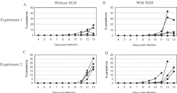
Effect of the SGH on avian infection - Two distinct experiments were conducted to analyze the effect of the saliva components in the infection of chickens with P. gallinaceum sporozoites (Fig. 1).
The parasitemia average among chickens that were infected by sporozoite inoculation in the presence of SGH was higher than those that did not receive SGH (Fig. 1). However, such tendency was not statistically different due to the significant deaths caused by malaria among chickens that were inoculated with parasites plus SGH.
711 711 711 711 711 Mem Inst Oswaldo Cruz, Rio de Janeiro, Vol. 99(7), Novem ber 2004
by the mosquito bites was 100% up to the 13 day after infection. Therefore, the mortality rate of chickens infected with sporozoites in the presence of SGH was observed after the 12 day after infection and those infected ones without SGH, only after the 15 day. It is important to point out that approximately 60% of chickens of the group in-fected by sporozoites inoculation (without SGH) and 40% of chickens of the group infected by sporozoites with SGH, controlled their parasitemia levels at the 18 day after infection (data not shown).
The \PPP among chickens naturally infected by mos-quito bites was 5 days. Therefore, the PPPs for chickens infected by subcutaneous sporozoite inoculations with or without SGH were around 10 days (data not shown)
showing no difference between the two groups.
Chicken infection after saliva immunization - Chick-ens that were immunized 7-fold with saliva by bites or inoculation of SGH showed similar PPP average values after a natural infection by mosquito bites. The average of the parasitaemia levels analyzed at the 9 day after in-fection of those immunized chicken groups showed to be lower than non-immunized chickens. Statistically signifi-cant differences of parasitaemia averages were observed at the 11day after infection among the groups of immu-nized chickens and the control group (non-immuimmu-nized chickens) (Table). The chickens that were immunized four times, either by the mosquito bites or by inoculation of SGH, showed to be more susceptible to the infection than
Fig. 1: the role of Aedes fluviatilis salivary gland homogenates (SGH) in individual parasitemias of Plasmodium gallinaceum infected chickens. Each group was inoculated with 103 sporozoites in the presence (B and D) or in absence (A and C) of SGH.
TABLE
The role of immunization of the chicken with Aedes fluviatilis saliva in the parasitaemia and pre-patent period of Plasmodium gallinaceum infection
Parasitemia (X ± d.p.) b
Groups a 7 day 8 day 9 day 10 day 11 day PPP c
4-fold immunizations by 2.54 ±2.19 4.02 ± 2.11 11.66 ± 6.22 4.66 ± 5.04 2.13 ± 2.14 5.00 ± 0.55 mosquito bites (n = 5)
4-fold immunizations by 3.04 ± 2.60 5.00 ± 3.08 8.06 ± 2.53 3.58 ± 2.25 4.87 ± 1.94 6.00 ± 0.55 SGH (n = 5)
4-fold inoculations with 2.12 ± 1.94 5.00 ± 5.27 12.60 ± 10.85 10.80 ± 12.66 6.6 ± 10.87 6.00 ± 0.89 PBS (control) (n = 5)
7-fold immunizations by 0.04 ± 0.04 0.80 ± 0.74 2.98 ± 3.32 4.28 ± 2.76 +2.94 ± 3.72 6.50 ± 0.71 mosquito bites (n = 10)
7-fold immunizations by 0.04 ± 0.007 1.26 ± 1.45 6.80 ± 7.15 4.76 ± 5.53 +3.61 ± 4.43 7.00 ± 0.83 SGH (n = 10)
7-fold inoculations with 0.02 ± 0.04 0.67 ± 0.85 5.59 ± 5.94 10.21 ± 8.38 11.43 ± 8,66 7.00 ± 0.42 PBS (control) (n = 10)
712 712 712 712
712 M osquito Saliva and Plasmodium Infection • Ana CVM da Rocha et al.
those chickens 7-fold immunized (data not shown). In a follow up of the infection rates during one month, the chickens naturally immunized by mosquito bites pre-sented an average of the mortality rates of 10.5%. Chick-ens from the SGH-immunized and non-immunized groups showed mortality rates of 12% and 13%, respectively. All the chickens that remained alive in these studied groups appeared to have controlled parasitaemia revealed by negative microscopy examination of blood smears.
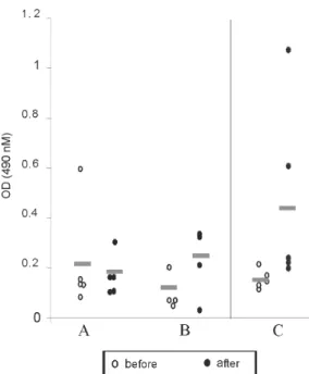
Anti-sporozoite antibodies in P. gallinaceum infected chickens - Anti-sporozoite antibodies in chicken sera be-fore and after infections were also evaluated. Anti-sporo-zoite antibodies were not detected by IFA in sera from one-week chickens infected with P. gallinaceum. Fig. 2 shows the individual and the average of sera absorbance values by ELISA verified in a representative experiment. A raise in average values can be observed comparing sera from chickens before and 10 days after infection, inde-pendent on the infection via. However, absorbance val-ues average among the experimental groups showed to be relatively low (lower than 0.4). Only two sera from chick-ens infected by sporozoites with SGH had individual ab-sorbance values higher than 0.6 (Fig. 2).
Anti-sporozoite antibodies in saliva-immunized chickens infected with P. gallinaceum -Presence of anti-sporozoite antibodies was not detected among non-in-fected saliva-immunized chickens. On the other hand, af-ter infection by mosquito bites, all groups of chicken showed anti-sporozoite antibodies including the non-im-munized control group. Anti-sporozoite IgG antibodies titles detected by IFA, ranged from 1:40 to 1:1280 in chick-ens 4-fold immunized reaching a high value (1: 2560) in 7-fold immunized ones. No statistical difference was ob-served for antibody titles detected by IFA. However,
anti-sporozoite antibody frequencies and proportions detected by ELISA showed a statistically significant raise in the average of the absorbance values, which were mainly observed 10 days after infection (Fig. 3).
Fig. 2: ELISA reactivity for Plasmodium gallinaceum sporozoite antigens to sera from 1-week old chicken before (!) and 10 days after infection (") using different via of parasite inoculation (A: mosquito bites; B: inoculation of sporozoites; C: inoculation of sporozoites plus SGE). Bars: average of the absorbance values.
Fig. 3: ELISA reactivity for Plasmodium gallinaceum sporozoite antigens to sera from chickens which were saliva-immunized by mosquito bites (A) or salivary gland homogenates inoculation (B) or non-immunized control (C). The experiments were done before (!) and 10 days after infection (") by mosquito bites. Bars: aver-age of the absorbance values.
Anti-saliva antibodies in P. gallinaceum infected chickens -Individual values of anti-antibody saliva ab-sorbancies from chickens infected by the mosquito bites or by sporozoite inoculation with or without SGH, showed to be low (lower than 0.4) with no statistically differences among groups (not shown). However, 10 days after infec-tion was observed significant increase in saliva anti-body levels among immunized chickens independently of the inoculation via. Such antibody raise were also veri-fied for the control group (Fig. 4). In spite of the number of previous immunizations no statistically significant dif-ference among groups was observed.
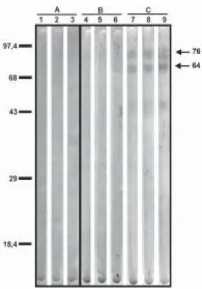
Recognition of antigenic proteins of P. gallinaceum sporozoite and salivary gland by Western blotting -Sera from chickens infected by P. gallinaceum bites, either from the group of previous saliva-immunized chickens (SGH or mosquito bites) or control group (non-immunized chickens), recognized sporozoite proteins with molecular weights of approximately 64 and 76 kDa (Fig. 5). Which correspond to the P. gallinaceum CS protein.
All those sera from chickens of the same groups rec-ognized a protein with a molecular weight of approximately 102 kDa that is presented in SGH (Fig. 6). In addition, proteins of 53 and 59 kDa present in SGH were recognized by the sera from immunized or non-immunized adult chick-ens infected by the mosquito bites.
DISCUSSION
713 713 713 713 713 Mem Inst Oswaldo Cruz, Rio de Janeiro, Vol. 99(7), Novem ber 2004
Fig. 4: ELISA reactivity against Aedes aegypti saliva antigens present in sera from chickens, which were saliva-immunized by mosquito bites (A) or salivary gland homogenates inoculation (B) or non-immunized, control (C). The experiments were done before the infection, 4-fold or 7-fold after immunizations and 10 days after infection by mosquito bites. The control group received only PBS.
Fig. 5: Western-blotting showing reactivity against Plasmodium gallinaceum sporozoites antigens using sera from different groups of chickens. A: one-week old chickens infected with inoculation of 103 sporozoites (1), 103 sporozoites plus salivary gland homogenates (SGH) (2) and mosquito bites (3); B: 4-week old chickens immu-nized with saliva by SGH inoculations (4); mosquito bites; (5) con-trol (inoculated with PBS) (6); C: 4-fold saliva-immunized chickens followed by infection with mosquito bites, which were immunized by salivary gland homogenates inoculations (7) or mosquito bites (8) and control (inoculated with PBS ) (9).
Fig. 6: Western-blotting showing reactivity against Aedes fluviatilis
saliva antigens using sera from different groups of chickens. A: one-week old chickens infected with inoculation of 103 sporozoites (1), 103 sporozoites plus salivary gland homogenates (SGH) (2), mos-quito bites (3), and PBS (control non-infection), (4); B: 4-week old chickens immunized four times with SGH inoculations (5), mos-quito bites (6), and control (inoculations with PBS) (7); C: 7-fold salivary gland homogenates immunized chickens; (8) or mosquito bites (9) which were respectively infected by mosquito bites (10 and 11) and control, non-immunized chickens but infected by mosquito bites (12).
related with the parasite-host interaction processes. In mental subcutaneous sporozoite inoculation as previously observed (Vaughan et al. 1999). In addition, it is important to consider that sporozoites isolated from salivary glands are considered an heterogeneous population. Moreover, sporozoites need to pass through several biological pro-cesses inside the salivary glands in order to reach the salivary duct (Pimenta et al. 1994). Although there are few thousands of sporozoites stocked up in the salivary gland, only a small number is ready to be injected by the mos-quito bites (Simonetti 1996). Certainly, not all sporozoites obtained from the salivary gland by the isolation proce-dure are able to stay alive and develop infection in the skin host.
714 714 714 714
714 M osquito Saliva and Plasmodium Infection • Ana CVM da Rocha et al.
the absence of deaths. These facts were only observed in infections caused by mosquito bites and not by subcuta-neous inoculation of sporozoites, once again, confirming the efficacy of the natural via of infection.
It is interesting to note that the presence salivary com-ponents in the sporozoite inoculum affect the parasitaemia levels and the mortality rates. An increase in parasitemia levels and in mortality rates was observed when chickens received sporozoites in association with SGH. However, prepatent period, was not affect by the saliva components present in the subcutaneous inoculum. Our results ob-tained for P. gallinaceum infection, corroborate literature data concerning other pathogens, which evidence the ef-fects of arthropod saliva in parasite-vector interactions (Ribeiro et al. 1985, Titus & Ribeiro 1988, Belkaid et al. 1998, Kamhawi et al. 2000).
In our experiments, anti-sporozoite antibodies were detected in chickens infected with P. gallinaceum by mosquito or subcutaneous inoculation. Lower levels of these antibodies were detected in young individuals com-paring with the adults. The chickens showed to produce anti-sporozoite antibodies with the same molecular (64 kDa and 76 kDa) weights of the well-known circums-porozoite protein family, which covers the surface of the
Plasmodium sporozoite (CS protein). The P. gallinaceum
CS protein is involved in parasite interaction with verte-brate and inverteverte-brate cell hosts and elicits a strong hu-moral response in chicks (Daher & Krettli 1987). In the present study, we demonstrated that adult chickens in-fected by the mosquito bites produced anti-SGH antibod-ies. During the blood meal, female mosquitoes deposit saliva into the host skin. Adult chickens sera recognized some SGH proteins including one that correspond to the molecular weight of apirase (64 kDa). Apirases are en-zymes that have been demonstrated in the saliva of sev-eral insect vectors and are recognized as playing a role in the insect feeding process avoiding the blood platelet aggregation (Ribeiro 1987). Previous work already showed that anti-apirase antibodies in saliva-immunized mice by successive bites of the mosquito Anhopheles stephensi
were able to inhibit apirase activity impairing the blood meal screening (Mathews et al. 1996).
The parasitaemia levels showed to be lower in immu-nized groups of chickens than in the control group. Alger and Harant (1976) also reported that immunized mice with mosquito salivary glands were protected against the P. berghei sporozoites challenge. Prior exposure of mice to bites of non-infected sand flies protects against Leishma-nia major (Kamhawi et al. 2000). However, the sand fly saliva components enhance the cutaneous lesion caused by the parasite. It appears that similar phenomena also occur in saliva-immunized chicken challenged with P. gallinaceum.
In conclusion, our results suggest that mosquito sa-liva components play an important role in the P. gallinaceum infection in chickens. Moreover, saliva also affects the course of P. gallinaceum infection in previ-ously immunized chickens controlling the parasitemia lev-els. Thus, the role of saliva and its possible use for vacci-nation against pathogens could be considered. Saliva also
can enhance transmission of parasites/pathogens by arthropods. As a result, vaccines that target the arthro-pod (e.g. salivary immunomodulators) should be consid-ered as one component of multi-subunit vaccines against arthropod-borne pathogens.
ACKNOWLEDGMENTS
To Denise Nacif Pimenta and Paola Seabra Eiras for cor-recting the English version.
REFERENCES
Alger NE, Harant JA, Willi LC, Jorgensen GM 1972. Sporozo-ite and normal salivary gland induced immunity in malaria. Nature 238: 341-342.
Alger NE, Harant JA 1976. Plasmodium berghei: protection against sporozoite by normal mosquito tissue vaccination of mice. Exp Parasitol 40: 269-272.
Belkaid YS, Kamhawi S, Modi G, Valenzuela J, Trauth NN, Rowton E, Ribeiro J, Sacks DL1998.Development of a natural model of cutaneous leishmaniasis: powerful effects of vector saliva and saliva pre-exposure on the long-term outcome of Leishmania major infection in the mouse ear dermis. J Exp Med 188: 1941-1953.
Coatney GR, Cooper WC, Miles VI 1944. Studies on Plasmo-dium gallinaceum Brumpt. I. The incidence and course of the infection in young chicks resulting from single mos-quito bites. Am J Trop Med Hyg 41: 109-118.
Consoli RA, Willians GBP 1978.Laboratory observation on the bionomics of Aedes fluviatilis (Lutz, 1904) (Diptera: Culicidae). Bull Entomol Res 68: 123-136.
Coulston F, Huff CG 1947. The morphology of cryptozoites and metacryptozoite of Plasmodium relictum and the rela-tionships of these stages to parasitemia in canaries and pi-geons. J Infect Dis 80: 209-213
Daher VR, Krettli AU 1987. Experimental vaccination of chikens with Plasmodium gallinaceum sporozoites. I. Circumsporo-zoite proteins are expressed by sporoCircumsporo-zoites recovered from both salivary glands and midguts of mosquitoes. J Protozool 34: 245-249.
Gillespie RD, Mbow ML, Titus RG 2000.The immunomodu-latory factors of blood feeding arthropod saliva. Parasit Immunol 22: 319-331.
Huff CG, Coulston F 1944.The development of Plasmodium gallinaceum from sporozoite to erytrocytic trophozoite. J Infect Dis 75: 231-249.
James AA, Rossignol PA 1991.Mosquito salivary glands: para-sitological and molecular aspects. Parasitol Today 7: 267-271.
Junes LD, Kaufman WR, Nuttali PA 1993.Modification of the skin-feeding site by tick saliva mediates virus transmission. Experientia 48: 779-782.
Kamhawi S, Belkaid Y, Govind M, Rowton E, Sacks DL 2000. Protection against cutaneous leishmaniasis resulting from bites of uninfected sand flies. Science 290: 1351-1354. Kogut MH, Lowry VK, Moyes RB, Bowden LL, Bowden R,
Genovese K, Deloach JR 1998. Lymphokine augmented activation of avian heterophils poult. Science 77: 964-971. Kogut M, Rothwell L, Kaiser P 2002. Differential effects of age on chicken heterophil functional activation by recombinant chicken interleukin-2. Dev Comp Immumol 26: 817-830. Laemmli UK 1970. Cleavage of structural proteins during the
assembly of the head of bacteriophage T4. Nature 227: 680-685.
715 715 715 715 715 Mem Inst Oswaldo Cruz, Rio de Janeiro, Vol. 99(7), Novem ber 2004
Lowry OH, Rosbrough NJ, Farr AL, Randall JR 1951.Protein measurement with the Folin phenol reagent. J Biol Chem 193:265-275.
Mathews GV, Sidjanski S, Vanderberg JP 1996. Inhibition of mosquito salivary gland apyrase activity by antibodies pro-duced in mice immunized by bites of Anopheles stephensi mosquitoes. Am J Trop Med Hyg 55: 417-423.
Mccutchan TF, Kissinger JC, Touray JC, Rogers MJ, Sullivan JL1M, Braga EM,Krettli AU, Miller LH 1996. Compari-son of circumsporozoite proteins from avian and mamma-lian malarias. Biological and phylogenetic implications. PNAS (USA) 93: 11889-11894.
Mccutchan T F, Dame JB, Miller LH, Barnwell J 1984. Evolu-tionary relatedness of Plasmodium species as determined by structure of DNA. Science 225: 808-811.
Meis JFGM, Runtjes PJM, Verhave JP, Ponnudurai T, Hollingdale MR, Smith JE, Sinden RE, Kap PHK, Meuwissen JHETH, Yap SH 1986. Fine structure of ma-laria parasite Plasmodium falciparum in human hepatocytes in vitro. Cell Tiss Res 244: 245-350.
Ozaki LS, Gwads RW, Godson GN 1984.Simple centrifugation method for rapid separation of sporozoites from mosqui-toes. J Parasitol 70: 831-833.
Pimenta PF, Touray M, Miller L 1994.The journey of malaria sporozoites in the mosquito salivary gland. Eukaryot
Microbiol 41: 608-624.
Paraense WL 1941.Observações preliminares sobre o ciclo exo-eritrocítário do Plasmodium juxtanucleare Versiani e Gomes. Mem Inst Oswaldo Cruz 45: 813-824.
Paraense WL 1943. Aspectos parasitários observados no local inoculado com esporozoítos de Plasmodium gallinaceum. Mem Inst Oswaldo Cruz 38: 352-360.
Simonetti AB 1996. The biology of malarial parasite in the mosquito – A review. Mem Inst Oswaldo Cruz91: 519-541. Ribeiro JMC 1987.Role of saliva in blood-feeding by arthopods.
Ann Rev Entomol 32: 463-478.
Ribeiro JMC 1989. Role of saliva in tick/host interactions. Exp Appl Acarol 7: 15-20.
Ribeiro JMC, Makoul G, Levine J, Robinson D, Spielman A 1985.Antihemostatic, antiinflamatory and immunosuppres-sive properties of the saliva of a tick, Ixodes dammini. J Exp Med 161: 332-344.
Ribeiro JMC, Rossignol P, Spielman A 1984.Role of mosquito saliva in blood vessel location. J Exp Biol 108: 1-9. Rosenberg R, Rungsiwongse J 1990.The number of
sporozoi-tes produced by individual malaria oocysts. Am J Trop Med Hyg 45: 574-577.
Rosenberg R, Wirtz RA, Schneider I, Burge R 1991.An estima-tionof the number of malaria sporozoites ejected by feed-ing mosquito. Trans R Trop Med Hyg 84: 7725-7727. Titus RG, Ribeiro JMC 1988.Salivary gland lysates from the
sand fly Lutzomyia longipalpis enhance Leishmania infec-tivity. Science 239: 1306-130.
Vanderberg JP 1977.Plasmodium berghei:quantification of sporozoites injected by mosquitoes feeding on a rodent host. Exp Parasitol 42: 169-181.
Vaughan JA, Scheller LF, Wirtz RA, Azad AF 1999. Compara-tive infectivity of Plasmodium berghei sporozoites deliv-ered by intravenous inoculation versus mosquito bite: im-plications for sporozoite vaccine trials. Infect Immun 67: 4285-4289.


