Paramyxovirus Serotypes 1–9 in Chickens and Ducks
Shin-Hee Kim1, Sa Xiao1, Heather Shive2, Peter L. Collins3, Siba K. Samal1*
1Virginia-Maryland Regional College of Veterinary Medicine, University of Maryland, College Park, Maryland, United States of America,2Experimental Transplantation and Immunology Branch, National Cancer Institute/National Institute of Health, Bethesda, Maryland, United States of America,3Laboratory of Infectious Diseases, National Institute of Allergy and Infectious Diseases, Bethesda, Maryland, United States of America
Abstract
Avian paramyxovirus (APMV) serotypes 1–9 have been isolated from many different avian species. APMV-1 (Newcastle disease virus) is the only well-characterized serotype, because of the high morbidity, mortality, and economic loss caused by highly virulent strains. Very little is known about the pathogenesis, replication, virulence, and tropism of the other APMV serotypes. Here, this was evaluated for prototypes strains of APMV serotypes 2–9 in cell culture and in chickens and ducks. In cell culture, only APMV-1, -3 and -5 induced syncytium formation. In chicken DF1 cells, APMV-3 replicated with an efficiency approaching that of APMV-1, while APMV-2 and -5 replicated to lower, intermediate titers and the others were much lower. Mean death time (MDT) assay in chicken eggs and intracerebral pathogenicity index (ICPI) test in 1-day-old SPF chicks demonstrated that APMV types 2–9 were avirulent. Evaluation of replication in primary neuronal cellsin vitroas well
as in the brains of 1-day-old chicks showed that, among types 2–9, only APMV-3 was neurotropic, although this virus was not neurovirulent. Following intranasal infection of 1-day-old and 2-week-old chickens, replication of APMV types 2–9 was mostly restricted to the respiratory tract, although APMV-3 was neuroinvasive and neurotropic (but not neurovirulent) and also was found in the spleen. Experimental intranasal infection of 3-week-old mallard ducks with the APMVs did not produce any clinical signs (even for APMV-1) and exhibited restricted viral replication of the APMVs (including APMV-1) to the upper respiratory tract regardless of their isolation source, indicating avirulence of APMV types 1–9 in mallard ducks. The link between the presence of a furin cleavage site in the F protein, syncytium formation, systemic spread, and virulence that has been well-established with APMV-1 pathotypes was not evident with the other APMV serotypes.
Citation:Kim S-H, Xiao S, Shive H, Collins PL, Samal SK (2012) Replication, Neurotropism, and Pathogenicity of Avian Paramyxovirus Serotypes 1–9 in Chickens and Ducks. PLoS ONE 7(4): e34927. doi:10.1371/journal.pone.0034927
Editor:Binu T. Velayudhan, Texas Veterinary Medical DIagnostic Laboratory-Amarillo, Texas A&M System, United States of America
ReceivedJanuary 30, 2012;AcceptedMarch 8, 2012;PublishedApril 30, 2012
This is an open-access article, free of all copyright, and may be freely reproduced, distributed, transmitted, modified, built upon, or otherwise used by anyone for any lawful purpose. The work is made available under the Creative Commons CC0 public domain dedication.
Funding:This research was supported by National Institute of Allergy and Infection Diseases (NIAID) contract no N01A060009 (85% support) and NIAID, National Institutes of Health Intramural Research Program (15% support). The views expressed herein do not necessarily reflect the official policies of the Department of Health and Human Services, nor does mention of trade names, commercial practices, or organizations imply endorsement by the United States Government. The funders had no role in study design, data collection and analysis, decision to publish, or preparation of the manuscript.
Competing Interests:The authors have declared that no competing interests exist.
* E-mail: [email protected]
Introduction
The family Paramyxoviridae consists of enveloped viruses with a nonsegmented, single-stranded, negative-sense RNA genome [1]. These viruses have been isolated from a great variety of mammalian and avian species around the world. Many members of the family cause important human and animal diseases, while the disease potential of many other members is not known. The family is divided into two subfamilies: Paramyxovirinae and
Pneumovirinae. The subfamilyParamyxovirinaecomprises five genera:
Rubulavirus, Respirovirus, Morbillivirus, Henipavirus, and Avulavirus. SubfamilyPneumovirinaeis divided into two genera:Pneumovirusand
Metapneumovirus. All paramyxoviruses isolated from avian species are classified into the genusAvulavirus, except avian metapneumo-viruses, which are classified in the genus Metapneumovirus. Avian paramyxoviruses (APMVs) have been divided into nine different serotypes (APMV 1 to 9) based on Hemagglutination Inhibition (HI) and Neuraminidase Inhibition (NI) assays [2]. APMV-1 comprises all strains of Newcastle disease virus (NDV) and has been well characterized because of its economic importance in poultry industry. As an initial step towards characterizing other
APMV serotypes, complete genome sequences of one or more representative strains of APMV serotypes 2 to 9 have been determined [3–10].
[6]. The HN protein possesses receptor-binding and neuramini-dase activity; whereas, the F protein is directly involved in membrane fusion which is necessary for the entry of the virus. Homotypic interactions between the HN and F proteins are hypothesized to control initiation of the fusion process for most paramyxoviruses [13,14]. The M protein forms the inner layer of the envelope and plays a key role in assembly by interacting with the HN and F proteins as well as ribonucleocapsid [15,16].
APMVs have been isolated from many different avian hosts [17]. APMV-1 is the only well-characterized serotype, because of the high morbidity, mortality, and economic loss caused by highly virulent strains. NDV isolates vary greatly in their pathogenicity for chickens, ranging from no apparent disease to severe respiratory and neurological disease causing 100% mortality [18]. NDV strains are categorized into three main pathotypes: lentogenic (avirulent), mesogenic (moderately virulent), and velogenic (virulent), based on their pathogenicity in chickens [19]. In contrast, the disease potential of APMV-2 to -9 is not well known because many of these viruses were isolated from birds dying in quarantine, hunter killed, trapped wild birds, apparently healthy poultry or exotic birds [20]. APMV-2 and -3 have been reported to cause significant disease in poultry, whereas the pathogenic potential of APMV-4 to -9 is generally unknown. In general, APMV-2 strains have been isolated from chickens, turkeys and wild birds across the globe and have been found to cause mild respiratory disease, decreases in egg production, and infertility [21–23]. APMV-3 strains have been isolated from wild and domestic birds and their infections have been associated with encephalitis and high mortality in caged birds [24]. APMV-5 strains have only been isolated from budgerigars (Melopsittacus undulatus) and cause depression, dyspnoea, diarrhea, torticollis, and acute fatal enteritis in immature budgerigars, leading to very high mortality [25]. Infections from APMV-4, -8, and -9 appear to be restricted to ducks and geese. APMV-6 and -7 infections in turkeys cause drops in egg production and induce respiratory disease. There are no reports of isolation of APMV-5, -8 and -9 from poultry [2]. But recent serosurveillance of commercial poultry farms in the U.S. indicated the possible prevalence of all APMV serotypes excluding APMV-5 in chickens [26]. The pathogenicity of APMV-2 and APMV-3 has been studied in experimentally infected chickens [27,28]. However, replication, pathogenicity, and neurovirulence of APMV serotypes 2 through 9 have not been comprehensively studied. Therefore, we characterized in vitro
replication of APMVs (growth kinetics and cytopatic effect in chicken fibroblast cells) and theirin vivoreplication and tropisms by infecting prototype strains of each serotype in two different ages of chickens (1-day-old and 2-week-old chickens) and 3-week-old ducks. Specifically, neurotropism of APMVs was evaluated in primary chicken neuronal cells and brain tissue of 1-day-old chicks.
Materials and Methods
2.1 Cells and Viruses
The chicken embryo fibroblast cell line (DF1, ATCC, Manassas, VA, USA) was grown in Dulbecco’s minimal essential medium (DMEM) with 10% fetal bovine serum (FBS) and maintained in DMEM with 5% FBS. The African green monkey kidney Vero cell line (ATCC, Manassas, VA, USA) was grown in Eagle’s minimum essential medium (EMEM) containing 10% FBS and maintained in EMEM with 5% FBS. Primary chicken neuronal cells were grown in Neurobasal medium with B-27 supplement (Invitrogen).
The viruses used in this study were nine prototype strains of APMV serotypes 1 to 9: APMV-1 (NDV), lentogenic strain LaSota/46 and mesogenic strain Beaudette C (BC); APMV-2, APMV-2/Chicken/California/Yucaipa/56; APMV-3, APMV-3/
PKT/Netherland/449/75; APMV-4,
APMV-4/duck/Hon-gKong/D3/75; APMV-5, APMV-5/budgerigar/Kunitachi/74;
APMV-6, APMV-6/duck/HongKong/18/199/77; APMV-7,
APMV-7/dove/Tennessee/4/75; APMV-8, APMV-8/goose/ Delaware/1053/76; and APMV-9, APMV-9/duck/New York/ 22/1978. All of the viruses were grown in 9-day-old embryonated specific-pathogen-free (SPF) chicken eggs inoculated by the allantoic route, except for APMV-5, which was grown in Vero cells. The ability of viruses to produce plaques was tested on Vero and DF1 cells under 0.8% methylcellulose overlay. Exogenous protease was supplemented into the cells for replication of APMV-1 LaSota, APMV-3 and -9 (APMV-10% allantoic fluid) and APMV-8 (1mg/ml of acetyl trypsin) (4, 9, 10). The monolayers were fixed with methanol and plaques were visualized by immunoperoxidase staining using virus specific antiserum raised against N protein. Virus titers forin vitro and in vivo replication were quantified by immunoperoxidase staining with N-specific antibodies on DF1 cells (APMV-1, -2, -3, -4, -6, and -9) or Vero cells (APMV-5, -7, and -8) [3–10].
2.2. Growth Characteristics of APMVs
The multicycle growth kinetics of the viruses was evaluated in DF1 cells. Duplicate wells of six-well plates were infected with each APMV at an MOI of 0.01 PFU/cell. After 1 h of adsorption, the cells were washed and then covered with DMEM containing 2% FBS at 37uC in 5% CO2. APMV-1 LaSota, APMV-3, -8, and -9
were supplemented with protease, as described above. Super-natants were collected and replaced with an equal volume of fresh medium at 12-h intervals until 72 h post-infection (hpi). Virus titers in the collected supernatants were quantified in DF1 cells or Vero cells by limiting dilution and immunostaining with N-specific antibodies [29–30]. Virus titers were expressed as 50% tissue culture infectious dose (TCID50/ml) by the end-point method of
Reed and Muench [31].
2.3. Mean Death Time and Intracerebral Pathogenicity Index Tests
IACUC, University of Maryland (protocol number R-09-81) and conducted following the guidelines.
2.4. Neurotropism of APMVsin vitroandin vivo
To evaluate neurotropism of APMVsin vitro, primary chicken neuronal cells were prepared from 9-day-old chicken embryos for virus infection. Briefly, the section of hippocampus was dissected from embryos, digested with trypsin, passed through a cell strainer (40mm nylon), seeded onto poly-L-lysine-coated plates, and then
infected with each virus [32]. Spread of viruses in neuronal cells was determined by confocal microscopy analysis. Briefly, at 48 hpi, the cells were fixed, permeabilized, stained with polyclonal antibodies against the respective N protein and a neuronal marker (anti-neuron specific beta III tubulin antibody, abcamH, Cam-bridge, MA) followed by anti-Alexa Fluor 488 and 594 antibodies, and then analyzed by confocal microscopy. In addition, virus replication in neuronal cells was determined by collecting super-natants at 12-h intervals until 72 hpi. Virus titers in the collected supernatants were quantified in DF1 or Vero cells by limiting dilution and immunostaining with N-specific antibodies as described above.
To evaluate the ability of APMVs to replicate in chicken brains, ten 1-day-old SPF chicks were inoculated with 0.05 ml of a 1:10 dilution of fresh infective allantoic fluid for each virus via the intracerebral route. Two birds were sacrificed daily until 5 days post-infection (dpi). Brain tissue samples were collected from the sacrificed birds and processed for immunohistochemistry as described later. Brain tissue samples also were homogenized and virus titers were determined by limiting dilution and immunoper-oxidase assay in DF1 cells or Vero cells using polyclonal antibodies against the respective N protein.
2.5. Replication of APMVs in 1-day-old Chicks, 2-week-old Chickens, and 3-week-old Ducks Following Intranasal Inoculation
Three 1-day-old chicks per group were inoculated with 100ml of each virus (256 HA units/bird) via the intranasal route. On day 3 post-infection, tissue samples (lung, trachea, spleen, and brain) were collected for virus titration by limiting dilution and immunoperoxidase assay as described above. To evaluate the tropism of APMVs in older birds, groups of ten 2-week-old SPF chickens were inoculated with 200ml of each
virus (256 HA units/bird) by the intranasal route. Three birds from each group were sacrificed at 4 dpi and tissues samples (lung, trachea, spleen, and brain) were collected and processed for immunohistochemistry as described later. Tissue samples also were homogenized for virus titration. Virus titers in DF1 or Vero cells were determined by limiting dilution as described above. To confirm the replication of viruses in infected birds, the tissue samples were also inoculated into 9-day-old embryonated chicken eggs. On 3 dpi, virus growth was determined by HA assay. The remaining birds were observed daily for 10 days for any clinical signs and then sacrificed for virus titration of various tissues as described above. To evaluate virus replication in different hosts, we further determined replication of APMVs in 3-week-old mallard ducks. Six birds each were infected with 500ml of individual APMVs (256 HA units/bird) via the combined intranasal and intratracheal routes and sacrificed at 4 dpi for collection of tissue samples (lung, trachea, spleen, and brain). Homogenates were prepared for virus titration in cell cultures and replication in eggs as described above. The remaining birds were observed daily for 10 days for any clinical signs.
2.6. Histopathology and Immunohistochemistry
From the experiments described above, brain tissue harvested 3 dpi from 1-day-old chicks infected by intracerebral route and various tissue samples harvested 4 dpi from 2-week-old chickens infected by the intranasal route were fixed in phosphate-buffered formalin (10%). Fixed tissues were embedded in paraffin and sectioned (Histoserv, Inc., Germantown, MD). Sections from mock-infected birds were used as controls. The tissues were deparaffinized, rehydrated, and subsequently, immunostained to detect viral N protein using the following protocol. Briefly, the sections were blocked with 1% BSA in PBS for 1 h at room temperature, incubated with a polyclonal antibody (1:200 dilution) against the respective N protein (29, 30) followed by horseradish peroxidase-conjugated goat anti-rabbit antibodies for 30 min, and then stained with AEC (3-amino-9-ethylcarbazole) substrate-chromogen.
Results
3.1. Cytopathic Effect of APMVs in Cell Culture
Syncytium formation is a hallmark of the cytopathic effect (CPE) caused by many paramyxoviruses, including APMV-1, in cell culture [1]. To investigate syncytium formation by APMV serotypes 2–9 and to compare their CPE, Vero cells were infected with mesogenic APMV-1 strain BC or with representatives of the other APMV serotypes at a multiplicity of infection (MOI) of 0.1 PFU/cell, incubated for 48 h, and visualized directly by photomicroscopy (data not shown) and following immunostaining with rabbit antiserum to the respective N protein (Fig. 1). Exogenous protease was supplemented into the culture medium for replication of APMV-3, 8, and 9, based on our previous studies [4,9,10]. APMV-1 strain BC was known to be independent of protease supplementation. APMV-3 and -5 produced distinctive CPE with syncytium formation similar to those of APMV-1. In contrast, the rest of APMVs produced single cell infections leading to cell rounding and detachment of infected cells but a lack of evident syncytia. Similar results were observed in chicken embryo fibroblast DF1 cells (data not shown).
3.2. Growth Characteristics of APMVs in DF1 Cells
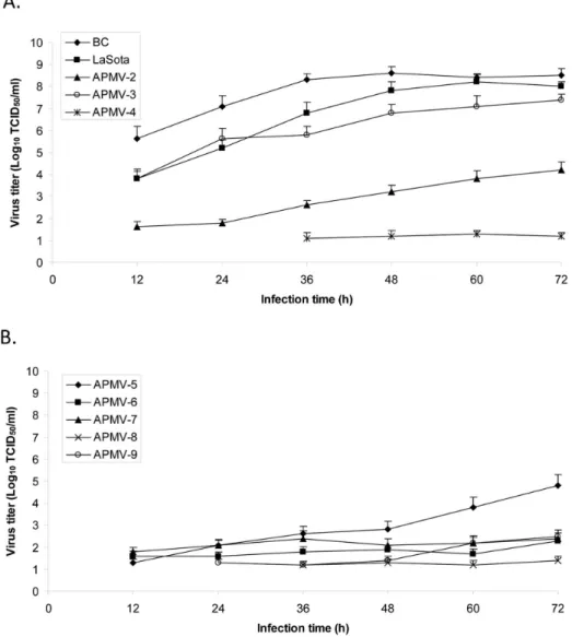
We further evaluated the multicycle replication of the APMVs in DF1 cells (Fig. 2). There was a great difference in the kinetics and magnitude of replication among different APMVs. APMV-1 strain BC and APMV-1 lentogenic strain LaSota grew better than any other APMVs and reached the highest titers (108TCID50/ml)
at 32 and 60 hpi, respectively. APMV-3 replicated relatively well, reaching 107TCID50/ml at 72 hpi. The highest titers of APMV-2
and -5 also were detected at 72 hpi, but their titers were 4.0 log10
lower than those of APMV-1. In general, replication of other APMV viruses was limited in DF1 cells (,102 TCID50/ml).
Inefficient virus replicationin vitro(i.e., APMV-4, 6, 7, 8, and -9) was correlated with the pattern of a single-cell infection as observed with CPE in infected cells (Fig. 1).
3.3. Evaluation of Pathogenicity of APMVs in Chickens by MDT and ICPI Tests
LaSota, regardless of their isolation sources. The one exception was APMV-3, which had a MDT value (117 h) that is similar to that of the LaSota strain, but had an ICPI value (0.53) that was higher than that of the LaSota strain (0.00), but nonetheless remained in the avirulent range. The MDT and ICPI values of other APMVs were.144 h and 0.00, respectively, consistent with being avirulent. Chicks infected with APMV serotypes 2–9 had no apparent clinical signs during the 8-day period of the ICPI test.
3.4. Replication of APMVs in Neuronal Cells and in the Brains of 1-day-old Chicks
To evaluate neurotropism of APMVs, virus replication was evaluated in primary chicken neuronal cells in vitro. We first evaluated infection (MOI of 0.1 PFU) in neuronal cells by confocal microscopy analysis of cells that had been immunostained at 72 hpi with N protein against each respective APMV (Fig. 3A). Expression of the N protein of APMV-1 strain BC was clearly detected in dendrites and axons. In contrast, APMV-1 strain LaSota failed to replicate in neuronal cells even after supplemen-tation with allantoic fluid as a source of protease. Among serotypes 2–9, APMV-3 was able to replicate well without the addition of
allantoic fluid, whereas the presence of APMV-7 and -8 antigens in infected cells was sporadically detected. Replication of the other APMVs was not detected up to 3 dpi. We also examined replication in the neuronal cell cultures by collecting supernatants from the cultures at 12 h intervals and assaying for infectious virus by titration. This confirmed the ability of APMV-3 to replicate in neuronal cells, although its titer was lower than that of neurovirulent APMV-1 strain BC (Fig. 3B). Titers of BC and APMV-3 in neuronal cells increased gradually and reached 5.5 and 3 log TCID50/ml, respectively, at 72 hpi. In contrast, other
APMVs, including APMV-1 strain LaSota, were not detected in the supernatants of neuronal cells, indicating their inability to replicate in these cultures of primary chicken neuronal cells.
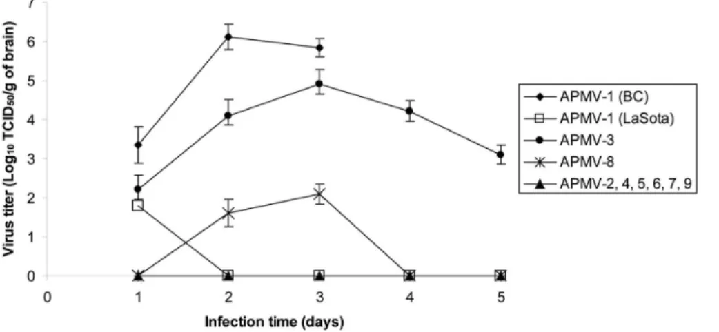
We further determined the ability of the APMV serotypes to replicate in chicken brains by inoculating 1-day-old chicks via the intracerebral route. The infected chickens were sacrificed daily and brain tissue was collected for virus titration (Fig. 4). In chicks infected with neurovirulent APMV-1 strain BC, the virus reached the highest titer (.6.0 log10 TCID50/g) in the brain at 2 dpi,
resulting in the death of all of the infected chicks on 3 dpi. The non-neurovirulent LaSota strain was detected at low titer on day 1, but was not detected on subsequent days and did not cause disease
Figure 1. Cytopathic effect in Vero cells infected with avian paramyxoviruses (APMVs).Vero cells in six-well plates were infected with each of APMVs at a multiplicity of infection (MOI) of 0.1 PFU/cell and incubated for 48 h. Exogenous protease was provided in the case of APMV-3, -8, and -9 (Materials and Methods). The viral plaques in the infected cells were visualized by immunoperoxidase staining using polyclonal antibodies raised against the N protein of the respective APMV.
or death during the 5 days of observation. APMV-3 strain reached a titer of 5 log10TCID50/g on 3 dpi, but did not cause disease or
death of any of the infected chicks during the 5 days of observation. We detected a low level of replication of APMV-8 (,2 log10 TCID50/g), with no observed disease or death.
Replication of the other APMVs was not detectable on any day, and there was no detectable disease or death. Thus, APMV-3 was identified as the only neurotropic virus among APMV serotypes 2– 9. However, despite the ability of APMV-3 to replicate to moderate titer in the brain, it did not cause discernable disease or death and thus was non-neurovirulent.
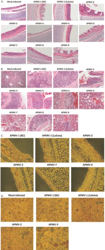
The neurotropism of the APMVs in 1-day old chicks also was evaluated by immunohistochemistry analysis of brain tissue harvested on day 3 (Fig. 5). Viral antigens were detected in the brain tissues that had been infected with APMV-1 strain BC and APMV-3, confirming the replication of the two viruses in chicken brain. Our result showed a more extensive distribution of viral antigens in brain tissue infected with BC compared to APMV-3. In contrast, presence of viral antigens was not detected in the brain tissues that had been infected with the other APMVs (Fig. 5).
3.5. Replication of APMVs in Chickens and Ducks
We next determined the replication and tissue tropism of APMVs in 1-day-old and 2-week-old chickens following intranasal inoculation. Chickens were sacrificed on 3 dpi for 1-day-old chicks and on 4 dpi for 2-week-old chickens, and the following tissues were harvested for quantitative virology: trachea, lungs, spleen, and brain (Fig. 6). In 1-day-old chicks, APMV-1 strain BC replicated to high titers (.5.0 log10 TCID50/g) in each of the
sampled tissues in each of the birds, whereas LaSota replicated in trachea, lung and spleen, but not the brain, and its titers were less than those of BC (Fig. 6A). APMV-3 replicated to moderate titers (.4.0 log10 TCID50/g) in all of the collected samples, including
the brain. However, replication of other APMVs was restricted to the trachea, and their titers were low, ranging from 1.5 to 3 log10
TCID50/g (data for APMV-2 are shown in Fig. 6A; the other
serotypes are not shown). Chicks infected with APMV-1 strain BC began to show clinical signs on 1 dpi and distinctive neurological signs on 2 dpi, whereas chicks infected with other APMVs had no apparent clinical signs during the 3-day post-infection.
Figure 2.In vitrogrowth characterization of APMVs in chicken embryo fibroblast DF1 cells.Part A shows APMV-1 to -4, and Part B shows APMV-5 to -9. The growth characteristics of the APMVs were determined by multicycle growth curve in DF1 cells infected with an MOI of 0.01 PFU/ cell. Exogenous protease was provided in the case of APMV-1 LaSota and APMV-3, -8, and -9 (Materials and Methods). The viral titers were determined by limiting dilution on DF1 or Vero cells and immunostaining with polyclonal antibodies raised against the respective N protein.
In 2-week-old chickens, virus replication was more restricted and viral titers were lower compared to 1-day-old chickens (Fig. 6B). APMV-1 strain BC was able to replicate in all of the collected tissues, but the virus titers in the brains of older birds (2.6 log10TCID50/g) were much lower than that of 1-day-old chickens
(6.5 log10 TCID50/g). APMV-1 strain LaSota replicated in the
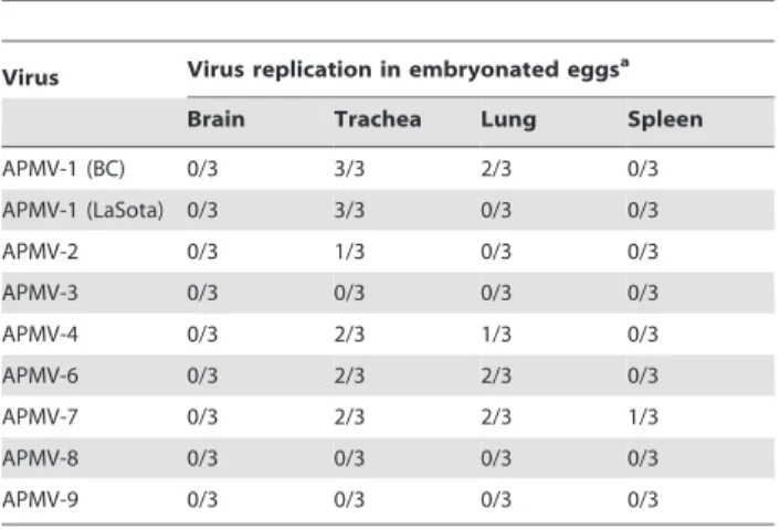
trachea, lungs, and spleen, but not the brain, as was observed in the 1-day-old chickens. Interestingly, APMV-3 replication was detected only in the trachea and the brain, indicating that it is neutrotropic in these older chickens despite this restricted replication. APMV-2 was also able to replicate in trachea of 2-week-old chickens. However, replication of the other APMVs was not detected in any of the harvested tissue samples. Since APMV 2–9 can replicate much better in embryonated chicken eggs than in cell culture [20], the collected tissue homogenates from this same experiment also were inoculated into 9-day-old embryonated chicken eggs to confirm the replication of viruses in various tissues (Table 2; APMV-5 was not assayed because it does not replicate in the allantoic cavity of chicken eggs). This approach with increased sensitivity enabled us to detect replication of all of the tested APMVs in the trachea of most of the birds. In addition, APMV-2, 3, 6, 7, and 9 were detected in the lungs of at least one of the three chickens in each group, and APMV-3 was detected in the spleen of two chickens. Among the tested APMVs, APMV-4 showed the least replication in chickens both in vitro and in vivo (Fig. 2 and Table 2), suggesting that it has a strong host range restriction in chickens. On 10 dpi, none of the APMVs were detected in any of the sampled tissues from any of the birds (not shown). Clinical signs of illnesses in any of the infected groups were not found up to 10 dpi.
Histopathological examinations of tissue samples collected on 4 dpi revealed similar microscopic findings in all APMVs. Specifically, the trachea showed mild lymphocytic tracheitis, mild to moderate multifocal mucosal attenuation, and loss of tracheal alveolar mucous glands (Fig. 7A). Lung sections exhibit moderate, multifocal, lymphocytic to lymphohistiocytic bronchitis with mild to moderate perivascular and peribronchial interstitial inflamma-tion and focal perivascular cuffing with varying severity (Fig. 7B). Minimal lymphoid depletion was detected in the spleen, and microscopic lesions were not found in any of the brain tissues (data
not shown). The presence of virus antigens in various tissues of infected chickens was further evaluated by immunohistochemistry analysis. Deparaffinized sections of the virus-infected and un-infected control tissue were immunostained using polyclonal antibodies against the N protein of the respective APMV. The presence of antigens for most APMVs was detected in the epithelial lining of trachea (Fig. 7C). However, no antigen was detected in any tissues harvested from chickens infected with APMV-5. In the lungs, the viral antigens were mostly localized in the epithelium surrounding the medium and small bronchi (Fig. 7D). Lung tissues showed extensive presence of antigens for APMV-1 strain BC followed by APMV-1 strain LaSota, APMV-3, and APMV-2. However, presence of antigens for other serotypes was not detected in lung tissues. Our results suggested that APMV 2–9 were avirulent, and their replication was mostly restricted to trachea and lungs of infected chickens.
Since several of the specific APMV strains under evaluation (i.e., APMV-4, -6, and -9) were isolated from other avian species including the mallard duck (see Table 1), their replication and pathogenicity were further evaluated in groups of 3-week-old ducks following intranasal inoculation. No clinical signs of illnesses in any of the infected groups were found up to 10 dpi. Three birds from each group were sacrificed on 4 dpi and the tissues were harvested for virus titration. None of the APMVs were detected in any of the tested tissues from any of the ducks by virus titration in DF1 cells (data not shown). However, APMVs were detected in some of the samples of trachea, lung, and spleens by inoculation into 9-day-old embryonated chicken eggs, although none of the APMVs (including APMV-1) were detected in any of the brain samples (Table 3; APMV-5 was not evaluated). APMV-1 strains BC and LaSota were the only APMVs that were detected in all three ducks in their respective groups, although replication was mostly restricted to the trachea. APMV-2 was found only in the trachea of a single duck, in contrast to its more efficient replication in chickens. APMV-3 was not detected in any samples from any duck, in contrast to its efficient replication and neurotropism in chickens. APMV-8 and -9 also were not detected in any duck. The other serotypes (APMV-4, -6, and -7) were each detected in the trachea of two ducks from its group, and in the lungs of one or two ducks from its group. APMV-4 replicated slightly better in ducks Table 1.Pathogenicity of avian paramyxoviruses (APMVs) in embryonated eggs and chicks.
Pathogenicity
Virus Strain Isolation source Fusion protein cleavage site MDTa ICPIb
APMV-1 BC Chicken RRQKRQF 58 h 1.55
APMV-1 LaSota Chicken GRQGRQL 112 h 0.00
APMV-2 Yucaipa Chicken KPASRQF .144 h 0.00
APMV-3 Netherlands Parakeet RPRGRQL 117 h 0.53
APMV-4 Hong Kong Duck DIQPRQF .144 h 0.00
APMV-5 Kunitachi Budgerigar KRKKRQF .144 h 0.00
APMV-6 Hong Kong Duck APEPRQL .144 h 0.00
APMV-7 Tennessee Dove LPSSRQF .144 h 0.00
APMV-8 Delaware Goose YPQTRQL .144 h 0.00
APMV-9 New York Duck IREGRQI .144 h 0.00
aMean embryo death time (MDT): the mean time (h) for the minimum lethal dose of virus to kill all of the inoculated embryos. Pathotype definition for APMV-1: virulent
strains,,60 h; intermediate virulent strains, 60 to 90 h; avirulent strains,.90 h.
bIntracerbral pathogenicity index (ICPI): evaluation of disease and death following intracerebral inoculation in 1-day-old SPF chicks. Pathotype definition: virulent strains,
than in chickens. APMV-7 was the only serotype detected in the spleen (of a single duck), whereas it had not been detected in the spleen of infected chickens. Thus, the pattern of infection in chickens and ducks was substantially different and could not be predicted by the isolation history.
Discussion
APMVs are frequently isolated from a wide variety of avian species and are grouped into nine serotypes based on antigenic analysis [33], with a likely tenth serotype recently described in
Figure 3. Replication of APMVs in primary chicken neuronal cells.Neuronal cells were infected with APMVs at an MOI of 0.1. (A) In 48 hpi, the cells were fixed with 4% paraformaldehyde, permeabilized with 0.2% Triton X-100, stained with a neuronal marker (anti-neuron specific beta III tubulin antibody) and polyclonal antibodies against the N protein followed by anti-Alexa Fluor 488 and 596, and then analyzed by confocal microscopy. The neuronal maker and viral N proteins stained red and green, respectively. (B) Virus replication in neuronal cells was determined by collecting the supernatants every 12 h. The viral titers were determined by limiting dilution on DF1 or Vero cells and immunostaining with polyclonal antibodies against the respective N protein.
penguins [34] that was not evaluated in the present study. APMV-1 (NDV) is the most extensively characterized member of the APMV serotypes. APMV-2 to -9 have been isolated from both wild and domestic birds, but their disease potential in wild or domestic birds was largely unknown. APMV-1 also has been shown to infect a number of non-avian species [35] and presently is being evaluated as a potential human vaccine vector for human
pathogens [36]. There is a possibility that APMV-2 to -9 could also be used as human vaccine vectors for human pathogens, providing multiple vectors with minimal cross-restriction from vector-specific immunity. Our previous studies demonstrated that APMV-2 to -9 can replicate in mammalian hosts, specifically in the hamster and mouse models [29,30]. However, their replication and pathogenicity in avian host has not been comprehensively
Figure 4. Growth kinetics of APMVs in the brains of infected 1-day-old chicks.Ten 1-day-old SPF chicks were inoculated with APMVs via the intracerebral route. Two birds in each group were sacrificed daily until 5 dpi. The virus titers in the collected samples were determined by limiting dilution in DF1 cells and immunostaining with polyclonal antibodies raised against the respective N protein.
doi:10.1371/journal.pone.0034927.g004
Figure 5. Detection of viral antigens of APMVs in the brains of infected 1-day-old chicks by immunohistochemistry.Chicks were inoculated with each virus by the intracerebral route, and brain tissue was harvested for immunohistopathology on 3 dpi. The tissues were fixed in phosphate-buffered formalin, and sections were prepared and stained using an antibody against the respective N protein (stained red).
evaluated and compared. Therefore, in this study, we evaluated
in vitro and in vivo replication and pathogenicity of APMVs in chickens and their in vivoreplication and pathogenicity in ducks and characterized differences in their tropisms, including neuro-tropism in chickens.
Virulent APMV-1 strains contain a multibasic cleavage site (with the general consensus sequence of RRQKRQF) with a polybasic furin motif (RX[R/K]RQ) that is readily cleaved in cell culture by intracellular furin or furin-like protease. Avirulent APMV-1 strains lack this polybasic site and depend on extracellular protease for cleavage, whichin vitrocan be supplied by exogenous protease andin vivois supplied by secretory trypsin-like protease found in the lumen of the respiratory and enteric tracts [20]. Our study showed that APMV-1 strain BC readily forms syncytia in cell culture and replicates more efficientlyin vitro
than any of the other APMVs, followed by the APMV-1 LaSota strain. Characterization of the CPE of APMV types 2–9
demonstrated that only APMV-3 and -5 induced syncytium formation in infected cells. The cleavage site of APMV-3 (RPRGRQL) lacks the furin motif, and efficient growth and syncytium formation depended on supplementation with exoge-nous protease, resembling an avirulent APMV-1 strain. The cleavage site of APMV-5 (KRKKRQF) is the only example from APMV2–9 to have a furin motif [6], and syncytium formation and growth were independent of added protease, resembling a virulent APMV-1 strain. Production of syncytia by APMV-3 and APMV-5 correlated with efficient multicycle replication in cell culture in the case of APMV-3 and a moderate level of replication in cell culture in the case of APMV-5. Although APMV-5 contains the greatest number of basic residues for any of the APMV except for some strains of APMV-1, its replication in DF1 cells was much less efficient compared to APMV-1 and -3. Interestingly, the level of replication of APMV-5 in cell culture was very similar to that of APMV-2, which lacks a furin motif (KPASRQF), does not form
Figure 6. Replication of APMVs in 1-day- and 2-week-old chickens.Groups of (A) 1-day- or (B) 2-week-old chickens were inoculated with each virus (256 HA units) by the intranasal route. Three birds from each group were sacrificed on 3 dpi (1-day-old chicks) or 4 dpi (2-week-old chickens), and virus titers in the collected tissues samples (brain, trachea, lung, and spleen) were determined by limiting dilution in DF1 cells and immunostaining with polyclonal antibodies raised against the respective N protein.
syncytia, and whose replication and lack of ability to form syncytia are independent of exogenous protease. Thus, for those two viruses (APMV-2 and -5), the growth phenotype in cell culture did not correlate well with the presence or absence of a furin motif in the cleavage sequence. The other APMVs (types 4, 6, 7, 8, and 9) have one or two basic amino acids in their cleavage site sequences and exhibited single cell infection and inefficient replicationin vitro. However, only in the case of serotypes 8 and 9 did the addition of exogenous protease increase the efficiency of replication, and the overall level of replication remained very low and syncytia were not observed. Thus, APMV types 2, 4, 6, 7, 8, and 9 have cleavage sites that seemed similar to those of avirulent APMV-1 strains, but the inclusion of protease had little effect on growth in cell culture. The pathogenicity of APMVs was first characterized by standard pathogenicity tests, namely the ICPI and MDT assays. We included two well-characterized strains of APMV-1, meso-genic BC and lentomeso-genic LaSota, and compared their pathoge-nicity with that of APMV serotypes 2–9. This showed that APMV serotypes 2–9 were avirulent by both assays. Of types 2–9, only APMV-3 was associated with embryo death in the MTD assay and disease in the ICPI assay, although these effects were modest and APMV-3 was categorized as avirulent by the standards of these assays. By the MDT assay, the other types (types 2, 4, 5, 6, 7, 8, and 9) lacked detectable virulence and thus were less virulent than the well-known avirulent vaccine strain APMV-1 LaSota, and by the ICPI assay these APMVs were equivalent to LaSota in lacking detectable virulence. Thus, possession of a furin cleavage site and the ability to form syncytia by APMV-5 did not correlate with virulence in vivo, nor did the ability of APMV-2 to replicate to moderate titers in cell culture without the addition of trypsin.
We investigated the neurotropism of APMVs in chicken neuronal cellsin vitroand in the brains of 1-day-old chicks infected via the intracerebral route. The neurovirulent APMV-1 strain BC was observed to replicate in chicken neuronal cells both on the basis of antigen expression and the production of infectious virus. In contrast, the non-neurovirulent LaSota strain did not detectably express viral antigens in these cells, and infectious virus was detected in the culture only at a low level on a single day early in
Table 2.Replication of APMVs in 2-week-old chickens.
Virus Virus replication in embryonated eggsa
Brain Trachea Lung Spleen
APMV-1 (BC) 3/3 3/3 3/3 3/3
APMV-1 (LaSota) 0/3 3/3 3/3 3/3
APMV-2 0/3 3/3 2/3 0/3
APMV-3 2/3 3/3 3/3 2/3
APMV-4 0/3 1/3 0/3 0/3
APMV-6 0/3 2/3 1/3 0/3
APMV-7 0/3 3/3 2/3 0/3
APMV-8 0/3 2/3 0/3 0/3
APMV-9 0/3 3/3 1/3 0/3
aGroups of 2-week-old chickens were inoculated with each virus by the
intranasal route. Three birds from each group were sacrificed on day 4, and tissues samples (brain, trachea, lung, and spleen) were collected and homogenized. To confirm the virus replication, each sample (100ml) in the
collected samples was inoculated into three eggs, and allantoic fluids were collected on 3 dpi. Virus replication was determined by hemagglutination assay. Note that APMV-5 was not analyzed because it does not replicate in the allantoic cavity of chicken eggs.
doi:10.1371/journal.pone.0034927.t002
infection. Among serotypes 2–9, expression of viral antigen in the neuronal cells was detected only with APMV-3, -7, and -8, and production of infectious virus was detected only with APMV-3. Thus, only APMV-1 strain BC and APMV-3 appeared to replicate productively in these neuronal cell cultures.
Following intracerebral inoculation of 1-day-old chicks, only APMV-1 strain BC, APMV-3, and (to a much lesser extent) 8 could be detected in brain tissue samples. With APMV-1, virus neurotropism depends on the presence of a furin cleavage site, since secretory proteases are unavailable in the brain [37]. The lack of neurotropism of the APMV-1 strain LaSota is consistent with this idea. However, APMV-3 appears to be an exception, since it lacks a furin site and is dependent on added protease in cell culture and thus presumably dependent on secreted protease in vivo, yet it replicated efficiently in the chick brains and neutronal cells in vitro. Interestingly, however, while APMV-3 was clearly neurotropic, it caused no disease or death and thus was not neurovirulent. These findings indicate that the simple paradigm that neurotropism depends on the presence of a furin cleavage site does not hold for APMV-3. Furthermore, it shows that neurotropism by an APMV does not necessarily confer neurovirulence. Our previous study with replacement of the APMV-2 F protein with that of APMV-1 BC suggested an important role for the BC F protein in virus neurotropism, neuroinvasiveness, and neurovirulence in 1-day-old chicks [38]. Based on the generally held model for APMV-1 pathogenesis, it would have been reasonable to suggest that the presence of the furin site in the BC F protein was the major determinant of the observed neurotropism, neuroinvasiveness, and neurovirulence. However, the present study challenges the predominant role for the cleavage site, and suggests that other features of the F protein may be involved in the observed neurotropism, neuroinvasiveness, and neurovirulence. Evaluating the difference in the roles of the F proteins in virus neurotropism versus neurovirulence may provide information on these activities and may help design safer vaccines for neurotropic pathogens.
We also evaluated replication, tropism, and pathogenesis following intranasal infection in 1-day-old chicks and 2-week-old chickens. In 1-day-old chickens, only the APMV-1 strain BC and APMV-3 were able to spread to the brain and replicate, demonstrating neuroinvasiveness and neurotropism. These two viruses, plus APMV-1 strain LaSota, also were detected in the spleen of 1-day-old chicks. Conversely, replication of the other APMVs was mostly detected the in trachea of chicks, and the virus titers were moderate.
In 2-week-old birds, at 4 dpi, infection of APMV-1 strain BC was found in all of the tested tissues, including the brain, and LaSota was detected in all of the tested tissues except the brain. APMV-3 also was detected in all of the tested tissues including the brain. Replication of the other APMVs was restricted to the trachea and lungs. In general, virus titers in the 2-week-old chickens were lower than in the 1-day-old chicks. By 10 dpi, no virus could be detected in any of the tissues in any of the infected chickens for any serotype by virus isolation, suggesting that the
virus was cleared from all tissues and disease was resolved, indicating the self-limited nature of the infections.
Histopathology analysis of APMV-infected 2-week-old chickens showed that infection by each of the APMVs, including APMV-5, produced tracheitis and mild pathology that was mainly restricted to the respiratory tract. Previous study with serologic assays in chickens infected with APMVs demonstrated a good humoral response on 14 dpi [39]. Thus, the birds indeed appeared to be infected even thought the recovery of infectious virus was often sporadic and low. A single APMV type, namely APMV-5, was not detected by virus isolation or by immunohistochemistry in any of the chickens in this study. However, the serologic assay of chickens that had been inoculated with APMV-5 also showed the development of virus specific antibodies detected by virus plaque reduction neutralization assay [39], suggesting that infection had occurred. This suggests a low level of virus replication in chickens. Thus, although APMV-5 bears a furin cleavage site and can cause 100% mortality in budgerigars [25], it was completely avirulent and replicated inefficiently in chickens. This is suggestive of a host range difference that was not greatly ameliorated by the presence of a multi-basic cleavage site.
Experimental infection of mallard ducks with APMVs indicated that all APMV serotypes, including duck isolates, are avirulent in ducks. Differences were observed in the replication of various APMVs between chickens and ducks. For example, replication of APMV-1 strain BC was low in ducks compared to its efficient replication chickens, although APMV-1 has shown to infect waterfowls, such as geese and Muscovy and Pekin ducks [40–42]. Several APMV serotypes (APMV-3, -8, and -9) did not replicate in ducks, even though APMV-3 replicated well in chickens and the APMV-9 strain had been isolated from ducks. In chickens, APMV-4 poorly replicated in both 1-day-old and 2-week-old chickens in our study. In addition, the virus has been shown to induce low HI titers in chickens compared to other APMVs, which was taken as evidence of a low level of replication of this virus in chickens [26,39]. However, we detected replication of APMV-4 in trachea and lungs of infected ducks, indicating its preference to ducks from which this strain had been isolated.
moderate multifocal mucosal attenuation, and loss of tracheal alveolar mucous glands. Lung sections (B) exhibit moderate, multifocal, lymphocytic to lymphohistiocytic bronchitis with mild to moderate perivascular and peribronchial interstitial inflammation and focal perivascular cuffing with varying severity. The presence of antigens for most APMVs was detected in the epithelial lining of trachea (C) and in the epithelium surrounding the medium and small bronchi of the lungs (D).
doi:10.1371/journal.pone.0034927.g007
Table 3.Replication of APMVs in 3-week-old ducks.
Virus Virus replication in embryonated eggsa
Brain Trachea Lung Spleen
APMV-1 (BC) 0/3 3/3 2/3 0/3
APMV-1 (LaSota) 0/3 3/3 0/3 0/3
APMV-2 0/3 1/3 0/3 0/3
APMV-3 0/3 0/3 0/3 0/3
APMV-4 0/3 2/3 1/3 0/3
APMV-6 0/3 2/3 2/3 0/3
APMV-7 0/3 2/3 2/3 1/3
APMV-8 0/3 0/3 0/3 0/3
APMV-9 0/3 0/3 0/3 0/3
aGroups of 3-week-old ducks were inoculated with each virus by the intranasal
route. Three birds from each group were sacrificed on day 4, and tissues samples (brain, trachea, lung, and spleen) were collected and homogenized. To confirm the virus replication, each sample (100ml) in the collected samples was
inoculated into three eggs, and allantoic fluids were collected on 3 dpi. Virus replication was determined by hemagglutination assay. Note that APMV-5 was not analyzed because it does not replicate in the allantoic cavity of chicken eggs.
In summary, our findings indicate that APMV serotypes 2–9 were avirulent in both chickens and ducks as well as in standard international assays. Each of the APMV serotypes replicated to low-to-moderate titers in the trachea of chickens, with APMV-3 having the highest titers. Among APMV types 2–9, only APMV3 replicated systemically and was neuroinvasive and neurotropic, although not neurovirulent. The sequence of the F protein cleavage site was not a reliable predictor of pathogenicity of APMVs in chickens, indicating incongruity with the well-known APMV-1 paradigm. For example, the systemic replication and neurotropism of APMV-3 is inconsistent with its lack of a furin cleavage site and dependence on exogenous trypsin for replication in cell culture. As another example, several of the APMV serotypes, including types 2, 4, 6, and 7, lacked a furin site, but replication of these viruses in cell culture was not improved by the addition of exogenous trypsin. As yet another example, APMV-5 was the only one of serotypes 2–9 to have a furin site, and yet it replicated to only moderate levels in cell culture, and replication in chickens could be detected only by seroconversion. Thus, the paradigm from studies with APMV-1 that virulence and systemic spread correlates with the presence of a furin site generally was not
observed with APMV serotypes 2–9. Reverse genetics for different pathotypes of APMV-1 and for prototype strains of APMV-2, 3, and -7 have been developed and used for studying determinants of virus pathogenesis [38,43–45]. Further characterization of virus replication and pathogenicity using reverse genetics will enhance our understanding of overall APMV pathogenesis.
Acknowledgments
We thank Daniel Rockemann, Girmay Gebreluul, Yonas Araya, and our laboratory members for excellent technical assistance. The views expressed herein do not necessarily reflect the official policies of the Department of Health and Human Services, nor does mention of trade names, commercial practices, or organizations imply endorsement by the U.S. Government.
Author Contributions
Conceived and designed the experiments: SHK PLC SKS. Performed the experiments: SHK SX. Analyzed the data: SHK SX HS. Contributed reagents/materials/analysis tools: PLC SKS. Wrote the paper: SHK PLC SKS.
References
1. Lamb R, Parks G (2007)Paramyxoviridae: the viruses and their replication. In: Knipe DM, Howley PM, Griffin DE, Lamb RA, Martin MA, Roizman B, Straus SE, eds. Philadelphia: Lippincott Williams & Wilkins. pp 1449–1496. 2. Alexander DJ (2003) Avian paramyxoviruses 2–9. In: Saif, Y.M. (Ed.), Diseases
of Poultry, 11th ed. Iowa State University Press, Ames, 88–92.
3. Subbiah M, Xiao S, Collins PL, Samal SK (2008) Complete sequence of the genome of avian paramyxovirus type 2 (strain Yucaipa) and comparison with other paramyxoviruses. Virus Res 137: 40–48.
4. Kumar S, Nayak B, Collins PL, Samal SK (2008) Complete genome sequence of avian paramyxovirus type 3 reveals an unusually long trailer region. Virus Res 137: 189–197.
5. Nayak B, Kumar S, Collins PL, Samal SK (2008) Molecular characterization and complete genome sequence of avian paramyxovirus type 4 prototype strain duck/Hong Kong/D3/75. Virology J 5: 124.
6. Samuel AS, Paldurai A, Kumar S, Collins PL, Samal SK (2010) Complete genome sequence of avian paramyxovirus (APMV) serotype 5 completes the analysis of nine APMV serotypes and reveals the longest APMV genome. PLoS One 5: e9269.
7. Xiao S, Subbiah M, Kumar S, De Nardi R, Terregino C, et al. (2010) Complete genome sequences of avian paramyxovirus serotype 6 prototype strain Hong Kong and a recent novel strain from Italy: evidence for the existence of subgroups within the serotype. Virus Res 150: 61–72.
8. Xiao S, Paldurai A, Nayak B, Subbiah M, Collins PL, et al. (2009) Complete genome sequence of avian paramyxovirus type 7 (strain Tennessee) and comparison with other paramyxoviruses. Virus Res 145: 80–91.
9. Paldurai A, Subbiah M, Kumar S, Collins PL, Samal SK (2009) Complete genome sequences of avian paramyxovirus type 8 strains goose/Delaware/ 1053/76 and pintail/Wakuya/20/78. Virus Res 142: 144–153.
10. Samuel AS, Kumar S, Madhuri S, Collins PL, Samal SK (2009) Complete sequence of the genome of avian paramyxovirus type 9 and comparison with other paramyxoviruses. Virus Res 142: 10–18.
11. Lamb RA, Paterson RG, Jardetzky TS (2006) Paramyxovirus membrane fusion: lessons from the F and HN atomic structures. Virology 344: 30–37. 12. Calain P, Roux L (1993) The rule of six, a basic feature for efficient replication of
Sendai virus defective interfering RNA. J Virol 67: 4822–4830.
13. Deng R, Wang Z, Mirza AM, Iorio RM (1995) Localization of a domain on the paramyxovirus attachment protein required for the promotion of cellular fusion by its homologous fusion protein spike. Virology 209: 457–469.
14. Melanson VR, Iorio RM (2004) Amino acid substitutions in the F-specific domain in the stalk of the Newcastle disease virus HN protein modulate fusion and interfere with its interaction with the F protein. J Virol 78: 13053–13061. 15. Teng MN, Whitehead SS, Collins PL (2001) Contribution of the respiratory
syncytical virus G glycoprotein and its secreted and membrane-bound forms to virus replication in vitro and in vivo. Virology 289: 283–296.
16. Waning DL, Schmitt AP, Leser GP, Lamb RA (2002) Roles for the cytoplasmic tails of the fusion and hemagglutinin-neuraminidase proteins in budding of the paramyxovirus simian virus 5. J Virol 76: 9284–9297.
17. Alexander DJ (1982) Avian paramyxoviruses-other than Newcastle disease virus. World’s Poul Sci J 38: 97–104.
18. Pedersen JC, Senne DA, Woolcock PR, Kinde H, King DJ, et al. (2004) Phylogenetic relationships among virulent Newcastle disease virus isolates from the 2002–2003 outbreak in California and other recent outbreaks in North America. J Clin Microbiol 42: 2329–2334.
19. Alexander DJ (1989) Newcastle disease, P.114–120. In: Purchase HG, Arp LH, Domermuth CH, Pearson JE, eds. A laboratory manual for the isolation and identification of avian pathogens, 3rd
ed The American Association of Avian Pathologists, Kendall/Hunt Publishing Company, Dubuque, IA.
20. Samal SK (2011) Newcastle disease and related avian paramyxoviruses. p. 69– 114. In Samal SK, editor. The biology of paramyxoviruses. Caister Academic Press, Norfolk, U.K.
21. Lipkind MA, Weisman Y, Shihmanter E, Shoham D, Aronovici A (1979) The isolation of yucaipa-like paramyxoviruses from epizootics of a respiratory disease in turkey poultry farms in Israel. Vet Rec 105: 577–578.
22. Bankowski RA, Almquist J, Dombrucki J (1981) Effect of paramyxovirus yucaipa on fertility, hatchability, and poult yield of turkeys. Avian Dis 25: 517–520. 23. Zhang GZ, Zhao JX, Wang HW, Yang AM, Bu CY, et al. (2006) Isolation,
identification, and comparison of four isolates of avian paramyxovirus serotype 2 in China. Avian Dis 50: 386–390.
24. Tumova B, Robinson JH, Easterday BC (1979) A hitherto unreported paramyxovirus of turkeys. Res Vet Sci 27: 135–140.
25. Nerome K, Nakayama M, Ishida M, Fukumi H (1978) Isolation of a new avian paramyxovirus from budgerigar (Melopsittacus undulatus). J Gen Virol 38: 293–301.
26. Warke A, Appleby L, Mundt E (2008) Prevalence of antibodies to different avian paramyxoviruses in commercial poultry in the United States. Avian Dis 52: 694–697.
27. Subbiah M, Xiao S, Khattar SK, Dias FM, Collins PL, et al. (2010) Pathogenesis of two strains of avian paramyxovirus serotype 2, Yucaipa and Bangor, in chickens and turkeys. Avian Dis 54: 1050–1057.
28. Kumar S, Dias FM, Nayak B, Collins PL, Samal SK (2010) Experimental avian paramyxovirus serotype-3 infection in chickens and turkeys. Vet Res 41: 72. 29. Samuel AS, Subbiah M, Shive H, Collins PL, Samal SK (2011) Experimental
infection of hamsters with avian paramyxovirus serotypes 1 to 9. Vet Res 42: 38. 30. Khattar SK, Kumar S, Xiao S, Collins PL, Samal SK (2011) Experimental infection of mice with avian paramyxovirus serotypes 1 to 9. PLoS One 6: e16776.
31. Reed LJ, Muench HA (1938) A simple method of estimating fifty percent endpoints. Am. J Hyg 27: 493–497.
32. Pasick JM, Kalicharran K, Dales S (1994) Distribution and trafficking of JHM coronavirus structural proteins and virions in primary neurons and the OBL-21 neuronal cell line. J Virol 68: 2915–2928.
33. Alexander DJ (2000) Newcastle disease and other avian paramyxoviruses. Rev Sci Tech 19: 443–462.
34. Miller PJ, Afonso CL, Spackman E, Scott MA, Pedersen JC, et al. (2010) Evidence for a new avian paramyxovirus serotype 10 detected in rockhopper penguins from the Falkland Islands. J Virol 84: 11496–11504.
35. Hofstad MS (1950) Experimental inoculation of swine and sheep with Newcastle disease virus. Cornell Vet 40: 190–197.
36. Bukreyev A, Collins PL (2008) Newcastle disease virus as a vaccine vector for humans. Curr Opin Mol Ther 10: 46–55.
38. Kim SH, Subbiah M, Samuel AS, Collins PL, Samal SK (2011) Roles of the fusion and hemagglutinin-neuraminidase proteins in replication, tropism, and pathogenicity of avian paramyxoviruses. J Virol 85: 8582–8596.
39. Nayak B, Dias FM, Kumar S, Paldurai A, Collins PL, et al. (2012) Avian paramyxovirus serotypes 2–9 (APMV-2-9) vary in the ability to induce protective immunity in chickens against challenge with virulent Newcastle disease virus (APMV-1). Vaccine 30: 2220–2227.
40. Shi SH, Huang Y, Cui SJ, Cheng LF, Fu GH, et al. (2011) Genomic sequence of an avian paramyxovirus type 1 strain isolated from Muscovy duck (Cairina moschata) in China. Arch Virol 156: 405–412.
41. Zou J, Shan S, Yao N, Gong Z (2005) Complete genome sequence and biological characterizations of a novel goose paramyxovirus-SF02 isolated in China. Virus Genes 30: 13–21.
42. Jinding C, Ming L, Tao R, Chaoan X (2005) A goose-sourced paramyxovirus isolated from southern China. Avian Dis 49: 170–173.
43. Subbiah M, Khattar SK, Collins PL, Samal SK (2011) Mutations in the fusion protein cleavage site of avian paramyxovirus serotype 2 that increase cleavability and syncytia formation but do not increase viral virulence in chickens. J Virol 85: 5394–5405.
44. Kumar S, Nayak B, Collins PL, Samal SK (2011) Evaluation of the Newcastle disease virus F and HN proteins in protective immunity using a recombinant avian paramyxovirus type-3 vector in chickens. J Virol 85: 6521–6534. 45. Xiao S, Khattar SK, Subbiah M, Collins PL, Samal SK (2012) Mutation of the