Universidade Nova de Lisboa
Instituto de Higiene e Medicina Tropical
Trypanosoma brucei peptidase inhibitors.
Immunolocalization, secretion and potential use as targets for
therapy
Raquel de Sá da Silva Laires
DISSERTAÇÃO PARA A OBTENÇÃO DO GRAU DE DOUTOR EM CIÊNCIAS BIOMÉDICAS,
ESPECIALIDADE DE BIOLOGIA CELULAR E MOLECULAR
ii
Universidade Nova de Lisboa
Insituto de Higiene e Medicina Tropical
Thesis: Trypanosoma brucei peptidase inhibitors. Immunolocalization, secretion and
potential use as targets for therapy
Author: Raquel de Sá da Silva Laires
Supervisor: Doctor Carlos Novo, IHMT, UNL
Co-supervisor: Professor Jeremy C. Mottram, (University of Glasgow)
Tutorial Committee:
Doctor Carlos Novo (IHMT, UNL) Doctor Luís Távora Tavira (IHMT, UNL) Professor Jorge Atouguia (IHMT, UNL)
iii
Universidade Nova de Lisboa
Insituto de Higiene e Medicina Tropical
Título da Tese: Inibidores de peptidases de Trypanosoma brucei. Imunolocalização,
secreção e potencial uso como alvo terapêutico
Autor: Raquel de Sá da Silva Laires
Orientador: Investigador Doutor Carlos Novo, IHMT, UNL
Co orientador: Professor Doutor Jeremy C. Mottram, (Universidade de Glasgow)
Comissão Tutorial:
Investigador Doutor Carlos Novo (IHMT, UNL) Investigador Doutor Luís Távora Tavira (IHMT, UNL) Professor Doutor Jorge Atouguia (IHMT, UNL)
Dissertação apresentada para cumprimento dos requisitos necessários à obtenção do grau de Doutor em Ciências Biomédicas, Especialidade de Biologia Celular e Molecular, realizada sob a orientação científica do Investigador Doutor Carlos Novo, do Professor Doutor Jeremy C. Mottram e do Investigador Doutor Luís Távora Tavira.
iv Dedico esta tese,
Ao meu pai e à minha mãe que sempre me encorajaram a ir mais longe Ao Zé que esteve sempre ao meu lado nesta longa caminhada Ao Henrique, a luz dos meus olhos, por quem eu aspiro sempre mais
“Há um tempo em que é preciso abandonar as
roupas usadas, que já tem a forma do nosso corpo, e esquecer os nossos caminhos, que nos levam sempre aos mesmos lugares. É o tempo da travessia: e, se não ousarmos fazê-la, teremos ficado, para sempre,
à margem de nós mesmos.”
v Para o meu filho Henrique uma importante lição, To my son Henrique, a valuable lesson,
“Sejam quais forem os resultados, com êxito ou não, o importante é que no final cada um possa dizer: ‘fiz o que pude’”
“Whether our efforts are, or not, favored by life, let us be able to say, when we come near the great goal, ‘I
have done what I could’”
vi
Acknowledgements
This journey wouldn't be possible without the support, scientific or moral, from several people to whom I am truly grateful.
To begin with, I would like to thank my supervisor, Dr. Carlos Novo for his constant support, guidance and friendship that made it easier to overcome all the obstacles inherent to such journey and added so much to my experience over the past 5 years.
I would also like to thank my co-supervisor, Prof. Jeremy C. Mottram for the opportunity to work in his lab in Glasgow and whose guidance and scientific support were valuable assets to my thesis.
Working in the “Mottram Lab” was definitely an amazing experience and I would like to express my gratitude to all the people that welcomed me and contributed for the success of my “Glasgow experience”, not only with scientific help but also with their friendship. Particularly, I would like to thank Elaine, Will, Daniela, Jim Scott, Nathaniel, Elmarie, Esther, Herbert, Tatiana, Alana and also Corinna, Tansy, Sophie and Glynn from the Hammarton group. It has been a pleasure to meet you all and I will sure be missing you.
To all the people that worked with me in the former UTPAM, Tiago, Bé, Sofia, Ângela, Maria, Ana Maria, Ana Domingos, Fernando; and the ones I met more recently in IHMT, Cátia, Pedro, Ana, Tiago, Isabel, Idalécia and so many others; thank you for all the support. You were all very important to the success of my PhD.
To my dear friend Rubina, a special thanks. You are partially responsible for my PhD. If it wasn’t for I wouldn’t have met all the people mentioned above. Thank you for being there for me, for your friendship and for caring.
vii Finally thanks to my “Maria” Cristina. She was my lab colleague, my roommate, my friend, my support when I was feeling down, my “clown” when I needed a laughter, my “sister” when we were away. Our times in Glasgow will never be forgotten and the Welcome Centre will never be the same!!!!
Gostava de agradecer a todos os meus amigos fora da esfera laboratorial. Não vou mencionar nomes porque são tantas as pessoas que corria o risco de me esquecer de alguém. Neste aspecto sou uma mulher rica, rica em boas amizades de pessoas que me acompanham há anos e sabem a luta que tem sido este meu percurso. Todos foram determinantes para o meu sucesso, cada um à sua maneira e a todos, um muito obrigado.
À minha irmã Claúdia e aos meus sobrinhos Andreia e Rodrigo que me acompanham sempre e que são para mim um motivo de orgulho. Obrigado por todo o amor e carinho!
Aos meus pais que sempre me encorajaram a seguir este caminho; que me apoiaram em todas as decisões; que me estimularam para ir sempre mais além; que me educaram para ser uma pessoa responsável e com valores; que fizeram de mim a pessoa que sou hoje e que desejo ser sempre!
ix
Trypanosoma brucei peptidase inhibitors. Immunolocalization, secretion and
potential use as targets for therapy
Raquel de Sá da Silva Laires
Keywords: Trypanosoma brucei, ISP1, ISP2, flagellum, flagellar pocket, endocytosis,
host-parasite interactions, monoclonal antibodies.
xi
Inibidores de peptidases de Trypanosoma brucei. Imunolocalização, secreção e potencial uso como alvo terapêutico
Raquel de Sá da Silva Laires
Palavras-chave: Trypanosoma brucei, ISP1, ISP2, flagelo, bolsa flagelar, endocitose, interacções parasita-hospedeiro, anticorpos monoclonais.
As peptidases encontram-se envolvidas em diversas funções biológicas possuindo um papel importante na patogenicidade de várias infecções parasitárias. Em mamíferos, a actividade peptidica é controlada por inibidores endógenos, como as cistatinas e as serpinas. Os genes que codificam para inibidores homólogos às cistatinas e serpinas de mamíferos, encontram-se ausentes do genoma de tripanossomatídeos. A ecotina é uma proteína de Escherichia coli, capaz de inibir uma grande variedade de peptidases serínicas da família S1A, tais como a tripsina. Existem duas proteínas do tipo da ecotina em T. brucei, ISP1 e ISP2. A ausência de peptidases sensíveis à acção dos ISPs no
genoma de Trypanosoma brucei, sugere que estes tenham como alvo as peptidases serínicas do hospedeiro. Linhas celulares de T. brucei deficientes em ISP1 (Δisp1), ISP2 (Δisp2) e em ambos os ISPs (Δisp1/2) foram produzidas com sucesso e a ausência dos inibidores comprovada com o uso de anticorpos monoclonais específicos contra o ISP1 e o ISP2, que reconhecem a proteina alvo em extractos de proteína total de parasitas selvagens mas não em extractos de parasitas mutantes. O efeito da ausência dos ISPs nas células foi avaliado in vitro e in vivo, verificando-se que a delecção individual de cada ISP não produz qualquer efeito nos parasitas que revelam um crescimento normal em cultura e padrões de infectividade em murganhos idênticos aos de parasitas selvagens. Em contraste, os parasitas Δisp1/2, embora possuam um crescimento normal
xii
Abbreviations
Amp Ampicillin
AP Alkaline phosphatase
Asp Aspartic acid
bp Base pair
BSA Bovine serum albumin
BSD Blasticidin
BSF Bloodstream form
CATT Card agglutination test for trypanosomiasis CDKs Cyclin-dependent kinases
CIP Calf intestinal alkaline phosphatase
CP Cysteine peptidase
CR3 Complement type-3 receptor DABCO 4-diazabicyclo[2.2.2]octane DAPI 4,6-diamidino-2-phenylindole DMEM Dulbecco’s Modified Eagle Medium
DMSO Dimethyl sulfoxide
DNA Deoxyribonucleic acid
DTT Dithiothreitol
EDTA Ethylene diamine tetra acetic acid ELISA Fnzyme-linked immunoabsorbent assay
FAZ flagellum attachment zone
FBS Fetal bovine serum
FC flagella connector
FCS Fetal calf serum
GPI Glycosylphosphatidylinositol GST Glutathione S-transferase
HAT Hypoxanthine-aminopterin-thymidine
HGPRT Hypoxanthineguanidinephosphoribosyltransferase
His Histidine
xiii
HT Hipozanthine-thymidine
HYG Hygromycin B
ICP Inhibitor of cysteine peptidase
IFA Indirect immunofluorescence antibody IFT Intraflagellar transport
IPTG Isopropyl-β-D-thiogalactopyranoside ISP1 Inhibitor of serine peptidase 1
ISP2 Inhibitor of serine peptidase 2
K Kinetoplast
Kan Kanamycin
Kb Kilo base
kDa Kilo Dalton
kDNA Kinetoplastid DNA
LB Luria bertani
Micro
m Milli
M Molar
MAb’s Monoclonal antibodies
MBP Maltose binding protein
N Nucleus
NE Neutrophil elastase
NEO Neomycin
NETs Neutrophil Extracellular Trap
OD Optical density
OH Hydroxyl
OPB Oligopeptidase B
ORF Open reading frame
PAC Puromycin
PBS Phosphate buffered saline
PCF Procyclic form
PCR Polymerase chain reaction
PEG Polyethylene glycol
xiv
PHLEO Phleomycin
POP Prolyl oligopeptidase
RNA Ribonucleic acid
RNAi Ribonucleic acid interference
SDS-PAGE sodium dodecyl sulphate polyacrylamide gel electrophoresis SEM Scanning electron microscopy
Ser Serine
SH Sulphydryl
SPF Specific pathogen free
T Threonine
TBS Tris buffered saline
TTBS Tween Tris buffered saline TLR4 Toll like receptor 4
UTR Untranslated region
UV Ultra violet
V Volts
VSG Variant surface glycoproteins
v/v Volume to volume
w/v Weight to volume
xv
Table of Contents
Acknowledgements ... vi
Abstract ... viii
Resumo ... x
Abbreviations ... xii
Table of Contents ... xv
Index of Figures ... xix
Index of Tables ... xxi
Chapter 1 – Introduction ... 1
1.1 Human African Trypanosomiasis ... 2
1.1.1 Epidemiology ... 2
1.1.2 Clinical features ... 5
1.1.3 Diagnosis ... 6
1.1.4 Treatment, control and potential targets for therapy ... 7
1.2 Trypanosomabrucei ... 8
1.2.1 T. brucei life cycle ... 9
1.2.1.1 Development of T. brucei in the mammalian host ... 9
1.2.1.2 Development of T. brucei in the tsetse fly vector ... 11
1.2.2 T. brucei cell biology ... 13
1.2.3 T. brucei Cell Cycle ... 21
1.3 Peptidases and their Natural Inhibitors ... 23
1.3.1 Peptidases ... 23
1.3.1.1 Cysteine Peptidases in Trypanosomatids ... 25
1.3.1.2 Serine Peptidases in Trypanosomatids... 29
1.3.2 Natural Inhibitors of Peptidases ... 33
1.3.2.1 Inhibitors of Cysteine Peptidases ... 34
1.3.2.2 Inhibitors of serine peptidases ... 38
xvi
2.1 Bacterial Cultures ... 43
2.1.1 Strains used ... 43
2.1.2 Bacterial culture and storage ... 43
2.1.3 Preparation of E. coli competent cells ... 43
2.2 Molecular Biology Techniques ... 44
2.2.1 Polymerase Chain Reaction (PCR) and oligonucleotides used ... 44
2.2.2 Agarose gel electrophoresis ... 45
2.2.3 Cloning of PCR products ... 46
2.2.4 Restriction endonuclease digestion ... 47
2.2.5 Ligation of DNA fragments ... 47
2.2.6 Transformation of DNA fragments in E. coli ... 48
2.2.7 Selection of transformants ... 48
2.2.8 Colony screening by PCR ... 48
2.2.9 Plasmid DNA purification ... 49
2.2.10 DNA sequencing ... 49
2.2.11 DNA preparation for transfection ... 49
2.2.12 Site directed mutagenesis ... 49
2.2.13 Plasmid generation ... 50
2.2.13.1 Mutant ICP construct ... 50
2.2.13.2 ISP knock out constructs ... 51
2.2.13.3 ISP1 and ISP2 re-expression constructs ... 51
2.2.14 Southern Blotting ... 52
2.3 Protein Biochemistry ... 53
2.3.1 Expression and purification of recombinant proteins ... 53
2.3.2 Determination of protein concentration ... 54
2.3.3 Polyacrylamide gel electrophoresis (SDS-PAGE) ... 54
2.3.4 Coomassie staining of SDS-PAGE ... 55
2.3.5 Western Blotting ... 55
2.4 T. brucei cell culture ... 56
2.4.1 Bloodstream form T. brucei culturing ... 56
2.4.2 BSF transfections ... 57
xvii
2.4.4 Isolation of T. brucei genomic DNA ... 59
2.4.5 Preparation of whole cell extracts ... 59
2.4.6 T. brucei animal infection and parasitemia determination ... 59
2.5 Monoclonal Antibodies ... 60
2.5.1 Generation of hybridomas ... 60
2.5.2 Screening for antibody producing Hybridomas... 61
2.5.3 Hybridomas culture and storage ... 62
2.5.4 Determination of antibody isotype ... 63
2.5.5 Antibody purification ... 63
2.6 Indirect Immnunofluorescence Analysis (IFA) ... 64
2.7 Scanning Electron Microscopy ... 64
2.8 In vitro growth inhibition assays ... 65
2.9 In vivo growth inhibition assays ... 65
2.9.1 Protection of T. brucei infected mice by ISP Immunization ... 65
2.9.2 Neutralization of T. brucei ininfected mice by ISP MAb’s ... 66
2.10 Statistical analysis ... 66
Chapter 3 – Results ... 67
3.1 Generation of Monoclonal Antibodies ... 68
3.1.1 Generation of MAb’s against ICP ... 68
3.1.2 Generation of MAb’s against ISP 1 and ISP2 ... 73
3.2 Localisation of ISP 1 and ISP2 in T. brucei cells ... 79
3.3 Deletion of T. brucei ISP1 and ISP2 by targeted gene replacement ... 81
3.3.1 Generation of ISP null mutant cell lines of T. brucei ... 81
3.3.2 Confirmation of null mutant and re-expression cell lines ... 86
3.3.2.1 PCR ... 86
3.3.2.2 Southern Blotting ... 92
3.3.2.3 Western Blotting ... 97
3.3.3 In vitro analysis of null mutant cell lines ... 98
3.3.4 In vivo analysis of null mutant cell lines ... 100
3.3.5 Electron Microscopy... 103
xviii
3.4 Growth Inhibition by Monoclonal Antibodies ... 105
3.4.1 In vitro growth inhibition assay ... 105
3.4.2 In vivo growth inhibition assay ... 107
3.5 Immuno-protection assay ... 109
Chapter 4 – Discussion and Conclusions ... 112
4.1 ISP1 and ISP2 have an intracellular function associated with the flagellum and the flagellar pocket……… 114
4.2 Δisp1/2 parasites have reduced virulence in mice... 121
4.3 Monoclonal antibodies against ISP1 and ISP2 do not confer protection against T. brucei infection in vivo ... 126
4.4 Conclusions and final considerations ... 131
xix
Index of Figures
Figure 1.1– Human african trypanosomiasis (HAT) transmission cycle ... 3
Figure 1.2– Geographical distribution of HAT and epidemiological status of the disease in the last decade. ... 4
Figure 1.3–Trypanosoma brucei life cycle. ... 10
Figure 1.4 – The basic structure of T. brucei. ... 13
Figure 1.5– The cell cycle of PCF T. brucei. ... 21
Figure 1.6 – The cell cycle of T.brucei trypomastigotes. ... 22
Figure 1.7– Representation of the ecotin-trypsin tetrameric complex ... 39
Figure 3.1– ICP serum titre determination by ELISA. ... 68
Figure 3.2– rICP and T31-T32 ICP Serum titre determination by ELISA. ... 70
Figure 3.3– Screening for anti-ICP antibody producing hybridomas by ELISA and Western Blotting. ... 71
Figure 3.4 – Western Blotting with anti-ICP MAb’s. ... 72
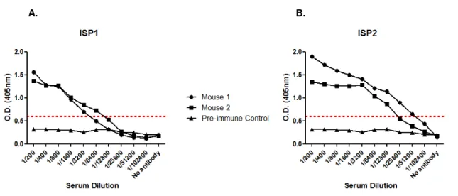
Figure 3.5– IS1 and ISP2 serum titre determination by ELISA. ... 73
Figure 3.6– Screening for anti-ISP1 antibody producing hybridomas by ELISA and Western Blotting. ... 75
Figure 3.7 – Screening for anti-ISP2 antibody producing hybridomas by ELISA and Western Blotting. ... 76
Figure 3.8– Western Blotting analysis of purified antibodies. ... 79
Figure 3.9 – Immunofluorescence analysis of ISPs in BSF T brucei. ... 80
Figure 3.10– Generation of ISP1 null mutants ... 83
Figure 3.11 – Generation of ISP2 null mutants ... 84
Figure 3.12 – Generation of ISP re-expression cell lines. ... 85
Figure 3.13– Confirmation of isp1 HYG/NEO by PCR. ... 87
Figure 3.14 – Confirmation of isp1 BSD/PAC by PCR. ... 88
xx
Figure 3.16 – Confirmation of isp1/2 by PCR. ... 91
Figure 3.17– Confirmation of ISP1 and ISP2 re-expression cell lines by PCR. ... 91
Figure 3.18– Southern Blotting analysis of isp1 HYG/NEO null mutants. ... 93
Figure 3.19– Southern Blotting analysis of isp1 BSD/PAC null mutants. ... 94
Figure 3.20– Southern Blotting analysis of isp2 null mutants. ... 95
Figure 3.21 – Southern Blotting analysis of isp1/2 null mutants. ... 96
Figure 3.22– Western Blotting analysis of isp1 cell lines. ... 97
Figure 3.23– Western Blotting analysis of isp2 cell lines. ... 98
Figure 3.24–In vitro growth analysis of ISP null mutant cell lines. ... 99
Figure 3.25– Mouse survival rates. ... 101
Figure 3.26– Mouse infection profile. ... 102
Figure 3.27 – Scanning Electron Microscopy analysis. ... 103
Figure 3.28– Analysis of T. brucei ISP1 and ISP2 expression by Western Blotting. 104 Figure 3.29–In vitro growth analysis of T. brucei. ... 106
Figure 3.30– Antibodies against ISP1 and ISP2 have no effect on T. brucei infection in vivo. ... 108
xxi
Index of Tables
Table 2.1 –Oligonucleotides used in this study ... 46
Table 2.2 –Plasmids generated and used in this study ... 50
Table 3.1 –Hybridomas screened by ELISA. ... 74
Table 3.2 –Isotype determination scheme. ... 77
Table 3.3 –Isotype determination results. ... 78
Chapter 1
2
1.1
Human African Trypanosomiasis
1.1.1 Epidemiology
African trypanosomiasis is a family of parasitic diseases affecting both humans and animals. It is caused by a flagellated protozoan parasite of the genus Trypanosoma
that lives and multiplies extracellularly in the blood and tissue fluid of its mammalian hosts. The parasite is transmitted by the blood feeding tsetse fly (Glossina sp.) and the geographical distribution of trypanosomiasis in Africa is directly linked to the vector’s suitable habitat, comprising a total of 10 million km2 of territory, between latitude 14° North and 29° South, from the southern edge of the Sahara and the north of Kalahari deserts, one third of Africa’s landscape (Brun et al., 2010, Barrett et al., 2003).
Trypanosoma brucei is divided in 3 subspecies, T. brucei gambiense and T. brucei rhodesiense causing the human form of the disease in West and East Africa, respectively and T. brucei brucei that, along with T. congolense, T. evansi and T.vivax, causes the animal form of African trypanosomiasis, also known as Nagana, a Zulu word meaning powerless/useless. Although these parasites cause relatively mild infections in wild animals, they cause a more severe form of the disease, often fatal in domestic animals. The symptoms begin with fever, eye discharge, oedema and anaemia and as the illness progresses, animals became more and more weak until paralysis and death eventually occur (Brun et al., 2010).
Domestic and wild animals can also be infected by T. brucei gambiense and T. brucei rhodesiense. Animals do not develop the disease when infected with these trypanosome species but they have a major epidemiological importance, acting as carriers or reservoirs of the infection (Figure 1.1).
3 transmission can occur directly from animals to humans, which is believed to be one of the causes of the long term maintenance of the disease in endemic areas (Malvy and Chappuis, 2011).
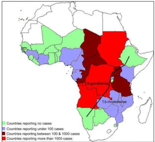
Figure 1.1–Human african trypanosomiasis (HAT) transmission cycle.
T. b. gambiense transmission is mostly human-to-human (anthroponotic infection) and transmission from domestic animals to human occurs occasionally. T. b. rhodesiense infection in humans is transmitted mainly between game animals (zoonotic infection) and sometimes between animals and humans. Adapted from (Simarro et al., 2011).
Human African Trypanosomiasis (HAT), also known as sleeping sickness disease is one of the “neglected diseases”, a group of diseases that includes schistosomiasis, visceral leishmaniasis and Chagas disease. This group of diseases is responsible for the death of hundreds of thousands of people in underdeveloped tropical regions. Nevertheless, current treatment of these diseases is often considered inadequate and ineffective and until recently the pharmaceutical industry as shown little interest in developing new drugs (Kennedy, 2008).
Human African trypanosomiasis is a potential fatal disease that affects mainly rural populations in 36 countries of sub-Saharan Africa (Figure 1.2). Along the past century, the prevalence has changed dramatically and three major epidemics have been reported. The first one affected equatorial Africa between 1896 and 1906 and killed an estimated 800,000 people (Brun et al., 2010, Steverding, 2008). A second major epidemic occurred between 1920 and 1940 and was controlled by the effort of mobile teams that organized active surveillance programs, screening millions of people at risk (Barrett, 2006).
re-4 emerged in the 1990s due to the collapse of the surveillance and control programs allied to civil conflicts in some of the most endemic countries, such as Angola, Uganda, Democratic Republic of Congo and Sudan (Brun et al., 2010, Stuart et al., 2008, Kennedy, 2008, Stuart et al., 2005). By that time, 40,000 cases were reported but an estimated number of 300,000 cases remained undiagnosed and therefore untreated (Simarro et al., 2011)(http://www.who.int/mediacentre/factsheets/fs259/en/).
In 2005, surveillance was reinforced and the number of reported cases decreased significantly. However, these numbers are known to be uncertain as a result of under-reporting, once that African Trypanosomiasis affects mainly remote rural populations with little or no access to health facilities (Brun et al., 2010).
Figure 1.2–Geographical distribution of HAT and epidemiological status of the disease in the last
decade.
5 Around 50 million people live now in HAT transmission areas and although the number of new cases per year reported to WHO has dropped below 10,000 (Simarro et al., 2011), thousands of HAT cases may remain unreported or undiagnosed especially in remote areas. HAT is part of a list of disabling conditions caused by parasitic and bacterial infections that affect the world’s poorest populations, often referred to as Tropical Neglected Diseases (Hotez et al., 2007) and along Animal African Trypanosomiasis, it is responsible for severe annual economic and human losses, causing a great socioeconomic effect on the affected populations, impairing development in sub-Saharan Africa (Brun et al., 2010, Antoine-Moussiaux et al., 2009, Kennedy, 2008, Hotez et al., 2007).
1.1.2 Clinical features
There are two different forms of sleeping sickness disease, the chronic form is transmitted by T.brucei gambiense in central and western Africa and the acute form of the disease is caused by T. brucei rhodesiense in East and South Africa (Brun et al., 2010, Kennedy, 2008).
In both forms, the disease appears in two stages and culminates in coma and death if left untreated. After the tsetse fly bite, parasites proliferate and spread to lymph nodes, reaching patient’s bloodstream, causing the first stage of the disease, the hemolymphatic stage. This early stage of the disease is characterized by non specific symptoms, such as intermittent fever, headache, pruritus, weight loss, fatigue, joint pain, lymphadenopathy, hepatosplenomegaly and is frequently misdiagnosed. At this stage, parasites can be detected in blood, lymph or tissue aspirates but are usually bellow detection levels, especially when the patient is infected with T.brucei gambiense
(Stuart et al., 2008, Barrett et al., 2003).
6 caused by dysregulation of the circadian rhythm (Stuart et al., 2008). Various severe neurological symptoms, such as psychiatric and mental features, motor and sensory disturbances and visual involvement accompany sleep disorders leading invariably to death in the absence of treatment (Kennedy, 2008).
T. brucei gambiense infection has a progressive course with an estimated average duration of 3 years, evenly divided between the two stages, whereas T.brucei rhodesienseinfection results in patient’s death within weeks or months (Checchi et al., 2008, Odiit et al., 1997).
1.1.3 Diagnosis
Diagnosis of HAT is divided in three distinct steps: screening, diagnostic confirmation and staging. Screening for potential infection is sometimes difficult, once the clinical features of the disease are not specific enough and lead to misdiagnose. The card agglutination test for trypanosomiasis (CATT) is considered the most efficient and best adapted screening method to use in endemic areas. It is a fast serological test that allows the screening of hundreds of individuals daily. However, it is only available for
T. bruceigambiense infections (Brun et al., 2010).
Diagnostic confirmation of trypanosomiasis is ideally done by microscopic identification of trypanosomes in peripheral blood samples or lymph node aspirates. This diagnostic approach is easier in rhodesiense infections due to the high parasitemia that generally persists in contrast with gambiense infections, where parasitemia tends to be irregular and low (Kennedy, 2008). Detection of parasite DNA by PCR techniques might be of valuable use in the future, as more sensitive approach, but existing tests need further standardization and validation (Brun et al., 2010).
7 The diagnostic approach for T. brucei rhodesiense infection is significantly different from the gambiense. There is no suitable serological screening method comparable to the CATT test used for T.bruceigambiense detection and the screening in the field is based mainly on clinical history and existing symptoms. As the two species do not co-exist in the same geographical areas, except in Uganda, the detection of parasites on blood samples has been enough to differentiate between the chronic and acute form of the disease. Nevertheless, there is evidence that this natural separation can be disrupted, as it has been shown that T. brucei rhodesiense is being carried by the movement of infected animals in Uganda towards areas of endemic gambiense form of the disease. The overlap of the chronic and acute forms of the disease can therefore represent a major problem for the control and treatment of HAT (Chappuis et al., 2005, Welburn and Odiit, 2002, Welburn et al., 2001).
1.1.4 Treatment, control and potential targets for therapy
8 Due to the high toxicity of the available drugs and the absence of vaccines against trypanosome infection, the only preventive measure that seems adequate is the reduction of the tsetse fly bites by disruption of the man/fly contact, but the main control strategies are the reduction of the disease reservoir and the control of the tsetse fly vector. In T. brucei gambiense infections, the most significant reservoir is the human, so the main control strategy is surveillance, population screening and treatment of infected individuals to reduce the reservoir. As the reduction of reservoir is very difficult in T. brucei rhodesiense infections, due to the high variety of animal hosts that can act as a reservoir, the main strategy in use is the vector control, usually done with traps or screens combined with insecticides and fly attracting odors (Brun et al., 2010, Kennedy, 2008).
Despite being one of the most neglected diseases in Africa, extensive research has been conducted. The sequencing of the genome (Berriman et al., 2005) and the use of powerful molecular biology techniques (Alsford et al., 2011, Clayton et al., 2005, Ngô et al., 1998) have given new insights on the parasite biology and allowed the identification of new potential targets for therapy, such as the glucose metabolism and the glycosome (Nowicki et al., 2008, Verlinde et al., 2001), the pentose phosphate pathway (Barrett, 1997), the thiol metabolism (Schlecker et al., 2005, Schmidt and Krauth-Siegel, 2002), the lipid and sterol metabolism (Paul et al., 2001, Buckner et al., 2000, Yokoyama et al., 1998), cell signaling and differentiation, protein degradation and peptidase inhibitors (Santos et al., 2007, Caffrey et al., 2000, Troeberg et al., 1999), membrane architecture, transporters and drug entry, regulation of nuclear gene expression, kinetoplastid and RNA editing, among others (Kennedy, 2008, Barrett et al., 2003).
1.2
Trypanosoma brucei
9 distinct DNA containing region in their single large mitochondrion, the kinetoplast (Stuart et al., 2008). As they exhibit rich and diverse cell biology, kinetoplastid parasites in general and Trypanosoma brucei in particular, provide an excellent means for studying and understanding many of the intriguing aspects of its cell biology, such as cell structure and morphology, organelle positioning, cell division and protein trafficking within the cell (Matthews, 2005, Gull, 2001).
1.2.1 T. brucei life cycle
T. brucei has a complex life cycle, in which the parasite must adapt to completely different and often hostile environments when transmitted between its mammalian host and arthropod vector or when travelling trough different compartments within the tsetse fly. During its life cycle, the trypanosome undergoes multiple changes in its cell biology, revealing numerous developmental forms, primary metabolism differences and gene expression changes, enabling the parasite to survive and escape the host’s immune system (Field and Carrington, 2009, Matthews, 2005).
During its developmental cycle, the trypanosome makes a series of transitions, colonizing 3 major environments: the mammalian bloodstream, the tsetse fly midgut and the tsetse fly salivary gland, alternating between proliferative stages and non-proliferative stages (Figure 1.3) (Stuart et al., 2008, Gull, 2001, Vickerman, 1985).
1.2.1.1 Development of T. brucei in the mammalian host
T. brucei is transmitted to the mammalian host by the tsetse fly during a blood meal. After the bite, metacyclic parasites are transmitted to the mammal, causing a local inflammatory reaction and forming a lesion at the site of the bite, a chancre.
10 As trypanosomes live extracellularly in the bloodstream, they undergo antigenic variation to evade the host’s immune system. The immune system’s evasion requires the sequential expression of distinct variant surface glycoproteins (VSGs) linked to the membrane’s surface by a glycosylphosphatidylinositol (GPI) anchor (Cross, 1975). Once the VSG coat is recognized by the host’s immune system and a specific antibody is raised, activating the complement, the parasitemia starts to decrease. However, a small number of trypanosomes survive by expressing a different VSG coat and thus escaping the immune system (Vickerman, 1978). This periodical switching of VSG coat and consequential evasion of the host’s immune system causes the cyclical waves of parasitemia characteristic of the chronic form of Human African Trypanosomiasis (Fenn and Matthews, 2007).
Figure 1.3–Trypanosoma brucei life cycle.
11 Dividing slender parasites are found in ascending parasitemia while non-dividing stumpy trypomastigotes can be observed when the parasitemia goes into remission (Fenn and Matthews, 2007, Vickerman, 1985). As the number of parasites increases, short stumpy trypomastigotes replace slender ones after proliferation in the mammalian bloodstream. This differentiation appears to be induced by a molecule secreted by the parasite, referred to as Stumpy Induction Factor, and may serve two purposes. First it limits the number of parasites in the blood, increasing host’s survival and maximizes the probability of disease transmission. Second, it ensures the coordination of morphological changes needed for the efficient transmission to the tsetse fly providing the continuation of the parasite’s life cycle (Matthews, 2005, Vassella et al., 1997).
When accumulated, the Stumpy Induction Factor triggers cell cycle arrest in G1 phase and the mitochondrial respiratory chain is activated, preparing the cell to glucose limited environment and pre-adapting the parasite for the drastic changes of environmental conditions and for efficient transmission to the tsetse fly vector (Vassella
et al., 1997, Priest and Hajduk, 1994).
1.2.1.2 Development of T. brucei in the tsetse fly vector
The trypanosome initially establishes in the tsetse fly midgut after a blood meal, before migrating to the salivary glands (Matthews, 2005). When the fly feeds from a mammalian host, the infected blood contains both long slender and short stumpy parasites. The transformation of this pleomorphic population of mammalian bloodstream forms of T. brucei to procyclic forms must rapidly occur to ensure parasite’s survival in the vector (Ziegelbauer et al., 1990).
12 The procyclic form (PCF) of T. brucei is extremely different from its mammalian counterpart: it uses proline as energy source instead of glucose, shows a different morphology and cell architecture and several changes in gene expression (Matthews and Gull, 1994).
After the differentiation of bloodstream forms into procyclic forms begins, the VSG coat is lost and replaced by a new surface protein coat, a procyclin coat resistant to tsetse fly midgut proteases (Matthews and Gull, 1994, Ziegelbauer et al., 1990). As approximately 99% of the parasites present in the ingested blood are eliminated by the fly, the trypanosome establishment in the midgut starts from a very small population of fully differentiated procyclic cells (Van Den Abbeele et al., 1999, Ziegelbauer et al., 1990).
That small population of PCF cells grows vigorously and as colonization progresses, from the posterior to the anterior midgut, parasites start to elongate and long trypomastigotes (mesocyclic trypomastigotes) can be found near the proventriculus, in the ectoperitrophic space in the anterior midgut. These cells invade the proventriculus lumen and migrate to the foregut and proboscis, where they undergo a complex differentiation to short epimastigotes (Roditi and Lehane, 2008, Van Den Abbeele et al., 1999).
To complete its life cycle, the parasite must colonize the tsetse fly salivary gland in order to generate metacyclic forms, infective for mammals. In the proventriculus, the long trypomastigote cells replicate their nuclear DNA and reposition their kinetoplast and basal body originating long epimastigote form parasites.
These cells undergo asymmetric division, generating short epimastigotes, presumed to be the ones colonizing the tsetse fly salivary gland and long epimastigotes whose fate is unknown.
13 Once in the salivary gland, these cells complete their division and the short epimastigotes are now able to attach to the wall and continue their development into mature metacyclic trypomastigote forms, infective for mammals (Roditi and Lehane, 2008).
1.2.2 T. brucei cell biology
Although T. brucei exists in two different developmental forms, perfectly adapted to survive in each of its hosts, both BSF and PCF parasites exhibit a similar morphology characterized by a cell body with a vermiform shape with tapered ends and a single flagellum emerging from the basal body, near the posterior end of the cell through a specialized plasma membrane invagination, the flagellar pocket (Figure 1.4) (Field and Carrington, 2009, Ralston et al., 2009).
Figure 1.4–The basic structure of T. brucei.
Schematic representation of T. brucei‘s cell morphology, showing the parasite’s major organelles and
their organization within the cell (Grünfelder et al., 2003).
14 In addition to its role in the parasite’s motility, the flagellum is essential for host -parasite interaction, once it is required for the attachment to epithelial cells in the tsetse fly salivary gland (Vaughan, 2010). The flagellum is also critical for cellular morphogenesis (Kohl et al., 2003, Moreira-Leite et al., 2001), organelle inheritance and positioning and cell division (Ralston et al., 2006, Kohl et al., 2003). Finally, the flagellum could be involved in cell signaling and sensory functions (Vaughan, 2010).
Electron microscopy analysis of the flagellum reveals the presence of many conventional flagellar structures, such as the axoneme and some electron dense particles resembling IFT particles, but also of unusual extra-axonemal structures such as the paraflagellar rod (PFR) and the flagellum attachment zone (FAZ) and the flagella connector (FC), unique to trypanosomes and a few related protozoa (Ralston et al., 2009, Kohl and Bastin, 2005, Landfear and Ignatushchenko, 2001).
The axoneme presents the typical 9+2 microtubule arrangement, with the 9 outer doublets surrounding a central pair of singlet microtubules (Ralston et al., 2009, Kohl and Bastin, 2005). It also possesses all the conventional components of other eukaryotic axonemes such as the inner and outer dynein arms, the radial spokes connecting the outer doublets to the central pair, the nexin links associated with the outer doublets and the projections from each central pair microtubules connecting them to one another, revealing a high structural degree of conservation (Kohl and Bastin, 2005, Bastin et al., 2000).
15 Along the length of the cell, the flagellum and the cell body are held by a network of cytoskeletal and membranous connections that together comprise the flagellar attachment zone (Ralston and Hill, 2008, Gull, 1999). Although the FAZ is not part of the flagellum, it is closely associated to the flagellum and its functions. When the flagellum emerges from the flagellar pocket, it bends over the cell body, attaching to the cell body membrane. The flagellar pocket and the cell body membrane remain in tight contact and the FAZ can be recognized from the flagellar pocket area, following the flagellum and ending at the anterior tip of the parasite (Kohl and Bastin, 2005, Kohl and Gull, 1998). Its components are mostly unknown, but the ultrastructure of FAZ is extensively characterized. It contains 2 distinct structures, the FAZ filament on the cytoplasmic side and a unique set of 4 microtubules associated with the membranous compartment. The FAZ filament is connected to both the axoneme and the PFR by a network of filaments promoting the contact of the cell body membrane with the flagellum (Ralston et al., 2009, Ralston and Hill, 2008). The flagellum and the FAZ together provide structural and positional support to the parasite, that influence cytokinesis and cell morphogenesis and the disruption of flagellum attachment results in failing to initiate cytokinesis (Ralston and Hill, 2006, Kohl et al., 2003).
Although it has critical roles in mobility, morphogenesis and cell division throughout the parasite’s life cycle, most studies of flagellum structure and function have been conducted in the procyclic form of T. brucei (Ralston and Hill, 2006). The new flagellum emerges from the flagellar pocket and extends along the cell body towards the anterior end of the parasite, following a left-handed helical path. As it elongates, the new flagellum is always located to the left of the old one, when looking at cell from the posterior end. In procyclic trypomastigotes, the distal tip of the new flagellum is connected to the side of the old one by the flagellar connector, a transmembrane complex of unknown composition, important in providing all the vital morphogenetic information for the daughter cell’s flagellum to assemble correctly (Field and Carrington, 2009, Davidge et al., 2006, Briggs et al., 2004).
16 During the assembly of the new flagellum, the transport of all flagellar precursors to the distal tip of the flagellum is dependent on intraflagellar transport (IFT), a bidirectional transport system of proteins in the flagella matrix. The IFT is driven by a two motor complexes, the heterotrimeric kinesin II that moves particles towards the flagellum tip and a dynein complex that moves the particles back to the base of the flagellum (Absalon et al., 2008b, Briggs et al., 2004). Functional studies reveal that IFT is required for the correct flagellar biogenesis in T. brucei and the presence of IFT particles in the old and new flagella of the same cell suggests that IFT operates in both and is not only required for the growth of the new flagellum but also for the maintenance of the mature one (Absalon et al., 2008b).
The silencing of IFT genes results in cells with shorter flagella and in some cases with no flagella, leading to progressively shorter cells (Absalon et al., 2008b). The absence of normal flagellum elongation does not affect the flagellar pocket formation, which remains present in these cells. Nevertheless, abnormally shorter parasites exhibit flagellar pocket with different shapes, suggesting that normal flagellum elongation is essential for the organization, orientation and function of the flagellar pocket (Absalon
et al., 2008a).
Once it exits the cytoplasm, through the flagellar pocket, the flagellum is surrounded by its own membrane and is attached to the cell body, along most of its length. The cell membrane, the flagellar pocket and the flagellar membrane comprise the three contiguous but morphologically and biochemically distinct domains of the parasite’s surface membrane, each one with unique functions (Ralston and Hill, 2008, Landfear and Ignatushchenko, 2001). All 3 domains are covered by the densely packed VSG coat in the bloodstream form parasites or during the metacyclic stage in the tsetse fly salivary gland or by a procyclin coat in the other procyclic forms (Bastin et al., 2000).
17 pocket defining it as a dynamic portal to host or vector environment. This dynamic interaction provides resistance to the host’s innate and acquired immune responses and although the flagellar pocket is a critical organelle for the trypanosome biology with important roles in immune evasion, very little is known about its architecture and how molecules get to and from the flagellar pocket (Lacomble et al., 2009, Gull, 2003).
The flagellar pocket has a complex organization and a precise positioning relatively to cytoskeletal elements and other organelles. It can be divided into several subdomains, with the basal body used to define one pole of this structure. The membrane and luminal volume of the flagellar pocket are asymmetric due to the association with the Golgi complex and the probasal body. Associated with the Golgi apparatus is the neck region which is located distal to the flagellum exit point. The neck of the flagellar pocket is a specialized membrane area also associated with the FAZ and where a microtubule quartet integrates into the subpellicular array, defining an axis for the entire flagellum and its associated structures (Field and Carrington, 2009, Lacomble
et al., 2009). The flagellar pocket has 2 boundary subdomains associated with organized structures connected across the membrane and cytoskeleton, the collar, defining the flagellum exit point, and the collarette, the point where it enters the flagellar pocket (Field and Carrington, 2009).
With the flagellar pocket as the only site of endocytosis and exocytosis, the T. brucei cell, and thus the T.brucei endomembrane system, is highly polarized. This high degree of polarization of the endomembrane system is reflected in the localization of the secretory and endocytic organelles, with the endoplasmic reticulum (ER) widely distributed throughout the cytoplasm and all the rest compactly organized in the posterior portion of the cell, between the nucleus and the kinetoplast and the single lysosome close to the nucleus (Field and Carrington, 2009, Morgan et al., 2002a).
The flagellar pocket has a distinct proteome with most of the protein families required for vesicle trafficking in other eukaryotic organisms represented in it, like the 16 Rab and Rab-related proteins encoded in T. brucei genome (Ackers et al., 2005, Berriman et al., 2005).
18 TbRAB5A, TbRAB5B and TbRAB11 as part of the early endosome and recycling arms of the endocytic pathway (Morgan et al., 2002b, Field et al., 1998).
Although trypanosomes have conventional endomembrane system, it has some distinct unusual features, such as the clathrin dependence of all endocytosis (Allen et al., 2003). These mechanisms seem to have an extreme importance in immune evasion and emphasize the high level of endocytic activity in BSF trypanosomes as a potential reason for the protection against the mammalian immune system (Natesan et al., 2010).
Trypanosomes can be efficiently eliminated by mammalian anti-VSG antibodies and complement activation. Nevertheless, the VSG-antibody complex doesn’t remain bound to the cell surface and is rapidly internalized, degraded and the VSG is recycled. This antibody uptake by the parasite appears to be mediated by hydrodynamic flow that drives the antibody bound to the highly abundant GPI-anchored VSG to the flagellar pocket where it is internalized (Natesan et al., 2011, Engstler et al., 2007). This process involves transport steps mediated by RAB5 and RAB11, both proteins regulated according to the developmental stage (Pal et al., 2003), suggesting that the endocytic system is activated in the bloodstream stage (Grünfelder et al., 2003, Pal et al., 2002).
Degradation of immunoglobulins inside the parasite is extremely selective, occurring with low proteolysis of the VSG molecules and the details the enzymes used in this selective process are unknown. There are metacaspases present in endosomal vesicles, co-located with RAB11. However they have no direct involvement in the anti-VSG antibody degradation or anti-VSG recycling since their knockdown and knockout produce no major effect on these processes(Helms et al., 2006).
It is not clear if there is any similar defensive role in PCF parasites or if there is an adequate explanation for the relatively low endocytic activity in insect stage parasites, although there are evidences that parasites do not require high endocytic activity in order to survive in the insect vector (Natesan et al., 2007).
19 composed by a structural mass of proteins and catenated circular DNA molecules, the kinetoplastid DNA (kDNA) (Stuart et al., 2005). The structure of the kDNA is unique, consisting of a network of thousands of circular DNA molecules, minicircles, topologically interlocked with a few dozen of larger DNA molecules, maxicircles (Chen
et al., 1995, Shapiro and Englund, 1995).
The kinetoplast and the basal body are connected by an attachment complex that crosses both the mitochondrial and cell membrane and through which the mitochondrial genome segregation and the basal body replication and segregation events are linked (Ogbadoyi et al., 2003).
The mitochondrion itself is an elongated structure that in BSF parasites is devoid of cristae, reflecting the absence of oxidative phosphorilation at this stage. Energy generation in BSF parasites is dependent on glycolytic reactions that take place within specialized organelles, the glycosomes (Parsons, 2004). Glycosomes are organelles belonging to the family of peroxisomes, but with an almost exclusively glycolytic role in T. brucei, with glycolytic enzymes comprising around 90% of protein content of these organelles in the BSF parasite. Compartmentation of glycolysis is unique and essential for BSF parasite to develop properly, enabling these organisms to overcome short periods of anaerobiosis, although without sustained growth (Michels et al., 2006).
When in the mammalian host, parasites encounter a large supply of glucose constantly available in the bloodstream or cerebrospinal fluid. The mitochondrion is largely repressed and all ATP is generated by a high rate of aerobic glycolysis inside the glycosome (Parsons, 2004). These parasites are not adapted to live in the insect vector and quickly die if ingested by the fly, unless they differentiate into short stumpy parasites which, although still glycolysis dependent, have a partially de-repressed mitochondrial system (Michels et al., 2006).
In the insect host, amino acids are the primary nutrients for parasites and even though glycosomes remain abundant, their enzymatic content changes.
20 This enzymatic content variation between different life-cycle stages of the parasite is extremely important for the rapid and efficient adaptation to the different environments and glycosomes may play a significant role in this metabolic adaptation (Michels et al., 2006, Parsons, 2004).
T. brucei also contains another distinct organelle involved in the storage of polyphosphates, calcium and other cations (Docampo et al., 2010). Acidocalcisomes are electrondense acidic organelles present in a wide variety of prokaryotic and eukaryotic organisms, but absent in mammalian cells (Moreno and Docampo, 2003). The membrane of acidocalcisomes has numerous pumps, exchangers and at least one channel and the matrix is filled with enzymes involved in polyphosphate metabolism. Acidocalsisomes have an important role not only in the storage of phosphorus and cations but also in calcium signaling, pH homeostasis and osmoregulation in response to environmental stress (Docampo et al., 2010).
This interesting and complex cellular organization of T. brucei is not constant and different morphological forms of the parasite can be observed depending on its life cycle stage (See section 1.2.1).
These morphological forms are distinguished according to the position of the kinetoplast and flagellar pocket in relation to the nucleus. In trypomastigotes, the kinetoplast and flagellar pocket are located on the posterior end of the cell with a centrally located nucleus and the flagellum emerging from the anterior end of the cell.
In epimastigotes the kinetoplast and flagellar pocket are anterior to the nucleus and the flagellum emerges approximately from the middle of the parasite (Field and Carrington, 2009).
Life cycle progression is closely connected to the cell cycle regulation with proliferative parasites alternating with non-proliferative ones, where the cell cycle is arrested as a form of pre-adaptation for transmission between hosts (McKean, 2003).
21 1.2.3 T. brucei Cell Cycle
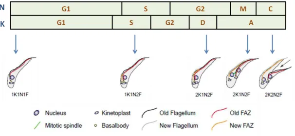
The cell cycle of trypanosomes has the same nuclear events characteristic of normal eukaryotic cells, consisting of four periods of nuclear activity – G0/G1, S, G2 and M. However, unlike other eukaryotes, duplication of nuclear and kinetoplastid DNA is coordinated but not simultaneous and trypanosomes exhibit a periodic S phase for the kinetoplast (SK) in addition to the nuclear S phase (SN) (Ploubidou et al., 1999, Woodward and Gull, 1990). Kinetoplast S phase (SK) begins immediately before the nuclear S phase (SN). It is much shorter and kinetoplast segregation occurs before the mitosis initiates (Hammarton, 2007, McKean, 2003).
Figure 1.5–The cell cycle of PCF T. brucei.
Diagram showing highly organized replication and organelle position of procyclic T. brucei, throughout
the stages of the parasite’s cell cycle. The number of nuclei (N) and kinetoplasts (K) is indicated for each
one of the cell cycle stages (G1/G2: gap phases; S: DNA synthesis; M: mitosis; C: cytokinesis; D: kinetoplast segregation; A: “apportioning phase”). Adapted from (McKean, 2003).
22 The timing and order of such events in T. brucei varies according to the life stage and subtle differences can be observed between procyclic and bloodstream trypomastigotes, specifically the relative position of the nucleus and the kinetoplast before cytokinesis occurs (Figure 1.5) (Briggs et al., 2004, McKean, 2003).
The first indicator of the cell cycle initiation is the elongation and maturation of the probasal body adjacent to the mature basal body and the single flagellum. Cell cycle progresses through the initiation and elongation of the new flagellum, along with the duplication of the basal body (Sherwin and Gull, 1989). After the basal body duplication, the Golgi apparatus begins to duplicate and stays located close the newly formed basal body and the endoplasmic reticulum exit point (He et al., 2004). Replication of kinetoplastid and nuclear DNA commences independently, with the segregation of the kinetoplast taking place before the mitotic division of the nucleus.
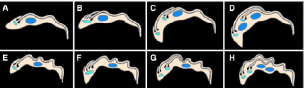
In procyclic form parasites, mitosis is then followed by the positioning of the nucleus (N) between the two kinetoplasts (K), in a linear arrangement referred to as KNKN, whereas in bloodstream form both nuclei remain next to each other and anterior to the kinetoplasts, in a KKNN formation (Figure 1.6).
Figure 1.6–Thecell cycle of T. brucei trypomastigotes.
Cartoon illustrating the main differences between cell cycle in procyclic (A – D) and bloodstream form (E
– H) parasites (Briggs et al., 2004).
23 Cell cycle regulation is essential for the parasite’s survival. Mis-regulation of cell cycle events can have severe consequences, so the cell developed a mechanism of checkpoints in which the integrity of the DNA is monitored ensuring the accuracy of DNA replication, the appropriate cell division and the production of two healthy daughter cells (McKean, 2003).
There are two major families implicated in the regulation of cell cycle progression in eukaryotes: cyclins and cyclin-dependent kinases (CDKs). The concentration of cyclins varies regularly during the cell cycle while CDKs are constantly expressed throughout the cycle but only become active when bound to a specific cyclin. Further regulation of CDKs activity is done by phosphorylation of conserved residues and the interaction with inhibitory proteins (CKIs) (Hammarton, 2007, Ploubidou et al., 1999).
There are 10 cyclins (CYC2–11) and 11 CDKs CRK1–4 and CRK6–12) homologous identified in T. brucei. Although the parasite’s cell cycle presents some typical features from eukaryotes, it shows some major differences in regulatory mechanisms and molecules that can also vary according to the parasite’s life cycle stage. This suggests that T. brucei has developed new cell cycle control mechanisms and exclusive checkpoints in order to coordinate the independent replication of the parasite’s two unit genome (Hammarton, 2007, McKean, 2003).
1.3
Peptidases and their Natural Inhibitors
1.3.1 Peptidases
24 The diversity of peptidases is also evident in their specificity. Most enzymes are non-specific and therefore can act on multiple substrates, cleaving different peptide bonds. In contrast, some peptidases are highly specific and catalyse the hydrolysis of a single peptide bond of a particular protein (López-Otín and Bond, 2008, Puente et al., 2003, Sajid and McKerrow, 2002).
Initially, peptidases were classified in endo and exopeptidases. The action of endopeptidases is directed to the internal peptide bonds, while exopeptidases act on peptide bonds at the amino or carboxyl terminus (López-Otín and Bond, 2008, Rosenthal, 1999). Currently, peptidases are classified according to the catalytic mechanism of proteolysis in: aspartic peptidases, serine peptidases, cysteine peptidases, glutamic peptidases, metallo-peptidases, threonine peptidases, asparagine peptidases and peptidases of unknown catalytic mechanism. Peptidases with significant similarity in amino acid sequence can be grouped into families and families with similar 3D structures can be assembled in clans (Rawlings et al., 2010).
This diverse group of enzymes has been identified in all biological systems, from viruses to vertebrates and is involved in several biological processes, playing essential roles in cell cycle progression, differentiation, morphogenesis, homeostasis, inflammation, immunity, angiogenesis, necrosis and apoptosis, amongst others (Quesada et al., 2009, López-Otín and Bond, 2008). Abnormal peptidase activity is also associated with numerous pathological conditions such as cancer, neurodegenerative disorders and cardiovascular diseases (Quesada et al., 2009, López-Otín and Bond, 2008, Puente et al., 2003).
Likewise, proteolytic enzymes seem to be central pieces in the life cycle of many parasitic protozoans causing diseases, such as malaria, leishmaniasis, amebiasis, cryptosporidiosis, toxoplasmosis and trypanosomiasis. Peptidases from all major classes have key roles in various functions, including host cell’s invasion and host’s immune system evasion (Rosenthal, 1999).
25 effective inhibitors such as the vinyl sulfone inhibitor K777, in late preclinical trials for Chagas disease treatment (Doyle et al., 2007).
In Plasmodium, the antimalarial effects of several peptidase inhibitors have been extensively studied. For example, the cysteine peptidase inhibitor E-64 and the aspartic peptidase inhibitor pepstatin have been shown to block the development of P. falciparum (Glushakova et al., 2009) and several peptide inhibitors of falcipain have been shown to inhibit P. falciparum growth and maturation (Korde et al., 2008).
Several studies have been focused on peptidases and their potential as targets for therapy and hundreds of peptidase inhibitors have been generated to be used for the treatment of several diseases, from cancer to auto-immune diseases, from infectious diseases caused by viruses to bacterial and parasitic infections (Coombs and Mottram, 1997).
1.3.1.1 Cysteine Peptidases in Trypanosomatids
In cysteine peptidases, the thiol group of the cysteine residue in the active site acts as the nucleophile in catalysis. The catalytic activity depends upon a catalytic dyad and is enhanced by the proximity of a histidine residue that acts as a proton donor.
The catalytic dyad is formed by the –SH group of the cysteine residue and the imidazole of the histidine and is often stabilized by the existence of a highly conserved asparagine residue. One other aminoacid can be found in the active site. Glutamine preceding the catalytic cysteine helps to form the oxyanion hole.
During hydrolysis, the enzyme is transiently bound to the substrate, forming an unstable tetrahedral intermediate (Rosenthal, 1999).
26 maintaining the enzyme in its inactive form until it reaches its destination, normally the lysosome (Rosenthal, 1999, Huete-Pérez et al., 1999).
Cysteine peptidases from trypanosmes are very different from their mammalian counterparts and exhibit several functions, some related to the host’s cells and tissues invasion and to the immune system evasion. Once they are highly immunogenic and exhibit significant structural and biochemical differences, cysteine peptidases have been exploited as potential targets for diagnostic therapy of parasitic diseases. In mammals, cysteine peptidases exhibit optimal activity at slightly acid or neutral pH, while in parasites they remain active through a wider range of pH, including alkali pH, are generally larger and more numerous than the mammalian ones and exhibit essential differences in substrate specificity, domain extensions and cellular localization (Sajid and McKerrow, 2002, North et al., 1990).
A great number of peptidases have been identified and characterized in parasitic protozoa. Although this vast group of peptidases includes members of all main classes, the majority of peptidases identified in parasitic organisms belongs to the cysteine class and are homologous to the cathepsin L in mammals (Rosenthal, 1999, Coombs and Mottram, 1997). Cathepsin L and cathepsin B–like peptidases belong to family C1, clan CA (papain-like peptidases), the largest cysteine peptidase clan, comprising 84% of all peptidases identified in parasitic protozoa (Atkinson et al., 2009, Sajid and McKerrow, 2002).
Apart from clan CA, cysteine peptidases can be classified in 8 additional clans (CD, CE, CF, CH, CL, CM, CN, CO) plus one unclassified clan. The classification of peptidases is done according to similarities in the amino acid sequence, presence of peptide loops and substrate specificity (Atkinson et al., 2009, Sajid and McKerrow, 2002).
27 As in other papain–like peptidases, cathepsin L and cathepsin B–like peptidases from kinetoplastids include a signal peptide, a pro-peptide essential for the activation of the catalytic domain and a PepC1 catalytic domain. Additionally, kinetoplastid cathepsin L–like peptidases have a C–terminal extension, which function is yet to be determined with accuracy. Although this C–terminal extension has no catalytic role, it is highly immunogenic and can be used for diagnostic purposes (Caffrey and Steverding, 2009, Caffrey et al., 2000).
The active site of these enzymes has a conserved cysteine, histidine and asparagines residue and is surrounded by highly conserved peptide motifs. Like in other cathepsin B–like peptidases, kinetoplastid ones also have an insertion of 20 aminoacids in the catalytic domain, the occluding loop, that contributes to the dipeptidyl carboxiyl peptidase activity of cathepsin B (Caffrey and Steverding, 2009).
The genome of kinetoplastids has several copies of cathepsin L–like peptidases genes. In T. brucei, there are more than 20 copies of the gene arranged in tandem array and in T. cruzi there are more than 130 polymorphic genes arranged in clusters located in different chromosomes. The expression of cathepsin B–like peptidases is much simpler and is normally restricted to one or two copies of the gene (Caffrey and Steverding, 2009, Mottram et al., 1998).
There is a total of 65 cysteine peptidases encoded in the genome of Leishmania major. Like in other parasitic protozoa, most studies are focused on family C1 peptidases, CPA and CPB, both cathepsin L–like peptidases and CPC, a cathepsin B– like peptidase. Leishmania’s cathepsin L peptidases are stage regulated and mainly expressed in amastigotes, where they are restricted to large lysosomes, also termed megasomes (Caffrey and Steverding, 2009).
28 enabling the parasite’s survival and proliferation inside the macrophages (Mottram et al., 2004, Mottram et al., 1998).
Cruzipain, the main cysteine peptidase of T. cruzi, can be found in all life cycle stages, although its expression is higher in the replicating forms of the parasite, particularly, in epimastigotes. Cruzipain localization is different in epimastigotes, where it is found in the endosomal/lysosomal system, and amastigotes, where it is localized on the cell surface (Caffrey and Steverding, 2009, Mottram et al., 2004). The enzyme seems to be involved not only in the host’s immune system evasion but also in metacyclogenesis (McKerrow et al., 2009). Additionally, cruzipain has significant roles in amastigote replication, intracellular development and cellular invasion (Caffrey and Steverding, 2009).
T. brucei has cysteine peptidases belonging to both cathepsin L and cathepsin B–
like peptidases. Brucipain, a cathepsin L–like peptidase, is the major CP of the parasite. It has been identified in all life cycle stages and its levels of expression is approximately 5 to 10 times higher in short stumpy forms than in long slender BSF or procyclic forms, where it has comparatively little activity. Its cellular localization is dependent on cell cycle stage. In BSF the enzyme is confined to the lysosomes while in PCF the localization of brucipain is yet to be determined (Caffrey and Steverding, 2009, Caffrey
et al., 2000). Cathepsin B–like peptidases of T. brucei (TbCatB) are expressed essentially in the bloodstream forms of the parasite, while in T. cruzi and Leishmania
these enzymes are expressed throughout the life cycle (Caffrey and Steverding, 2009).
Both cathepsin L and cathepsin B–like peptidases are essential for the parasite successful survival within the mammal host. RNAi of TbCatB resulted in engorged lysosome, growth arrest and death of parasites in vitro. Similar studies involving brucipain RNAi in vitro showed no perceptible phenotype. In in vivo studies, the knockdown of TbCatB cured the infection whereas the RNAi against brucipain led to an extended life time of infected mice (Caffrey and Steverding, 2009, Abdulla et al., 2008).