In vitro culture, PCR , and nested PCR for the detection of Theileria equi in horses submitted to exercise
[Cultivo in vitro, PCR e nested PCR na detecção de Theileria equi em eqüinos submetidos a exercícios]
C.D. Baldani, P.A. Canola, J.C.L. Neto, R.Z. Machado*
Faculdade de Ciências Agrárias e Veterinária - UNESP Via de Acesso Prof. Paulo Donato Castellane, s/n
14884-900 – Jaboticabal, SP
ABSTRACT
This study compared the usefulness of in vitro culture, PCR, and nested PCR for the diagnosis of
Theileria equi in horses submitted to stress during exercise. Blood samples from 15 apparently healthy horses, previously conditioned to a high-speed equine treadmill, were taken prior to and after exercise. The animals were divided into two experimental groups: 30-day training schedule (G1) and 90-day
training schedule (G2). Statistical analysis was performed using a chi-square test and kappa statistic was
used in order to assess agreement. No significant difference was observed between samples collected at
resting or after exercise. In G1, merozoites of T. equi were detected in the blood smears of four horses
before in vitro culture, whereas 14 samples were positive, confirmed by culture. In G2, five and 11 horses
were positive before and after culture, respectively. No PCR amplified product was observed in any of the
tested animals although the PCR system based on the 16S rRNA gene of T. equi detected DNA in blood
with an equivalent 8x10⁻⁵% parasitaemia. The nested PCR based on the T. equi merozoite antigen gene
(EMA-1) allowed the visualization of amplified products in all the horses. Therefore, nested PCR should
be considered as a means of detection of sub-clinical T. equi infections and in vitro culture could be used
as a complement to other methods of diagnosis.
Keywords: horse, Theileria equi, diagnosis, in vitro culture, PCR
RESUMO
Comparou-se a utilização do cultivo in vitro, PCR e nested PCR no diagnóstico de Theileria equi em eqüinos submetidos ao estresse induzido por exercícios. Amostras de sangue foram obtidas de 15 eqüinos submetidos a treinamento em esteira rolante de alto desempenho, sendo as amostras colhidas antes e após os exercícios. Os animais foram divididos em dois grupos experimentais: 30 dias de treinamento (G1) e 90 dias de treinamento (G2). O teste do qui-quadrado foi empregado para as análises estatísticas e o índice kappa utilizado para avaliar a concordância. Não houve diferença significativa entre as amostras obtidas em repouso e após exercícios. Merozoítas de T. equi foram detectados em apenas quatro eqüinos do G1 pela microscopia direta realizada antes do cultivo, enquanto 14 animais apresentaram-se positivos pelo cultivo in vitro. No G2, cinco e 11 eqüinos foram positivos antes e após o cultivo, respectivamente. Nenhum produto de amplificação foi observado pela técnica de PCR, apesar do PCR baseado no gene 16S rRNA de T. equi ter detectado DNA em sangue com parasitemia equivalente a 8x10
-5
%. O nested PCR, baseado na seqüência do gene do antígeno de merozoíta de T. equi (EMA-1,) permitiu a visualização de produtos de amplificação em todos os eqüinos testados. O nested PCR deve ser considerado para a detecção de infecções subclínicas de T. equi e o cultivo in vitro pode ser utilizado como uma ferramenta complementar a outros métodos de diagnóstico.
Palavras-chave: eqüino, Theileria equi, diagnóstico, cultivo in vitro, PCR
Recebido em 21 de novembro de 2007 Aceito em 27 de abril de 2008
INTRODUCTION
Theileria equi is a tick-transmitted protozoan parasite and one of the causative organisms of equine piroplasmosis. The disease is characterized by fever, anemia, icterus, hepatomegaly, splenomegaly, intravascular hemolysis, petechial hemorrhages of the mucuous surfaces; hemoglobinuria, and death can occur in some cases (Schein, 1988). Equine piroplasmosis has worldwide distribution, being endemic in most tropical and subtropical areas of the world as well as in some temperate climatic zones (Schein, 1988; De Waal, 1992; Bruning, 1996). Low level carriers animals or ticks, which may act as reservoirs, represent a risk of these parasites being introduced to disease-free areas such as Japan, Australia, and North America (except in the state of Florida, USA). Therefore, the detection of sub-clinical infections has become very important, with special relevance to the horse-racing industry in which the geographical movement of presumably healthy
horses may aid the spread of T. equi or where
sub-clinical infection may negatively affect the performance of the animal (Rampersad et al., 2003). It has been shown that strenuous exercise, such as that experienced with horse-racing or endurance, may predispose clinical manifestation of the disease (Hailat et al., 1997).
Equine piroplasmosis can be diagnosed by means of several ‘methods. Direct microscopic identification of the parasite in stained blood smears can confirm diagnosis, but it is usually difficult to find the organism in carrier animals since parasites are generally present in very low numbers in the blood. Therefore, despite the high specificity, microscopic examination of blood smears has low sensitivity for the detection of carrier animals. Serological methods, such as complement fixation test (CFT) and the indirect fluorescent antibody test (IFAT), are commonly
used for detecting T. equi infections. However,
these tests are generally restricted by antibody detection limits and cross reactivity (Schein, 1988; Bruning, 1996). Besides the CFT and IFAT, the enzyme-linked immunosorbent assay (ELISA) has also been used for the detection of Theileria infection. Various researchers have suggested this serological test as an alternative for increased specificity and sensitive detection of acute and latent infections, especially with the
use of recombinant antigens (Tanaka et al., 1999; Xuan et al., 2001ab; Hirata et al., 2003).
Although regulatory control relies on serological tests to identify infected animals whose movement should be restricted, several other
techniques including in vitro cultures, DNA
probes, and polymerase chain reaction (PCR)
have been used for the diagnosis of T. equi.
Specific and sensitive molecular tools have helped the diagnosis of tick-borne diseases and contributed towards the elucidation of their epidemiology. A PCR test based on the ribossamal 16S RNA sequence has been used for
the diagnosis of T. equi infection in horses
showing 0.000083% parasitemia (Bashiruddin et al., 1999). Nested PCR has also been proven to
be more sensitive in the detection of T. equi
parasites than traditional microscope procedures, as it is able to detect the parasite in blood with an equivalent 0.000006% parasitemia (Nicolaiewsky et al., 2001). Additionally, it has
been shown that in vitro techniques are capable
of successfully detecting the carrier status of
horses suspected of harboring T. equi (Holman et
al., 1993; Zweygarth et al., 1997; Holman et al.,
1998). Results from these studies suggests that in
vitro culture could contribute to the identification of carrier animals and complement other methods of parasite detection, such as microscopy, serology, or PCR. The objective of this study was to investigate the effectiveness of
PCR, nested PCR, and in vitro culture in the
diagnosis of T. equi in horses exercised under
controlled conditions and submitted to exercise-induced stress.
MATERIALS AND METHODS
aseptically obtained from the jugular vein of each horse. The blood samples were collected in the presence of the EDTA anticoagulant and either used immediately for blood smears stained
with Giemsa and in vitro culture or held at –20ºC
for later use in PCR and nested PCR.
In vitro culture of parasites was carried out as previously described by Zweygarth et al. (1997). Blood samples were washed four times by centrifugation (700 x g, 10 minutes, at room temperature) and resuspended in a modified Vega y Martinez phosphate-buffered saline solution (Vega et al., 1985). The white blood cell layer overlaying the horse red blood cells (HRBC) was removed after each wash. After the final wash, the supernatant was removed and aliquots of 100µl of the cell pellet of each sample were distributed in a 24-well culture plates containing
900µl H-Y medium1, supplemented with 20%
normal adult horse serum, 100U penicillin per liter, 100µg streptomycin per milliliter, and
0.25µg amphotericin B2 per milliliter. The
cultures were incubated at 37ºC in a 2% oxygen, 5% carbon dioxide, and 93% nitrogen atmosphere. The medium was daily changed and 25µl of uninfected HRBC suspension was added to each well every three days. Giemsa-stained thin smears were performed every two days to
monitor the appearance of T. equi in the cultures.
Cultures were discontinued after 10 days and erythrocytes were stored in sterile microtubes at –20ºC until be used in molecular assays.
PCR assays were performed as previously described by Bashiruddin et al. (1999), with minor modifications. A DNA template was prepared from 200µl of each blood sample, which was washed three times by centrifugation with TE (10mM Tris-HCl pH 7.4, 1mM EDTA). The resulting pellets were resuspended in 200µl of water and incubated at 37ºC for 90 minutes with 0.1mg of proteinase K, 1% SDS, and 1% sarcosine. Then, DNA was extracted with phenol-chloroform-isoamilicum alcohol and precipitated in cold ethanol with 3M sodium acetate pH 5.2 at –70ºC for two hours. The final pellet was dissolved in 25µl of water and 3µl were used in a 50µl PCR mixture. All blood samples were also prepared using the Puregene
1Sigma - St. Louis, USA.
2Gibco - Gaithersburg, USA.
kit3 according to the protocol of the
manufacturer. The specific BEQF (5'-CATCGTTGCGGCTTGGTTGG-3') and BEQR (5'-CCAAGTCTCACACCCTATTT-3') primers,
designed from the 16S rRNA sequence of T. equi
(GenBank accession number Z15105), were used. PCR products were detected by electrophoresis on ethidium bromide stained 1% agarose gel.
Nested PCR was carried out according to Nicolaiewsky et al. (2001), in which nucleic acid
was prepared as previously outlined The
EMAE-F (5’-CCGCCCTTCACCTCGTTCTCAA-3’)/EMAE-R (5’-TCTCGGCGGCATCCTTGACCTC-3’) and EMAI-F (5’-CCGTCTCCGTTGACTTGGCCG-3’)/EMAI-R (5’-GGACGCGCTTGCCTGGAGCCT-3’) primers sets were chosen to flank the 396 and 102bp regions of the EMA-1 gene sequence (GenBank accession number L13784), respectively.
The detection threshold test of T. equi DNA in
blood sampleswith the specific 16S rRNA based
PCR and EMA-1 nested PCR was carried out
according to Nicolaiewsky et al. (2001). Briefly,
DNA (160ng) prepared from the blood of a T.
equi experimentally infected horse (Jaboticabal,
Brazil strain, GenBank accession number DQ250541) was serially diluted with blood from an uninfected horse. The initial parasitemia was 80% as measured in Giemsa stained thin blood smears. Serial dilutions were carried out in 10-fold steps to give final percentages of parasitized
erythrocytes from 80 to 8.0x10⁻⁸%.
Statistical analysis was performed using a
chi-square test (χ2 test), comparing positive rates
obtained for T. equi by direct microscopic
identification of the parasite in stained blood
smears, in vitro culture, PCR, and nested PCR,
before and after strenuous exercise. Agreement
between the assays was assessed by kappa
statistic. Kappa ranges from 1 (complete
agreement) to 0 (agreement is equal to that expected by chance), whereas negative values indicate agreement less than that expected by
chance. Benchmarks for interpreting kappa
values were defined according to Everitt (1989): >0.81: almost perfect agreement; 0.61-0.80: substantial agreement; 0.41-0.61: moderate agreement; 0.21-0.40: fair agreement; 0-0.20: slight agreement; <0: poor agreement.
RESULTS
Merozoites of T. equi were detected in blood
smears of four horses under stress condition, from the 15 blood samples collected from G1
horses and examined before in vitro culture,
while only two of these were also positive at rest. The results were similar in G2, except for a horse which was no longer positive by microscopic examination when at rest. Additionally, a horse negative at the first collection was deemed to be
positive for T. equi after exercise (Tab. 1).
After culture, T. equi parasites were identified in
14 animals from G1. The time span required for
culture diagnosis of T. equi varied, being most of
the samples (n=12) positive within two days. A higher positive rate was observed in samples collected immediately after exercise, when compared to samples collected at rest. Parasites could be detected in all the samples taken during
stress, which were positive by in vitro culture,
whereas four of these samples were negative at rest. Similar results were obtained for samples collected from G2, in which 12 horses were
positive for T. equi by culture diagnosis (Tab. 1).
In only one of the samples which were positive before culture, no parasite could be detected until the end of the experiment. Similar to G1, the
time span required for culture diagnosis of T.
equi in G2 varied.
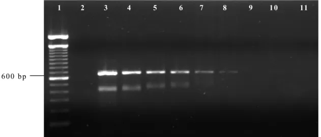
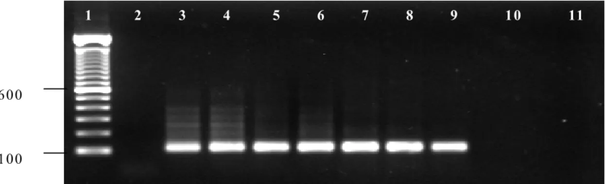
The experiment to determine the threshold of parasite DNA detection by PCR and nested PCR are shown in Fig. 1 and Fig. 2, respectively. The
16S rRNA based PCR detected T. equi DNA in
blood with an equivalent 8x10⁻⁵% parasitemia,
while nested PCR based on the EMA-1 gene
detected 8x10⁻⁶% parasitemia. Extra bands can
be seen, but only at higher parasite DNA concentrations. No differences in the sensitivity of the PCR and nested PCR assays were observed when DNA extraction was performed either by phenol-chloroform or the Puregene kit. Additionally, no amplification was observed with DNA obtained from uninfected horse blood.
No amplified product was observed in any of the animals tested by 16S rRNA PCR. On the other
hand, T. equi DNA was detected by nested PCR
in all the horses, except for one which was negative at rest (Tab. 1). In relation to the samples collected after exercise, 13 horses were positive including the animal which was negative at rest. When DNA template was prepared from
samples obtained after in vitro cultures, the
nested PCR demonstrated that seven and five
horses were negative for T. equi before and after
exercise, respectively. No significant difference was observed in G1 and G2, considering the
molecular detection of T. equi (Tab. 1).
Figure 1. Detection threshold of B. equi DNA in infected equine blood using the 18S rRNA PCR. The
PCR was performed on total DNA prepared from serially diluted blood of an infected horse (Jaboticabal strain, GenBank DQ250541) with initial parasitemia of 80%. Lane 1: 100 bp ladder DNA marker; lane 2: DNA from uninfected horse (negative control); lane 3: PCR with DNA from equine erythrocytes with
parasitemia of 80%; lane 4: 8x10⁻¹%; lane 5: 8x10⁻²%; lane 6: 8x10⁻³%; lane 7: 8x10⁻⁴%; lane 8: 8x10⁻⁵%;
lane 9: 8x10⁻⁶%; lane 10: 8x10⁻⁷%; lane 11: 8x10⁻⁸%.
1 2 3 4 5 6 7 8 9 1 0 11
Figure 2. Detection threshold of B. equi DNA in infected equine blood using the EMA-1 nested PCR. The PCR was performed on total DNA prepared from serially diluted blood of an infected horse (Jaboticabal strain, GenBank DQ250541) with initial parasitemia of 80%. Lane 1: 100 bp ladder DNA marker; lane 2: DNA from uninfected horse (negative control); lane 3: PCR with DNA from equine erythrocytes with
parasitemia of 80%; lane 4: 8x10⁻¹%; lane 5: 8x10⁻²%; lane 6: 8x10⁻³%; lane 7: 8x10⁻⁴%; lane 8: 8x10⁻⁵%;
lane 9: 8x10⁻⁶%; lane 10: 8x10⁻⁷%; lane 11: 8x10⁻⁸%.
Table 1. Comparison of microscopic, in vitro culture, and molecular tests for the detection and diagnosis
of T. equi in blood samples of naturally infected horses submitted to exercise after a 30-day (G1) and 90-day (G2) training schedules
PCR
Microscopy Before culture
In vitroculture
(16S rRNA
gene) Nested PCR (EMA-1 gene) 2th dc 4th dc 6th dc 10th dc Before culture After in vitro culture
Rest Exercise Rest Exercise Rest Exercise Rest Exercise Rest Exercise Rest/Exercise Rest Exercise Rest Exercise Horse
G1 G2 G1 G2 G1 G2 G1 G2 G1 G2 G1 G2 G1 G2 G1 G2 G1 G2 G1 G2 G1 G2 G1 G2 G1 G2 G1 G2 G1 G2 1 - - + + + + + + - - - + + - - - - - - + + + + - - + + 2 - - - - + + + - - - - + - - - + - - - - - - + + + + + + + + 3 - - + + - - + + - - - + - - + - - - + + - - - - + + - - + + 4 + + + + - + + + - - + + - - - + - - - - - - + + + + - - + + 5 - - - - + - + - - - + + - - - + - - - - - - + + + + - - - - 6 - - - - + + + + - - + - - - + - - - - - + + + + - - + + 7 - - - - + - + - - - - - - - - - - - - - - - + + + + + + + + 8 - - - - - - - - - - - - - - + - - - - - - - + + - - + + - - 9 - - - - + - + - + + + + + - + + - - - + - - + + + + - - - - 10 - - - - + - - - + + + + - - - - - + + + + + + + + 11 - - - - + + + + - - - + - - - - - + + + + + + - - 12 - - - + + + + + + + - + - - - - - + + + + + + - - 13 - - - + - + - - + + + + + - + + + - - - - - + + + + + + + + 14 - - - + + - - + + - - + + - - + - - - + + + + - - + + 15 + - + + - - - - - - - - - - - - - - - - - - + + - - + + + + Positive
Rate 2 1 4 5 7 5 11 7 5 5 8 9 3 0 8 7 2 0 2 2 0 0 14 14 13 13 8 8 10 10
dc = days of culture, (+) = positive animals, (-) = negative animals.
The following differences were significant (χ2 test): positive nested PCR (before or after culture) reactions vs. positive microscopy
(P<0.01), positive nested PCR (after culture) vs. positive microscopy (P<0.05), and positive microscopy vs. positive in vitro culture (P<0.01) for T. equi infections.
The detection and diagnosis of T. equi in blood
samples of naturally infected horses submitted to exercise were compared. The differences were not significant among the samples collected at rest or after strenuous exercises in all the diagnostic methods performed in the present study. The following differences were significant
(χ2 test): positive nested PCR reactions (before or
after culture) versus positive microscopy (P<0.01), positive nested PCR reactions (after culture) versus positive microscopy (P<0.05),
and positive microscopy versus positive in vitro
culture (P<0.01) for T. equi infections (Tab. 1).
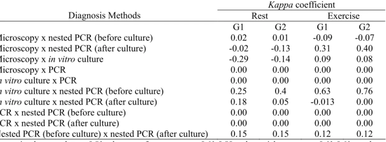
Assessment of agreement among the assays by
method of determining kappa statistic is shown
in Tab. 2. The kappa coefficient ranged from –
0.029 (microscopy and in vitro culture),
indicating agreement less than is expected by chance, to 0.76 (culture and nested PCR before culture), suggesting fair agreement. After
strenuous exercises (G1/G2), the kappa
coefficient ranged from –0.09/–0.07 between microscopy and nested PCR (before culture), to 0.63/0.76 between culture and nested PCR (before culture).
1 0 0 6 0 0
Table 2. Concordance among microscopic, in vitro culture, PCR, and nested PCR for the detection and
diagnosis of T. equi in blood samples of naturally infected horses submitted to exercise after a 30-day
(G1) and 90-day (G2) training schedules demonstrated by values of kappa statistic
Kappa coefficient
Diagnosis Methods Rest Exercise
G1 G2 G1 G2
Microscopy x nested PCR (before culture) 0.02 0.01 -0.09 -0.07
Microscopy x nested PCR (after culture) -0.02 -0.13 0.31 0.40
Microscopy x in vitro culture -0.29 -0.14 0.09 0.08
Microscopy x PCR 0.00 0.00 0.00 0.00
In vitro culture x PCR 0.00 0.00 0.00 0.00
In vitro culture x nested PCR (before culture) 0.25 0.4 0.63 0.76 In vitro culture x nested PCR (after culture) 0.18 0.05 -0.013 0.00
PCR x nested PCR (before culture) 0.00 0.00 0.00 0.00
PCR x nested PCR (after culture) 0.00 0.00 0.00 0.00
Nested PCR (before culture) x nested PCR (after culture) 0.15 0.15 0.12 0.12
Interpreting kappa values: >0.81: almost perfect agreement; 0.61-0.80: substancial agreement; 0.41-0.61: moderate agreement; 0.21-0.40: fair agreement; 0-0.20: slight agreement; <0: poor agreement.
DISCUSSION
The low positive rate for T. equi observed in the
present study on the direct microscopic identification of the parasite by blood smears could be especially due to the low sensitivity of the method. Bose et al. (1995) reported the low sensitivity of this technique and suggest its use only in acute cases of the disease. Holman et al. (1998) suggested that negative results in stained blood smears could be due to time delay and handling between blood collection and slide preparation. Therefore, it should be mentioned therefore, that in the present study, the blood was immediately processed after the return from the treadmill, implicating in intervals of approximately three hours, a fact that would not justify the occurrence of false-negative results.
Theileria equi parasites were observed in cultured erythrocytes from 14 horses of G1 and 12 of G2. Such results demonstrated that the sensibility of this method is much higher than that related for blood smears and are in agreement with those recorded by Holman et al. (1998). Statistical analysis demonstrated
significant differences (P<0.01) and kappa
coefficient between culture and microscopy ranged from –0.29 to 0.09, showing poor
agreement (Tab. 2). Although T. equi was
observed in cultured erythrocytes from most of the sampled animals, the cultures did not become continuous and lower positive rates were observed toward the end of the experiment. It is
possible that the parasitemia was simply too low, or the parasites were not robust enough to allow in vitro establishment, or both. Holman et al. (1998) suggested that the successful cultivation of the parasite may be due to selection of a population that adapts to imposed conditions, and that some field sample parasites may be less
adaptable to certain in vitro conditions than
others. Considering that the samples of the present study were collected from Brazilian field horses and that very little information about the T. equi strains from Brazil are available, further studies should be carried out in order to elucidate such aspects. Unknown factors during field collection, handling, and shipping may also have affected the viability of the parasites (Holman et al., 1998). Zweygarth et al. (1997) demonstrated that although parasites are present in the blood, the sample volume to initiate culture may be too small to give rise to positive cultures.
Thus, the culture diagnostic technique used in the present study has been shown to be suitable for
the diagnosis of T. equi, permitting the detection
of several animals with positive results, especially carrier horses which frequently have extremely low circulating parasitemia. The differences observed in the time span for a
positive diagnosis of T. equi could be related to
from two to 15 days. Additionally, the high cost of the reagents and the possibility of bacterial contamination makes this method unsuitable routine diagnosis or for epidemiological studies.
The occurrence of higher positive rates in stained
blood smears and in vitro culture with samples
collected at stress (Tab.1) could be explained by marked increases in erythrocyte numbers, packed cell volume, and mean corpuscular volume in horses submitted to training programmes and attributed to spleen contraction (Allen and Powell, 1983). The spleen of most mammals contracts during stress, resulting in a marked increase in blood erytrocytes, platelets and, to a lesser extent, blood leucocytes (Smith et al.,1989). Additionally, the effects of excitement or exercise seems to be most dramatic in horses, in which the packed cell volume can increase as much as 40% under stressful conditions (Kramer, 2000). Rose and Hodgson (1982) suggest that exercise programmes involving a large component of maximum exercise may stimulate increased erythrocyte production in order to transport oxygen more efficiently to the tissues.
The 16S PCR system was able to detect 8x10⁻⁵%
parasitaemia, equivalent to eight infected cells
out of 107 erythrocytes. Bashiruddin et al. (1999)
reported similar results in the molecular
detection of T. equi, being able to detect an
approximate 0.000083% parasitaemia in naturally infected horse blood. On the other hand, nested PCR using oligonucleotides
designed on the sequence of a T. equi merozoite
antigen gene (EMA-1) was able to detect the
parasite in blood with an equivalent 0.000008% parasitaemia. Similar results were obtained by Nicolaiewsky et al. (2001) which reported detection limits of 0.000006% in a mock
infection, equal to six infected cells out of 108
erythrocytes. The enhanced sensitivity of nested PCR observed in the present study when compared to the single-round 16S PCR could be explained by the fact that two separate rounds of amplification are required on a nested PCR and that the primers used codes for a highly
intra-species conserved equi merozoite antigen (
EMA-1) (Knowles et al., 1991; 1997).Rampersad et al.
(2003) showed that nested PCR detected 3.6
times more T. equi infections than microscopic
analysis and 2.2 times more than primary PCR.
All horses were negative for T. equi in 16S PCR
tests, including animals which were positive in
stained blood smears and in vitro culture (Tab.1).
No positive results were also observed in the
samples collected after in vitro culture. However,
species-specific amplified products from positive control were visualized by agarose gel electrophoresis, demonstrating that both the nucleic acid preparation and the amplification
conditions were correct for T. equi; therefore,
validating the results of the present study. Canola et al. (2002) used this same PCR system for the
detection of T. equi in naturally infected horses
and also did not report positive results in any of the 18 horses evaluated. Although Bashiruddin et
al. (1999) detected T. equi DNA in 22 horses
using the 16S rRNA gene, animals had very high parasitaemia as confirmed by direct microscopic examination of stained blood smears.
Fourteen horses at rest and 13 animals after strenuous exercise tested positive with the nested
PCR for T. equi. These results demonstrated that
the nested PCR can easily detect T. equi DNA in
blood from chronically infected horses, especially those which are negative by other direct diagnosis methods. It should be mentioned
that all horses that tested negative by in vitro
culture were positive by nested PCR, both at rest and stress, except horse number 15, which was positive only at rest.
The results obtained by nested PCR with samples
collected after in vitro culture showed that only
one of the horses with negative results turned out to be positive. Additionally, a lower number of
samples were positive for T. equi when compared
No statistical differences were observed between
nested PCR (before culture) and in vitro culture.
The best agreement (kappa coefficient: 0.25/rest
- 0.76/exercise) was observed between these diagnostic methods. These findings suggest that the nested PCR described herein is a useful tool
for the detection of sub-clinical T. equi infections
and that in vitro culture could be used as a
supplement for other methods of parasite detection. Both methods should be considered for the diagnosis of equine piroplasmosis and could greatly aid in controlling the importation of infected animals, avoiding performance problems in racehorses. Additionally, the results of the present study have provided evidence that samples may be collected at rest or after strenuous exercises, not interfering in positive rates, although stress seems to have a positive
effect on the detection of T. equi using blood
smears and in vitro culture.
ACKNOWLEDGEMENTS
This study was supported by grants from the Fundação de Amparo à Pesquisa do Estado de São Paulo (FAPESP, n. 02/06126-1;02/13561-6), Brazil.
REFERENCES
ALLEN, B.V.; POWELL, D.G. Effects of training and time of day of blood sampling on the variation of some common hematological parameters in normal thoroughbred racehorses.
In: SNOW, D.H. (Ed). Equine exercise
physiology. Cambridge: Granta, 1983. p.328.
BASHIRUDDIN, J.B.; CAMMA, C., REBELO,
E. Molecular detection of Babesia equi and
Babesia caballi in horse blood by PCR
amplification of part of the 16S rRNA gene. Vet.
Parasitol., v.84, p.75-83, 1999.
BOSE, R.; JORGENSEN, W.K.; DALGIESH, R.J. et al. Current state and future trends in the
diagnosis of babesiosis. Vet. Parasitol., v.57,
p.61-74, 1995.
BRUNING, A. Equine piroplasmosis an update
on diagnosis, treatment and prevention. Br. Vet.
J., v.152, p.139-151,1996.
CANOLA, P.A.; OLIVEIRA, R.; AQUINO, L.P.C.T. et al. Uso da reação em cadeia da polimerase (PCR) na detecção de Babesia equi e
Babesia caballi. In: CONGRESSO BRASILEIRO
DE PARASITOLOGIA VETERINÁRIA, 12., 2002,
Rio de Janeiro. Anais… Rio de Janeiro: Colégio
Brasileiro de Parasitologia Veterinária, 2002. CD-ROM. Resumo.
DE WAAL, D.T. Equine piroplasmosis: a review. Br. Vet. J., v.148, p.6-14, 1992.
EVERITT, R.S. Statistical methods for medical
investigations. London: Oxford University, 1989
HAILAT, N.Q.; LAFI, S.Q.; AL-DARRAJI, A.M. et al. Equine babesiosis associated with strenuous exercise: clinical and pathological
studies in Jordan. Vet. Parasitol., v.69, p.1-8,
1997.
HIRATA, H.; XUAN, X.; YOKOYAMA, N. et al. Identification of a specific antigenic region of
the P82 protein of Babesiaequi and its potential
use in serodiagnosis. J. Clin. Microbiol., v.41,
p.547-551, 2003.
HOLMAN, P.J.; FRERICHS, W.M.; CHIEVES, L. et al. Culture confirmation of the carrier status of Babesia caballi-infected horses. J. Clin. Microbiol., v.31, p.698-701,1993.
HOLMAN, P.J.; BECU, T.; BAKOS, E. et al. Babesia equi field isolates cultured from horse
blood using a microcentrifuge method. J.
Parasitol., v.84, p.696-699, 1998.
KNOWLES, D.P.; KAPPMEYER, L.S.;
PERRYMAN, L.E. Genetic and biochemical analysis of erythrocyte-stage surface antigens belonging to a family of highly conserved
proteins of Babesia equi and Theileria species.
Mol. Biochem. Parasitol.,v.90, p.69-79, 1997.
KNOWLES, D.P.; PERRYMAN, L.E.; GOFF, W.L. et al. A monoclonal antibody defines a geographically conserved surface protein epitope of Babesia equi merozoites. Infect. Immunol., v.59, p.2412-2417, 1991.
KRAMER, J.W. Normal hematology of the
horse. In: SCHALM, O. (Ed). Veterinary
hematology. Philadelphia: Lippincott Williams & Wilkins, 2000. p.1069-1074.
LOPAREV, V.N.; CARTAS, M.A.; MONKEN, C.E. et al. An efficient and simple method of DNA extraction from whole blood and cell lines
to identify infectious angents. J. Virol. Methods,
NICOLAIEWSKY, T.B.; RICHTER, M.F.;
LUNGE V.R. et al. Detection of Babesia equi
(Laveran, 1901) by nested polymerase chain
reaction. Vet. Parasitol., v.101, p.9-21, 2001.
RAMPERSAD, J.; CESAR, E.; CAMPBELL, M.D. et al. A field evaluation of PCR for the
routine detection of Babesia equi in horses. Vet.
Parasitol., v.114, p.81-87, 2003.
ROSE, R.J.; HODGSON, D.R. Haematological and plasma biochemical parameters in endurance
horses during training. Equine Vet. J., v.14,
p.144-148, 1982.
SCHEIN, E. Equine babesiosis. In: RISTIC, M.
(Ed.). Babesiosis of domestic animals and man.
Boca Raton: CRS, 1988. p.197-208.
SMITH, J.E.; ERICKSON, H.H.; DEBOWES, R.M. Changes in circulating equine erythrocytes
induces by brief, high-speed exercise. Equine
Vet. J., v.21, p.444-446, 1989.
TANAKA, T.; XUAN, X.; IKADAI, H. et al.
Expression of Babesia equi merozoite antigen-2
by recombinant baculovirus and its use in the
ELISA. Int. J. Parasitol., v.29, p.1803-1808,
1999.
XUAN, X.; IGARASHI, I.; TANAKA, T. et al.
Detection of antibodies to Babesia equi in horses
by a latex agglutination test using recombinant EMA-1. Clin. Diagn. Lab. Immunol., v.8, p.645-646, 2001a.
XUAN, X.; NAGAI, A.; BATTSETSEG, B. et al. Diagnosis of equine piroplasmosis in Brazil by serodiagnostic methods with recombinant
antigens. J. Vet. Med. Sci., v.63, p.1159-1160,
2001b.
VEGA, C.A.; BUENING, G.M.; GREEN, T.J. et al. In vitro cultivation of Babesia bigemina. Am. J. Vet. Res., v.46, p.416-420, 1985.
ZWEYGARTH, E.; JUST, M.C.; DE WAAL,
D.T. In vitro cultivation of Babesia equi:
detection of carrier animals and isolation of
parasites. Onderstepoort J. Vet. Res., v.64,