Is There Any Difference between the
In Situ
and Systemic IL-10 and IFN-
γ
Production
when Clinical Forms of Cutaneous
Sporotrichosis Are Compared?
Fernanda N. Morgado1¤a, Armando O. Schubach2, Maria Inês Pimentel2, Marcelo R. Lyra2, Érica C. F. Vasconcellos2, Claudia M. Valete-Rosalino2,3¤b, Fátima Conceição-Silva1
*
1Laboratório de Imunoparasitologia, Instituto Oswaldo Cruz/FIOCRUZ, Rio de Janeiro, RJ, Brazil, 2VigiLeish-Serviço de Infectologia, Instituto Nacional de Infectologia Evandro Chagas/FIOCRUZ, Rio de Janeiro, RJ, Brazil,3Departamento de Otorrinolaringologia-Oftalmologia/Faculdade de Medicina/ Universidade Federal do Rio de Janeiro, Rio de Janeiro, RJ, Brazil
¤a Current address: Laboratório de Pesquisa em Leishmaniose, Instituto Oswaldo Cruz/FIOCRUZ, Rio de Janeiro, RJ, Brazil
¤b Current address: Hygiene and Tropical Medicine Institute (IHMT), New University of Lisbon (UNL), Lisbon, Portugal
Abstract
Fungus of theSporothrix schenckiicomplex can produce skin lesions in humans, commonly lymphocutaneous (LC) and fixed (F) forms of sporotrichosis. Some authors have suggested that clinical forms are influenced by differences in virulence and genetic profile of isolates. But little is known about the role of immune response in determining the clinical outcome of sporotrichosis. To verify the profile of systemic and in situ IFN-γand IL-10 expression in
sporotrichosis patients, and consequently to detect any difference between the two com-partments and/or clinical presentation, we quantified the number of IFN-γand IL-10
pro-ducer peripheral blood mononuclear cells stimulated withS.schenckiiantigen (Ss-Ag) by Elispot, and quantified cytokines expression byin situimmunohistochemistry in the same patient. Three groups were formed: 1- LC (n = 9); 2- F (n = 10); 3- healthy individuals (n = 14). All sporotrichosis patients produced high amounts of systemic IFN-γwhen compared
to uninfected individuals. No differences were observed between LC and F groups. Regard-ingin situIL-10 expression, a difference between LC and F groups was observed: LC lesions presented higher amounts of IL-10 than F lesions differently from systemic IL-10 which showed similarities. Our data suggests that LC lesions present higher IL-10 expres-sion which could be related to regulatory mechanisms for compensating the tissue injury, however favoring fungal persistence in the lesions. Surprisingly, there were no differences in systemic andin situIFN-γexpression between CL and F patients, although it was
signifi-cantly higher expressed in these patients than in healthy individuals. a11111
OPEN ACCESS
Citation:Morgado FN, Schubach AO, Pimentel MI, Lyra MR, Vasconcellos ÉCF, Valete-Rosalino CM, et al. (2016) Is There Any Difference between theIn Situ and Systemic IL-10 and IFN-γProduction when
Clinical Forms of Cutaneous Sporotrichosis Are Compared? PLoS ONE 11(9): e0162764. doi:10.1371/journal.pone.0162764
Editor:Hiroshi Shiku, Mie University Graduate School of Medicine, JAPAN
Received:March 19, 2016
Accepted:August 30, 2016
Published:September 13, 2016
Copyright:© 2016 Morgado et al. This is an open access article distributed under the terms of the Creative Commons Attribution License, which permits unrestricted use, distribution, and reproduction in any medium, provided the original author and source are credited.
Data Availability Statement:All relevant data are within the paper and its Supporting Information file.
Introduction
Sporotrichosis is a subcutaneous mycosis caused by fungus of theSporothrix schenckiicomplex [1–3]. Infection occurs by traumatic inoculation of the fungus in individuals working with soil
and plants [4] or through the bite or scratch of sick cats [4–7].
After fungus inoculation, the individual may develop different clinical forms, varying from localized skin lesions (fixed—F) or lymphocutaneous (LC) forms, to systemic disease
(extracu-taneous form) [4]. About 75% of patients present skin lesions, mainly LC and F forms [4]. The F form is characterized by one lesion (verrucous, ulcerated or plaque-like) on the site of the fungus inoculation without lymphatic involvement. But, the most common form is the LC which is characterized by an involvement of the lymphatic system, accompanied by the occur-rence of subcutaneous nodules that may progress to necrosis, liquefaction of their content and ulceration, showing the aspect known as sporotrichoid [4–8]. Although some authors have
sug-gested that clinical forms are influenced by differences in virulence [9] and genetic profile of isolates [10], little is known about immune response in human SP.
Previously, our group [11] had verified differences in thein situimmune response between LC and F lesions suggesting that the clinical presentation could be influenced by the different profile of thein situimmune response. However, asSporothrixsp can spread all over the body, the role of systemic immune response in immunopathology of sporothrichosis, particularly cytokine profile, cannot be neglected. Based on these facts and aiming at understanding some aspects of the IFN-γ/IL-10 profile in the different clinical presentations and tissue compart-ment of human sporotrichosis, we quantified the systemic andin situIFN-γand IL-10 expres-sions by immunohistochemistry and Elispot assay.
Materials and Methods
Ethics statement
An informed written consent was obtained from all individuals. In the case of minor/children, the written consent was obtained from the next of kin, caretakers or guardians. Ethical approval was obtained from the institutional Ethics Committee on Human Research—
FIO-CRUZ (CEP-INI protocol 014/2001).
Patients
Nineteen sporotrichosis patients treated at the Instituto Nacional de Infectologia Evandro Cha-gas—INI/FIOCRUZ and were grouped as follows: 1- lymphocutaneous form (LC; n = 9); and
2- fixed cutaneous form (F; n = 10). Fourteen healthy donors were included in this study. Peripheral venous blood was collected in heparin from all analyzed individuals and lesion frag-ments were collected from 14 patients. All samples used in this study were obtained at the moment of diagnosis, so the patients were free of medication. After the sample uptake, the diagnosis was confirmed by fungal isolation in appropriate culture media and according to the diagnosis, they were treated by the medical staff of Instituto Nacional de Infectologia Evandro Chagas—Fiocruz.
Antigen preparation (Ss-Ag)
The yeast-like phase of theSporothrix schenckii(Ss) strain 17629, kindly provided by Dr Zan-copé-Oliveira (INI-FIOCRUZ) was cultured in brain heart infusion (BHI) medium as described by Brito et al. [9]. Fungal cells were adjusted to 108cells/mL and disrupted by cycles of freezing and thawing, followed by ultra-sonication (Lab-line Ultra-Lip Labsonic Systems IL, USA).
Fortalecimento dos Laboratórios Credenciados e das Áreas de Apoio à Pesquisa do Instituto Oswaldo Cruz-Fundação para o desenvolvimento Científico e Tecnológico em Saúde (IOC-008-FIO-15), obtained by FCS; and Fundação Carlos Chagas Filho de Amparo à Pesquisa do Estado do Rio de Janeiro (E26/111.230/2014), obtained by FCS. AOS is a recipient of fellowships from CNPq and FAPERJ. CMVR is a recipient of FAPERJ fellowship. The funders had no role in study design, data collection and analysis, decision to publish, or preparation of the manuscript.
Separation of peripheral blood mononuclear cells (PBMC)
PBMC were obtained as previously described [12]. Briefly, PBMC cells were separated by cen-trifugation over a Ficoll-Hypaque gradient (Histopaque 1077, Sigma, MO, USA) and adjusted to 2x10⁶cells / mL and 1x10⁶cells / mL.
IFN-
γ
and IL-10 Elispot
The Elispot assay was performed as previously described [13]. Briefly, 96-well microplates (Multiscreen Millipore, France) were coated with 5μg/mL of antibodies anti-IFN-γor anti-IL-10 (Mabtech, OH, USA). A total of 2xanti-IL-105and 1x10⁵PBMC were added to the ELISPOT plates in the presence of medium alone (spontaneous cytokine production), Ss-Ag (the equivalent of 106disrupted fungal cells/well) or Concanavalin A (ConA) (4μg/mL–Sigma). After
stimula-tion, the plates were washed and incubated with either 1μg/mL biotinylated anti-IFN-γor anti-IL-10 (MabTech). The plates were incubated with streptavidin-alkaline phosphatase (Mab-Tech) and revealed with BCIP/NBT (Sigma). The spots were counted in an Immunospot reader [Cellular Technology (CTL) Ltd, OH, USA] using the Immunospot Software Version 3 (CTL).
The results were expressed as spots/10,000 PBMC in the antigen stimulated wells subtracted of the spontaneous expression.
Immunohistochemistry
Thein situinflammation was evaluated by immunoperoxidase staining as described [14]. The primary antibodies used were: rat anti- human IL10 (clone JES3-12G8) and rat anti- human IFN-γ(clone XMG1-2) (BD Biosciences, CA, USA), diluted 1:100 in PBS (phosphate buffered saline). Suppressed primary antibodies was used as negative control. The images were acquired using the Motic Images Plus2.0 program (Motic China Co., China) and light microscopy (Nikon eclipse E200, Japan). The staining intensity was scored in ten microscopic fields (200x magnification) as rare (at least 1 positive area / field), discrete (2–3 positive areas / field),
mod-erate (4–5 positive areas / field) and intense (>5 positive areas / field).
Statistical analysis
The SPSS16 for windows (SPSS, Inc., IL, USA), was used for statistical analysis. Data were reported as median and range and the nonparametric Mann-Whitney test and Fisher`s exact test were used to compare the LC, F and healthy groups. Spearman test was used for nonpara-metric correlations. P values0.05 were considered as positive.
Results
Patients
Clinical data are described inTable 1andS1 table.
Histopathology
Suppurative granulomatous inflammation was the predominant finding (7/9) in fixed lesions; the remaining (2/9) presented only granulomatous inflammation. LC lesions presented suppu-rative granulomatous inflammation (4/8) and necrotic granulomatous inflammation (4/8). Yeast cells compatible withSporothrix schenckiicomplex were observed in three LC and in four F cases. No differences were observed between groups (p>0.05). Histopathological
IFN-
γ
expression by PBMC
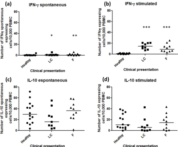
The amount of spontaneous IFN-γproducing cells was lower in the healthy group (median 0.11, 0–3.84) than in LC (median 0.5, 0.1–5.5; p = 0.013) and F patients (median 0.83, 0–5.92;
p = 0.026) (Fig 1aandS1 Table). All subjects presented positive response to ConA stimulation (data not shown). Regarding Ss-Ag stimulation, LC (median 14.1, 7–20.3) and F (median 8.8,
3.1–25) patients presented similar quantities of IFN-γ-producer cells (p>0.05), but they were
higher than in the healthy group (median 0.65, 0–2, p = 0.0001 and 0.0001, respectively) (Fig
1b). An inverse correlation between spontaneous IFN-γ-producer cells and duration of lesions was observed (r = -0.578; p = 0.008).
IL-10 expression by PBMC
Healthy subjects showed substantial amounts of spontaneous IL-10 expression (median 29.45, 3.3–71.6) and they were similar to LC (median 15.7, 3.5–55.0) and F patients (median 36.45,
1.6–58.7; p>0.05) (Fig 1candS1 Table).
Ss-Ag was able to stimulate IL-10 expression by PBMC from the majority of subjects (LC: median 5.4, 0–42.7; F: median 14.35, 0–43.4; Healthy: median 10.45, 0–39) (p>0.05) (Fig 1d).
An inverse correlation between spontaneous IL-10 producer cells and the number of lesions was observed (r = -0.542; p = 0.017).
IFN-
γ
in situ
expression
The distribution of thein situIFN-γin LC was as follows: three patients with rare, one with dis-crete, one with moderate and one patient with intense expression. The distribution in F was: three patients with rare, one with discrete and four patients with moderate expression. No
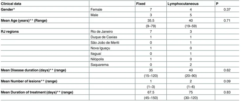
Table 1. Clinical data of patients with lymphocutaneous (LC) and fixed (F) forms of sporothrichosis.
Clinical data Fixed Lymphocutaneous P
Gender* Female 7 4 0.37
Male 3 5
Mean Age (years)**(Range) 35.5 40 0.71
(9–79) (19–59)
RJ regions Rio de Janeiro 7 3
Duque de Caxias 1 1
São João de Meriti 0 1
Nova Iguaçu 1 0
Itaguaí 0 1
Nilópolis 1 0
Saquarema 0 2
Mean Disease duration (days)**(range) 35 40 0.62
(15–120) (20–90)
Mean Number of lesions**(range) 1 2 0.09
(1–3) (1–6)
Mean Duration of treatment (days)**(range) 67.5 75 0.83
(45–150) (30–120)
*Fisher`s exact test
**Mann-Whitney test
RJ regions = municipalities of Rio de Janeiro state A p value0.05 was considered to be significant
difference was observed when groups were compared (p = 0.627; OR = 2.00) (Fig 2a and 2b
andTable 2andS1 Table).
IL-10
in situ
expression
Thein situIL-10 expression was distributed in LC as follows: one patient with rare, two with moderate and three patients with intense expression. The distribution in F was: two patients with rare, five with discrete and one patient with moderate expression. The LC group presented higher IL-10 expression than the F patients (p = 0.03; OR = 0.028) (Fig 2c and 2dandTable 3
andS1 Table). In addition, an association between higher IL-10in situexpression by cells in LC lesions and necrotic granulomatous inflammation was observed.
Discussion
In the present study, to detect differences in IFN-γ/IL-10 profile in different clinical presenta-tions of human sporotrichosis, we quantified spontaneous and antigen stimulated IFN-γand IL-10 producer cells from peripheral blood and in skin lesions of patients with lymphocuta-neous (LC) and fixed (F) forms of sporotrichosis. Although relevantly expressed, IFN-γand IL-10 systemic expression cells (peripheral blood) was similar when the two clinical forms were
Fig 1. Number of IFN-γand IL-10 producer cells/2 x 105cells from peripheral blood.(A) Spontaneous IFN-γ producing cells and (B) IFN-γproducing cells after Ss-Ag stimulation. (C) Spontaneous IL-10 producing cells and (D) IL-10 producing cells after Ss-Ag stimulation. The spontaneous values were subtracted from total spots detected in stimulated cultures. LC—lymphocutaneous sporotrichosis; F- fixed form of sporotrichosis;*p-value =
Fig 2.In situIFN-γand IL-10 expression in lymphocutaneous and fixed lesions of sporothrichosis.Thein situ IFN-γand IL-10 expression was detected by immunohistochemistry. The arrows point positive areas (red/AEC– 3-amino-9-ethylcarbazole). The intensity of staining was scored in ten microscopic fields (200x magnification) as rare (at least 1 positive area / field), discrete (2–3 positive areas / field), moderate (4–5 positive areas / field) and intense (>5 positive areas / field). Scale bar = 10μm.
doi:10.1371/journal.pone.0162764.g002
Table 2. In situIFN-γexpression in lymphocutaneous (LC) and fixed (F) forms of sporotrichosis.
Clinical presentation Rare to discrete Moderate to intense Total
Lymphocutaneous 4 2 6
Fixed 4 4 8
Total 8 6 14
p>0.05 Fisher’s exact test.
doi:10.1371/journal.pone.0162764.t002
Table 3. In situIL-10 expression in lymphocutaneous (LC) and fixed (F) forms of sporotrichosis.
Clinical presentation Rare to discrete Moderate to intense Total
Lynphocutaneous 1 5 6
Fixed 7 1 8
Total 8 6 14
p = 0.03; OR = 0.028 (Fisher’s exact test).
compared. On the other hand, we could detect a higher IL-10in situexpression by cells in LC lesions, associated with necrotic granulomatous inflammation and a tendency of higher num-ber of lesions, longer duration and longer treatment duration. Our previous data showed that, when compared with F group, LC lesions presented stronger inflammatory profile with higher number of lesions, longer duration and required longer treatment duration, as well as morein situCD4+cells, neutrophils, and more intense NOS2 expression [11]. Macrophage concentra-tion was similar and comprised about one third of all cells in sporothrichosis lesions. In this context, the high IL-10 expression now verified could play a role in regulatory mechanisms to control/modulate the tissue injury observed in these lesions. IL-10 can be produced by alterna-tively activated macrophages (M2) [15]. These diverse physiological functions result from the remarkable plasticity of macrophages, which allows these cells to dramatically change their form and function in response to local environmental signals or tissue damage [16]. Further-more, the subtype M2b (IL-10 and TNF-αproducer) was correlated to increased susceptibility to gastrointestinal candidiasis in murine model [17]. Recently, thein vitroinduction of M2 by
Sporothrix schenckii’s cell wall molecules was demonstrated [18]. Mice experimentally infected presented high IL-10 which was associated with the apoptotic process induced by the fungal infection [19].
The detection of systemic andin situIFN-γproduction cells in LC and F patients suggested a role of IFN-γin the inflammation of cutaneous sporotrichosis. The induction of Th1 response along with abundant IFN-γproduction in anin vitrostudy [20] in mice was achieved by fungal isolates from patients presenting cutaneous manifestations. In addition, the resis-tance of NOS2 knockout mice was related to a higher IFN-γproduction and a reduced sponta-neous and antigen stimulated IL-10 expression in wild type mice [21]. Altogether, these data suggested that the role of IFN-γin fungal burden control and development of benign clinical presentations could not be related to macrophage activation via NOS2. Furthermore, the authors also suggested that although NO was an essential mediator to thein vitrokilling ofS.
schenckiiby macrophages, NOin vivocould contribute to the immunosuppression and cyto-kine balance during the early phases of infection [21].
In experimental sporotrichosis andin vitrostudies using isolates from visceral and cutane-ous origins it was established that Th1 response mostly IFN-ƴ, which is produced by different cells including CD4 T cells, strongly activates macrophages and thereby innate and adaptive immune responses and perhaps determines its clinical manifestations[20,22–24]. Herein, we
observed a markedly IFN-ƴexpression in active lesions that did not differ between two differ-ent cutaneous manifestations, probably suggesting that Th1 cells were predominant at the lesions site. However, other factors could be taking place in LC lesions favoring fungal persis-tence and dissemination. In this context, intrinsic regulations, as the elevated IL-10 expression observed herein could favor the development of extensive lesions in lymphocutaneous patients. In addition, the possibility of extrinsic regulations such as the use of anti-TNF-alpha mono-therapy favoring the lesion severity was described[25].
Zhang et al [26] showed large IL-10 expression induced by components ofS.schenckiiin rats experimentally infected. An induction of a Th2 profile with IL-10 and IL-4 production by
induce high liberation of NO and TNF-αin macrophage cultures [28]. Furthermore, the lipid extract could be recognized by TLR4 which induces IL-10 and TNF expression by peritoneal macrophages, as well as NO production, all associated with clinical worsening [30]. This sug-gests thatS.schenckiicould be able to modulate the immune response also by inducing IL-10 production by human cells, probably as a mechanism of immune escape. Maia et al. [19] dem-onstrated that the ExoAntigen was able to stimulate IL-10 production inin vitroinfected mice. We observed that Ss-Ag stimulated IL-10 expression by PBMC from both healthy and patient groups. Thus, IL-10 could influence fungal clearance allowing the antigen persistence and con-sequently favoring the worsening of the lesion. On the other hand, we cannot exclude the possi-bility of a cross reaction between Ss-Ag and antigens from other fungal agents, particularly those that compose the normal microbiota like those with similar PAMPs [31,32]. The influ-ence ofCandidasp cell wall in the expression of inflammatory and regulatory cytokines has been described [33,34].β-glucans are frequently observed in cell walls of several fungi [35] includingCandidasp andS.schenckii[36] and can induce IL-10 expression causing subversion of immune response [35]. Moreover, the possibility of lipopolysaccharide (LPS) contamination of Ss-Ag can be rejected since PBMC from healthy subjects produced insignificant amounts of IFN-spots under Ss-Ag stimulation.
Spontaneous IFN-γ-producing PBMC from patients were higher than from healthy sub-jects. The present data supports previous results which indicate that despite the inflammatory activity being concentrated at the lesion site, the immune response can be reflected at the peripheral system as observed in the peripheral blood compartment, and it can be specifically measured. Systemic reactions like erythema nodosum have already been verified in sporothri-chosis patients presenting only cutaneous lesions, along with fever, arthralgia and malaise [37]. As fungal cells were not verified in tissues from erythema nodosum, Gutierrez-Galhardo et al. [37] suggested that these systemic reactions could play a protective role, leading to a more benign evolution of the disease.
Although the small number of cases could be considered as a limitation of this study, this is the first publication comparing thein situand systemic cytokine expressions in human sporo-trichosis. Moreover, studies with similar numbers of human cases have been published in the literature[11,14,38–41].
In conclusion, our data suggests that: 1- LC lesions present higher IL-10 expression which can be related to regulatory mechanisms for compensating the tissue injury, however favoring fungal persistence in the lesions. 2- Surprisingly, there were no differences in systemic andin situIFN-γexpression between CL and F patients, although it was significantly higher expressed in these patients than in healthy individuals. 3-S.schenckiiis able to modulate the immune response also by inducing IL-10 production by human cells probably using this as a mechanism of immune escape.
Supporting Information
S1 Table. Raw data of patients and healthy donors evaluated.
(DOCX)
Acknowledgments
Tools for Health—PDTIS-FIOCRUZ for use of its facilities. A.O.S is a recipient of fellowships
from CNPq and FAPERJ. C.M.V.R. is a recipient of FAPERJ fellowship.
Author Contributions
Conceived and designed the experiments:FCS FNM.
Performed the experiments:FNM AOS MIP MRL ECFV CMVR FCS.
Analyzed the data:FNM FCS.
Contributed reagents/materials/analysis tools:FNM AOS FCS.
Wrote the paper:FNM AOS MIP MRL ECFV CMVR FCS.
References
1. Rodrigues AM, de Hoog Gs, Zhang Y, de Camargo ZP. Emerging sporotrichosis is driven by clonal and recombinant Sporothrix species. Emerg Microbes Infect. 2014 May; 3(5):e32. doi:10.1038/emi.2014. 33PMID:26038739
2. Rodrigues AM, de Hoog S, de Camargo ZP. Emergence of pathogenicity in the Sporothrix schenckii complex. Med Mycol. 2013 May; 51(4):405–12. doi:10.3109/13693786.2012.719648PMID:
22989196
3. Oliveira MME, Almeida-Paes R, Muniz MM, Gutierrez-Galhardo MC, Zancope-Oliveira RM. Phenotypic and molecular identification of Sporothrix isolates from an epidemic area of sporotrichosis in Brazil. Mycopathologia. 2011 Oct; 172(4):257–67. doi:10.1007/s11046-011-9437-3PMID:21701792
4. Barros MB de L, Schubach A de O, do Valle ACF, Gutierrez Galhardo MC, Conceição-Silva F, Schu-bach TM, et al. Cat-transmitted sporotrichosis epidemic in Rio de Janeiro, Brazil: description of a series of cases. Clin Infect Dis. 2004 Feb 15; 38(4):529–35. PMID:14765346
5. Schubach TM, Valle AC, Gutierrez-Galhardo MC, Monteiro PC, Reis RS, Zancopé-Oliveira RM, et al. Isolation of Sporothrix schenckii from the nails of domestic cats (Felis catus). Med Mycol. 2001 Feb; 39 (1):147–9. PMID:11270404
6. Schubach A, Barros MB de L, Wanke B. Epidemic sporotrichosis. Curr Opin Infect Dis. 2008 Apr; 21 (2):129–33. doi:10.1097/QCO.0b013e3282f44c52PMID:18317034
7. Schubach TMP, Schubach A, Okamoto T, Barros MB, Figueiredo FB, Cuzzi T, et al. Evaluation of an epidemic of sporotrichosis in cats: 347 cases (1998–2001). J Am Vet Med Assoc. 2004 May 15; 224 (10):1623–9. PMID:15154732
8. Lopes-Bezerra LM, Schubach A, Costa RO. Sporothrix schenckii and sporotrichosis. An Acad Bras Cienc. 2006 jun; 78(2):293–308. PMID:16710567
9. Brito MMS, Conceição-Silva F, Morgado FN, Raibolt PS, Schubach A, Schubach TP, et al. Comparison of virulence of different Sporothrix schenckii clinical isolates using experimental murine model. Med Mycol. 2007 Dec; 45(8):721–9. PMID:17885952
10. Almeida-Paes R, de Oliveira MME, Freitas DFS, do Valle AC, Zancopé-Oliveira RM, Gutierrez-Gal-hardo MC. Sporotrichosis in Rio de Janeiro, Brazil: Sporothrix brasiliensis is associated with atypical clinical presentations. PLoS Negl Trop Dis. 2014 Sept 18; 8(9):e3094. doi:10.1371/journal.pntd. 0003094PMID:25233227
11. Morgado FN, Schubach AO, Barros MBL, Conceição-Silva F. The in situ inflammatory profile of lym-phocutaneous and fixed forms of human sporotrichosis. Med Mycol. 2011 Aug; 49(6):612–20. doi:10. 3109/13693786.2011.552532PMID:21254963
12. Conceição-Silva F, Dórea RC, Pirmez C, Schubach A, Coutinho SG. Quantitative study of Leishmania braziliensis braziliensis reactive T cells in peripheral blood and in the lesions of patients with American mucocutaneous leishmaniasis. Clin Exp Immunol. 1990 Feb; 79(2):221–6. PMID:2311299
13. Lima-Junior JC, Tran TM, Meyer EVS, Singh B, De-Simone SG, Santos F, et al. Naturally acquired humoral and cellular immune responses to Plasmodium vivax merozoite surface protein 9 in Northwest-ern Amazon individuals. Vaccine. 2008 Dec 2; 26(51):6645–54. doi:10.1016/j.vaccine.2008.09.029
PMID:18832003
15. Martinez FO, Sica A, Mantovani A, Locati M. Macrophage activation and polarization. Front Biosci. 2008 Jan 1; 13:453–61. PMID:17981560
16. Ferrante CJ, Leibovich SJ. Regulation of Macrophage Polarization and Wound Healing. Adv Wound Care (New Rochelle). 2012 Feb; 1(1):10–6.
17. Lefèvre L, Galès A, Olagnier D, Bernad J, Perez L, Burcelin R, et al. PPARγligands switched high fat diet-induced macrophage M2b polarization toward M2a thereby improving intestinal Candida elimina-tion. PLoS ONE. 2010 Sep 20; 5(9):e12828. doi:10.1371/journal.pone.0012828PMID:20877467
18. Alegranci P, de Abreu Ribeiro LC, Ferreira LS, Negrini Tde C, Maia DC, Tansini A, et al. The predomi-nance of alternatively activated macrophages following challenge with cell wall peptide-polysaccharide after prior infection with Sporothrix schenckii. Mycopathologia. 2013 Aug; 176(1–2):57–65. doi:10. 1007/s11046-013-9663-yPMID:23686275
19. Maia DCG, Gonçalves AC, Ferreira LS, Manente FA, Portuondo DL, Vellosa JC, et al. Response of Cytokines and Hydrogen Peroxide to Sporothrix schenckii Exoantigen in Systemic Experimental Infec-tion. Mycopathologia. 2015 Apr; 181(3–4):207–15. doi:10.1007/s11046-015-9966-2PMID:26603044
20. Uenotsuchi T, Takeuchi S, Matsuda T, Urabe K, Koga T, Uchi H, et al. Differential induction of Th1-prone immunity by human dendritic cells activated with Sporothrix schenckii of cutaneous and visceral origins to determine their different virulence. Int Immunol. 2006 Dec; 18(12):1637–46. PMID:
17035348
21. Fernandes KSS, Neto EH, Brito MMS, Silva JS, Cunha FQ, Barja-Fidalgo C. Detrimental role of endog-enous nitric oxide in host defence against Sporothrix schenckii. Immunology. 2008 Apr; 123(4):469–
79. doi:10.1111/j.1365-2567.2007.02712.xPMID:18194265
22. Carlos IZ, Sassá MF, da Graça Sgarbi DB, Placeres MC, Maia DC. Current research on the immune response to experimental sporotrichosis. Mycopathologia. 2009; 168(1):1–10. doi:
10.1007/s11046-009-9190-zPMID:19241140
23. Tachibana T, Matsuyama T, Mitsuyama M. Involvement of CD4+ T cells and macrophages in acquired protection against infection withSporothrix schenckiiin mice. MedMycol.1999; 37(6):397–404
24. Maia DC,Sassá MF,Placeres MC,Carlos IZ. Influence of Th1/Th2 cytokines and nitric oxide in murine systemic infection induced bySporothrix schenckii. Mycopathologia.2006; 161(1):11–9. PMID:
16389479
25. Ursini F, Russo E, Leporini C,Calabria M,Bruno C,Tripolino C, et al. Lymphocutaneous Sporotrichosis during Treatment with Anti-TNF-Alpha Monotherapy.Case Rep Rheumatol.2015; 2015:614504 doi:
10.1155/2015/614504PMID:25755904
26. Zhang X, Zhang J, Huang H, Xue R, Hu X, Li M, et al. Taenia taeniaeformis in rat favors protracted skin lesions caused by Sporothrix schenckii infection: Dectin-1 and IL-17 are dispensable for clearance of this fungus. PLoS ONE. 2012; 7(12):e52514. doi:10.1371/journal.pone.0052514PMID:23285072
27. Carlos IZ, Sgarbi DB, Placeres MC. Host organism defense by a peptide-polysaccharide extracted from the fungus Sporothrix schenckii. Mycopathologia. 1998–1999; 144(1):9–14.
28. Carlos IZ, Sgarbi DBG, Santos GC, Placeres MCP. Sporothrix schenckii lipid inhibits macrophage phagocytosis: involvement of nitric oxide and tumour necrosis factor-alpha. Scand J Immunol. 2003 Mar; 57(3):214–20. PMID:12641649
29. Nascimento RC, Almeida SR. Humoral immune response against soluble and fractionate antigens in experimental sporotrichosis. FEMS Immunol Med Microbiol. 2005 Feb 1; 43(2):241–7. PMID:
15681154
30. Sassá MF, Saturi AET, Souza LF, Ribeiro LC, Sgarbi DB, Carlos IZ. Response of macrophage Toll-like receptor 4 to a Sporothrix schenckii lipid extract during experimental sporotrichosis. Immunology. 2009 Oct; 128(2):301–9. doi:10.1111/j.1365-2567.2009.03118.xPMID:19740386
31. Martínez-Álvarez JA, Pérez-García LA, Flores-Carreón A, Mora-Montes HM. The immune response against Candida spp. and Sporothrix schenckii. Rev Iberoam Micol. 2014 Jan-Mar; 31(1):62–6. doi:10. 1016/j.riam.2013.09.015PMID:24252829
32. Hearn VM, Wilson EV, Latgé JP, Mackenzie DW. Immunochemical studies of Aspergillus fumigatus mycelial antigens by polyacrylamide gel electrophoresis and western blotting techniques. J Gen Micro-biol. 1990 Aug; 136(8):1525–35. PMID:2175766
33. Ghosh S, Howe N, Volk K, Tati S, Nickerson KW, Petro TM. Candida albicans cell wall components and farnesol stimulate the expression of both inflammatory and regulatory cytokines in the murine RAW264.7 macrophage cell line. FEMS Immunol Med Microbiol. 2010 Oct; 60(1):63–73. doi:10.1111/ j.1574-695X.2010.00717.xPMID:20618847
34. Gow NAR, Netea MG, Munro CA, Ferwerda G, Bates S, Mora-Montes HM,et al. Immune recognition of Candida albicans beta-glucan by dectin-1. J Infect Dis. 2007 Nov 15; 196(10):1565–71. PMID:
35. Nisini R, Torosantucci A, Romagnoli G, Chiani P, Donati S, Gagliardi MC, et al. beta-Glucan of Candida albicans cell wall causes the subversion of human monocyte differentiation into dendritic cells. J Leukoc Biol. 2007 Nov; 82(5):1136–42. PMID:17656653
36. Lopes-Bezerra LM. Sporothrix schenckii Cell Wall Peptidorhamnomannans. Front Microbiol. 2011 Dec 21; 2:243. doi:10.3389/fmicb.2011.00243PMID:22203817
37. Gutierrez Galhardo MC, de Oliveira Schubach A, de Lima Barros MB, Moita Blanco TC, Cuzzi-Maya T, Pacheco Schubach TM, et al. Erythema nodosum associated with sporotrichosis. Int J Dermatol. 2002 Feb; 41(2):114–6. PMID:11982651
38. Koga T, Duan H, Urabe K, Furue M. Immunohistochemical localization of activated and mature CD83+ dendritic cells in granulomas ofsporotrichosis.Eur J Dermatol.2001; 11(6):527–9. PMID:11701401
39. Koga T, Duan H, Furue M. Immunohistochemical detection of interferon-gamma-producing cells in granuloma formation of sporotrichosis.Med Mycol.2002; 40(2):111–4. PMID:12058722
40. Ferraz R, Cunha CF, Gomes-Silva A, Schubach AO, Pimentel MIF, Lyra MR, et al. Apoptosis and fre-quency of total and effector CD8+T lymphocytes from cutaneous leishmaniasis patients during antimo-nial therapy. BMC Infect Dis. 2015; 15: 74. doi:10.1186/s12879-015-0799-xPMID:25870976