Quality of the interpretation of diagnostic mammographic
images*
Qualidade da interpretação do diagnóstico mamográfico
Hilton Koch1, Melissa Vieira Koch e Castro2
OBJECTIVE: To demonstrate the knowledge of mammogram readers working in the public healthcare system in the State of Rio de Janeiro, RJ, Brazil, and to evaluate their progress in the early diagnosis of breast cancer after a training course specifically developed for medical professionals . MATERIALS AND METHODS: A group of 53 physicians with experience in mammography reports were invited. A pre-test was given to assess their initial knowledge level. Afterwards, they were trained by experts mammographers, and for final conclusion, requested to take a post-test for comparison and evaluation of gained knowledge. RESULTS: The course, with emphasis on theoretical classes, has not resulted in a significant improvement on the quality of mammogram reading, highlighting the persistence of errors in morphological description of fundamental lesions of the breast, in the classification of such lesions according to the BI-RADS®, besides the lack of coherence between the BI-RADS classification and follow-up recommendation as observed in both the pre- and post-test. CONCLUSION: The authors conclude that the mammogram readers have demonstrated insufficient knowledge in relation to early imaging diagnosis of breast cancer, and that the theoretical training has not resulted in a significant improvement on the quality of mammogram reading.
Keywords: Quality; Mammographic diagnosis; Technical parameters.
OBJETIVO: Demonstrar o conhecimento mamográfico dos médicos interpretadores que trabalham na rede de saúde pública do Estado do Rio de Janeiro e avaliar o conhecimento adquirido após um curso elaborado com o objetivo de capacitar profissionais médicos no diagnóstico precoce do câncer de mama. MATERIAIS E MÉTODOS: Foram convidados 53 médicos que laudam exames mamográficos para o treinamento. Esses médicos eram submetidos a um pré-teste, no qual se avaliava o grau de conhecimento inicial. Depois, foram lecionadas aulas previamente elaboradas por mamografistas experientes, e para conclusão do curso esses médicos eram submetidos a um pós-teste para avaliação do conhecimento adquirido. RESULTADOS: O curso de capacitação de profissionais médicos, com ênfase em aulas teóricas, não mostrou aumento significativo na qualidade da interpretação mamográfica, destacando-se a persistência do erro na descrição morfológica das lesões fundamentais da mama, erro da classificação pelo sistema de padronização das lesões mamárias (BI-RADS®), falta de coerência entre a classificação BI-RADS adotada e a recomendação de conduta, tanto no pré-teste como no pós-teste. CONCLUSÃO: Concluiu-se que os médicos interpretadores mostram conhe-cimento insuficiente em relação ao diagnóstico precoce por imagem do câncer de mama e que o curso teó-rico não mostrou aumento significativo na qualidade da interpretação mamográfica.
Unitermos: Qualidade; Diagnóstico mamográfico; Parâmetros técnicos. Abstract
Resumo
* Study developed at Universidade Federal do Rio de Janeiro (UFRJ), Rio de Janeiro, RJ, Brazil.
1. PhD, Titular Professor and Head of the Department of Ra-diology at Faculdade de Medicina da Universidade Federal do Rio de Janeiro (UFRJ), Head of the Division of Radiology at Santa Casa da Misericórdia do Rio de Janeiro, Rio de Janeiro, RJ, Brazil.
2. PhD, Responsible by the Unit of Breast Diagnosis at Divi-sion of Radiology at Santa Casa da Misericórdia do Rio de Janeiro, Rio de Janeiro, RJ, Brazil.
Mailing address: Dr. Hilton Koch. Estrada de Itajuru, 425, Itanhangá, Barra da Tijuca. Rio de Janeiro, RJ, Brazil, 22641-190. E-mail: [email protected]
Received July 17, 2009. Accepted after revision February 26, 2010.
ment and accessories had been certified by CBR(3,4). As regards the mammographic interpretation, specialized and training courses in mammography may enhance the effectiveness of the interpreting physi-cian(5). The CBR has approved the stan-dardization of mammographic reports in accordance with the standards set by the Breast Imaging Reporting and Data System – BI-RADS®(6) to improve diagnosis; how-ever, it was observed that only 15% of the mammography centers applied the BI-RADS in their reports(7). It is interesting to observe that even with unsatisfactory data, the courses organized throughout the
coun-Koch HA, Castro MVK. Quality of the interpretation of diagnostic mammographic images. Radiol Bras. 2010;43(2):97–101.
of mammography as a method for screen-ing breast diseases in asymptomatic pa-tients has intensified the mammographic diagnosis complexity and has highlighted the need to evaluate the different factors that may influence the variability of such diagnosis, such as the definition of techni-cal parameters and the level of learning of the observer interpreting the images(1,2). In 1991, Colégio Brasileiro de Radiologia (CBR) launched its Mammography Qual-ity Control Program, offering radiologists the conditions to improve the quality of their studies, which reached a peak in 1997, when 75% of the mammography equip-INTRODUCTION
try emphasize innovative methods, as for example, the use of magnetic resonance imaging, without highlighting the impor-tance of baseline diagnosis achieved by means of mammography. Therefore, there is a gap between what the interpreting phy-sicians need to know to perform an early diagnosis of breast disease and what they really know.
Objective
The main objectives of the present study were the evaluation of the knowledge on mammography possessed by practitioners reading such images, and later, the assess-ment of their level of learning after under-going a specific course on breast studies by means of mammography, with the objective of improving the breast diseases diagnosis.
MATERIALS AND METHODS
The mentioned course was initially ide-alized in a consensus meeting attended by experienced specialized radiologists and mastologists, public organs officers and representatives of the professional council. In this meeting, the course curriculum and schedule were defined. The schedule com-prised nine classes basically aimed at the
learning on BI-RADS classification. Classes were to be one hour long at most, and after class the students would have time as long as needed to solve their doubts. On a later step, the classes were standard-ized and a manual was developed.
A pre-course test was applied in order to assess the attendees’ degree of knowl-edge and, upon the course conclusion, an-other test was applied to evaluate their level of learning through a review of the same cases presented on the first test along with new cases.
The mammographic studies presented on both tests were collected at the Unit of Breast Diagnosis – Division of Radiology of Santa Casa da Misericórdia do Rio de Janeiro, RJ, Brazil, selected from the period comprised between September 20, 2005 and December 15, 2005. The images were digitized and saved by means of a computer software.
The cases were presented by means of a datashow device and attendees were asked to fill out a form that followed the standardized BI-RADS system. Such forms were compared in order to assess the level of learning.
The selected physicians participating in this project were radiologists and
masto-logists responsible for the preparation of mammographic reports for centers of the public health system (Sistema Único de Saúde – SUS) in the State of Rio de Janeiro, Brazil.
Five courses were given, four of them for physicians from the SUS network and one for residents and trainees, in a total of 53 evaluated participants, as follows: course 1 (Avon) with 10 physicians; course 2 (Avon) with 12 physicians; course 3 (resi-dents and trainees) with 17 physicians; course 4 (Avon 3) with 7 physicians; and course 5 (Avon 4) with 7 physicians. How-ever, other physicians were not submitted to the post-course test, as some attended only part of the course, and others could not make the test for different reasons, so that for these cases, it was not possible to com-pare the results.
The selected cases covered breasts with no radiological alteration, operated breasts (with silicone implants and mammoplasty), breasts with benign calcifications, breasts with typically malignant masses, breasts with typically benign masses, breasts with architectural distortions, besides studies with technical errors (processing) (Table 1). Note: Suspicious microcalcifications were not evaluated in the tests, because of the
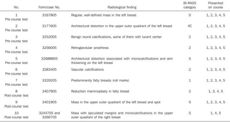
Table 1 Cases valuated in the pre- and post-course tests.
No.
1 Pre-course test
2 Pre-course test
3 Pre-course test
4 Pre-course test
5 Pre-course test
6 Pre-course test
7 Pre-course test
8 Post-course test
9 Post-course test
10 Post-course test
Form/case No.
3167805
3177605
3252005
3256005
32688805
3283405
3329205
3407905
3401905
3244705 and 3266705
Radiological finding
Regular, well-defined mass in the left breast
Architectural distortion in the upper outer quadrant of the left breast
Benign round calcifications, some of them with lucent center
Retroglandular prosthesis
Architectural distortion associated with microcalcifications and skin thickening on the left breast
Vascular calcifications
Predominantly fatty breasts (roll marks)
Reduction mammoplasty in fatty breast
Mass in the upper outer quadrant of the left breast and spot
Mass with spiculated margins and microcalcifications in the upper outer quadrant of the right breast
BI-RADS category
0
4C
2
2
5
2
1
2
0
5
Presented on course
1, 2, 3, 4, 5
1, 2, 3, 4, 5
1, 2, 3, 4, 5
1, 2, 3, 4, 5
1, 2, 3, 4, 5
1, 2, 3, 4, 5
1, 2, 3, 4, 5
1, 3, 4, 5
1, 2, 3, 4, 5
poor definition and sharpness of images when presented with the datashow device. The capability of identifying the lesions, classification in accordance with BI-RADS, and coherence with recommended approach were evaluated.
RESULTS
The present study was aimed at improv-ing the quality of mammographic diagno-sis in three categories: lesion description, BI-RADS categories, and recommended approach. The average grades obtained between these three categories before and after the course did not show significant improvement in the learning, ranging from 5.6 to 5.8 in relation to lesion description, from 5.1 to 5.3 in relation to BI-RADS categories and from 5.5 to 5.9 in relation to recommended approach.
In relation to BI-RADS classification, categories assignment in the pre- and post-course tests for each mammographic study were compared with the following results: 1 – In post-course test a higher number of answers on the categories expected for those cases was observed. The only excep-tion was observed for category 4, in which most students selected category 0, with no significant alterations, as the pre- and post-course testes were compared.
2 – Except for the correct category that should be chosen, the second most referred category in the cases was the same, as the pre- and post-course tests were compared (Table 2).
3 – As regards the correctness rate, there was only a small improvement in catego-ries 2 and 4. In all the other categocatego-ries, there was a reduction in the correctness rate as the pre- and post-course tests were com-pared.
4 – Category 3 was not utilized in the cases, considering that according to
BI-RADS the three alterations comprised in this category are: punctate, isodense and clustered microcalcifications (the course did not have appropriate viewing resources for microcalcifications evaluation), regular and well-defined masses, and focal asym-metry corresponding to confluent and nonpalpable breast tissue (these two alter-ations require additional mammographic evaluation, such as focal compression and magnification, which are not part of the baseline mediolateral oblique and cranio-caudal views, and were not among the im-ages presented in the course)
Besides the evaluation of the classifica-tion according to the standardized system, the consistency regarding the recom-mended approach was also evaluated. In spite of the clarity of such system, primary inconsistencies were observed, for ex-ample:
– category 5 with six-month radiological follow-up;
– category 4 with six-month radiological follow-up;
– category 3 with recommendation for fur-ther evaluation with anofur-ther imaging method;
– category 3 with 12-month radiological follow-up;
– category 3 with further pathological in-vestigation;
– category 2 with recommendation for fur-ther evaluation with anofur-ther imaging method;
– category 2 with six-month radiological follow-up;
– category 1 with recommendation for fur-ther evaluation with anofur-ther imaging method;
– category 1 with recommendation for pro-ceeding with pathological investigation; – category 0 with recommendation for pro-ceeding with pathological investigation. In spite of such inconsistencies, it was observed that after the courses, many stu-dents only mentioned the BI-RADS cat-egory, without recommended approaches, or vice-versa.
Some frequent errors, such as descrip-tion of axillary lymph nodes with usual radiological aspect as lymphadenopathies, diminished after the courses, particularly after the second one, considering that as the first group had already been analyzed, such
errors could be highlighted to the later groups.
As regards the analysis of breasts with silicone implants, there was a high rate of disagreement even in the post-course tests. Five attendees kept on selecting category 0, recommending further evaluation with ultrasonography, two attendees selected category 1 with a recommendation for 12-month radiological follow-up, one selected category 1 and another selected category 2 with recommendation for further evalua-tion with ultrasonography, and finally one attendee selected category 0, with recom-mendation for proceeding with pathologi-cal investigation.
DISCUSSION
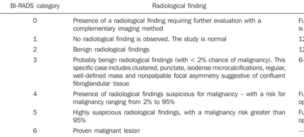
The BI-RADS classification system, that is aimed at the standardization of ra-diological alterations and recommenda-tions for approach to be adopted, has been well established and determined (Table 3). In the evaluation of the capacity of iden-tifying a mammographic lesion, its clas-sification in compliance with BI-RADS and the consistency with recommended approach, the following points were ob-served:
1 – Regular, well-defined mass (3167805): There was an improvement in the identification and characterization of the lesion, however there was no signifi-cant improvement in relation to BI-RADS classification and its consistency with the recommended approach. Some observed examples were: category 2 for mass; cat-egory 3 for mass with recommendation for further evaluation with other imaging method, or category 3 with 12-month ra-diological follow-up. It is important to note that one attendee did not correctly describe the alteration as a mass, but as an asymme-try; however he classified the lesion as cat-egory 0 with recommendation for further evaluation with another imaging method. In another case, a regular mass with par-tially obscured limits (3401905), whose evaluation is a little more difficult than the first one, a greater difficulty in the identi-fication was indeed observed; however, those who identified it continued to clas-sify it into category 3, recommending six-month radiological follow-up. Other
ex-Table 2 Error in classification of BI-RADS cat-egories in the presented cases.
Category
0
1 2
4 5
Second most selected category
3
2 1
amples were category 4 with recommended histopathological investigation or category 0 with recommendation for further inves-tigation with other imaging method.
2 – Highly suspicious high-density spiculated mass (3244705 and 3266705): The mass was correctly described by the attendees, however some of them did not mention the associated findings, but most classified the lesion as category 5 and some as category 4, with recommendation for proceeding with histopathological investi-gation. This is a relevant fact, as in such case what really matters is that a highly suspi-cious mass be histologically evaluated.
3 – Architectural distortion (3177605 and 32688805): In the first case the archi-tectural distortion was more subtle, with slight improvement when the pre-course and post-course tests were compared, in-cluding the description of other lesions that were not present and that guided the clas-sification in compliance with BI-RADS, and the recommended approach, which in most cases was not consistent; in the sec-ond case, in which the distortion was more noticeable, with associated findings in-creasing the suspiciousness of the lesion, the descriptions remained practically un-changed in the pre- and post-course tests (not all attendees described the architec-tural distortion, some described it as a mass or as a distortion associated with a mass), as well as in the BI-RADS categorization; however, the identification of the lesion malignancy characteristics was more evi-dent, as there was a significant improve-ment with respect to recommendation for
proceeding with histopathological investi-gation.
4 – Benign calcifications – with lucent center, round calcifications (3252005) and vascular calcifications (3283405): In the first case, no significant improvement was observed in spite of the fact that the calci-fications were in general mostly identified and described correctly; inexistent alter-ations were described, which conditioned the BI-RADS classification and approach recommendations that were not always consistent. On the other hand, in the case of vascular calcifications, no improvement was observed in the characterization, with many attendees persisting in the same de-scription, which included inexistent alter-ations, consequently with wrong categories and recommended approach.
5 – Operated breasts (silicone implants-3256005 and reduction mammoplasty-3407905): All the attendees identified the implant, with the exception of one that only described it in the post-course test. No sig-nificant improvement was observed with regards to BI-RADS classification and rec-ommended approach. Two facts should be highlighted: some attendees identified in-existent lesions which compromised the BI-RADS classification and recommended approach, and the fact that some believed that breasts with silicone implants required other imaging methods for a thorough study. With respect to the case with reduc-tion mammoplasty, only four attendees identified the case as operated breasts, and only these attendees continued describing the reduction mammoplasty in the
post-course test, with no comparative improve-ment in the pre- and post-course tests; thus, for this particular case, the BI-RADS clas-sification and recommendation for ap-proach were based on alterations that were not present. It should also be mentioned that in many cases the BI-RADS classifi-cation was not consistent with the recom-mended approaches.
6 – Normal studies presenting process-ing errors – roll marks (3229205): There was no significant improvement with the course. Only the same attendees that iden-tified the study as normal in the pre-course test, continued to do so in the post-course test; however, in most groups inexistent lesions were identified, which obviously misdirected the BI-RADS classification, which moreover, was many times not con-sistent with the recommended approach. Roll marks were poorly observed, and only in the pre-course test.
Even after some changes in the course structure, with direct approaches and use of repetitive methods, no significant improve-ment in level of learning was observed.
It was observed that many times the at-tendees visualized the radiological alter-ation, but could not describe it correctly; frequently, confusion between focal asym-metry and mass was observed, as well as the description of architectural distortion as a mass with malignancy characteristics. It should be pointed out that the most relevant aspect is the identification of the radiologi-cal alteration and the approach to be adopted by the assisting physician for the patient.
Table 3 BI-RADS classification system.
BI-RADS category
0
1
2
3
4
5
6
Radiological finding
Presence of a radiological finding requiring further evaluation with a complementary imaging method
No radiological finding is observed. The study is normal
Benign radiological findings
Probably benign radiological findings (with < 2% chance of malignancy). This specific case includes clustered, punctate, isodense microcalcifications, regular, well-defined mass and nonpalpable focal asymmetry suggestive of confluent fibroglandular tissue
Presence of radiological findings suspicious for malignancy – with a risk for malignancy ranging from 2% to 95%
Highly suspicious radiological findings, with a malignancy risk greater than 95%
Proven malignant lesion
Recommended approach
Further evaluation with other imaging method is required
12-month radiological follow-up is suggested
12-month radiological follow-up is suggested
6-month radiological follow-up is suggested
Further diagnostic investigation is required. Bi-opsy is recommended
Further diagnostic investigation is required. Bi-opsy is recommended
Many times, in cases classified as cat-egory 0, it is possible to define that a mass is probably benign after all the additional views required for the study show a nonpalpable regular mass.
Categories 1 and 2 correspond to mam-mographic studies with recommendation for a 12-month follow-up, considering that such studies with results negative for ma-lignancy with no harm for the patient.
Categories 5 and 4 indicate respectively highly suspicious and suspicious mammo-graphic findings; in such cases, recommen-dation of histopathological investigation of the described finding is mandatory. There-fore, when category 5 is confused with category 4, the patient will not suffer any harm, since in both categories the recom-mended approach will be proceeding with histopathological diagnosis. However, when a radiologically suspicious lesion is classified as category 0, the correct diagno-sis will be delayed, as a further imaging study, ultrasonography for example, would not be necessary to confirm a suspicious finding that could have been referred for biopsy after the initial mammographic study.
Berg et al.(8) have developed a study to determine whether training on BI-RADS improves observer performance and interobserver agreement among experi-enced breast radiologists, taking into con-sideration the analysis of mammographic characteristics and the final evaluation. Fifty-four lesions were selected: 28 nod-ules (9 malignant) and 26 microcalcifi-cations (10 malignant). The observer per-formance was evaluated before and after the BI-RADS training course and there was improvement in agreement regarding the description of mass margins. A similar improvement was observed in the descrip-tion of morphologic characteristics of microcalcifications. No improvement was observed in the description of calcifications distribution. The final evaluations were more consistent after the training.
Recom-mendations for biopsy increased from 73% to 88%, with a subtle increase in the num-ber of recommendations for biopsy in be-nign lesions (from 43% to 51%).
Sickles et al.(9) have observed that radi-ologists specialized in breast imaging un-dergo courses with a six-times higher fre-quency than generalist radiologists, and interpret a ten times higher number of mammograms per year, being capable of diagnosing a greater number of cases of early stage breast cancer, recommending more biopsies and presenting lower rates of patient recall than generalist radiologists.
CONCLUSIONS
1 – Breast radiologists in the public health system of the Rio de Janeiro State demonstrate insufficient knowledge on early imaging diagnosis of breast cancer.
2 –The course for training medical prac-titioners in the early detection of breast cancer, with emphasis on theoretical classes, did not produce a significant im-provement in the quality of mammography interpretation, with persistent mistakes in morphological description of critical breast lesions and in their classification by the BI-RADS, as well as poor consistency be-tween the BI-RADS classification and the recommended approach.
Recommendations
1 – The training courses must empha-size the practice by means of mammo-graphic cases in which the repeated dem-onstration of critical breast lesions, their BI-RADS classification and recommended approaches are thoroughly covered and trained.
2 – Diagnosis errors can be minimized when the characteristics of typically benign or malignant lesions are well defined, re-ducing the uncertainty in mammographic diagnosis.
3 – The knowledge acquired in theoreti-cal courses is important. However, one
must bear in mind that advanced technolo-gies (such as magnetic resonance imaging in the evaluation of breast diseases) are not widely utilized in Brazil. However, pro-grams of theoretical courses for breast can-cer diagnosis emphasize such novel diag-nosing methods.
4 – It is necessary to promote the gath-ering of professionals involved in the teaching of mammographic diagnosis in order to reach a consensus on a course cur-riculum and the most appropriate teaching method.
REFERENCES
1. Elmore JG, Carney PA. Does practice make per-fect when interpreting mammography? J Natl Cancer Inst. 2002;94:321–3.
2. Elmore JG, Nakano CY, Koepsell TD, et al. Inter-national variation in screening mammography interpretations in community-based programs. J Natl Cancer Inst. 2003;95:1384–93.
3. Eiras AL, Koch HA, Peixoto JE. Parâmetros en-volvidos na qualidade da imagem mamográfica – revisão dos fundamentos teóricos. Rev Imagem. 2000;22:143–8.
4. Koch HA. Diagnóstico por imagem mamária no Brasil. Rio de Janeiro: Academia Nacional de Medicina; 2004.
5. Linver MN, Paster SB, Rosenberg RD, et al. Im-provement in mammography interpretation skills in a community radiology practice after dedicated teaching courses: 2-year medical audit of 38,633 cases. Radiology. 1992;184:39–43.
6. Colégio Brasileiro de Radiologia, Sociedade Bra-sileira de Mastologia, Federação das Sociedades Brasileiras de Ginecologia e Obstetrícia. I Reu-nião de Consenso da Radiologia – Padronização dos laudos mamográficos. 28ª Jornada Paulista de Radiologia, São Paulo, abril de 1998. 7. Luna MM. Avaliação dos laudos mamográficos:
padronização de recomendação de conduta para um programa de detecção precoce do câncer de mama por meio da mamografia [tese de douto-rado]. Rio de Janeiro: Universidade Federal do Rio de Janeiro; 2001.
8. Berg WA, Campassi C, Langenberg P, et al. Breast Imaging Reporting and Data System: inter- and intraobserver variability in feature analysis and final assessment. AJR Am J Roentgenol. 2000; 174:1769–77.