LONG-TERM EVOLUTION OF PAPILLEDEMA IN
IDIOPATHIC INTRACRANIAL HYPERTENSION
Observations concerning two cases
Elcio Juliato Piovesan, Marcos Cristiano Lange, Liciane do Rocio Maia Piovesan,
Sergio Monteiro de Almeida, Pedro André Kowacs, Lineu Cesar Werneck
ABSTRACT - Chronic headaches, associated with papilledema and pulsatile tinnitus without any neuroradiologic, cytobiochemical or cerebrospinal fluid abnormalities are suggestive of idiopathic intracranial hypertension (IIH). However the absence of the papilledema does not rule out this diagnosis. The reason why some patients do not develop papilledema in IIH is ignored, however there are some hypotheses concerning the structure of the optical nerve. In this study we described two female patients that presented diagnosis of IIH with papilledema, with subsequent resolution of papilledema without the due resolution of intracranial hypertension. The long-term behavior of the optic nerve (ON) facing an increased intracranial pressure was evaluated through repeated measurements of the intracranial pressure. We concluded that the ON submitted to high intracranial pressure for a certain lenght of time can adapt itself with subsequent disappearance of the papilledema. The presence or not of papilledema in IIH can be related to the period in which the diagnosis is accomplished.
KEY WORDS: headache, intracranial pressure, papilledema, pseudotumor cerebri.
Avaliação do comportamento do papiledema na hipertensão intracraniana idiopática: a propósito de Avaliação do comportamento do papiledema na hipertensão intracraniana idiopática: a propósito deAvaliação do comportamento do papiledema na hipertensão intracraniana idiopática: a propósito de Avaliação do comportamento do papiledema na hipertensão intracraniana idiopática: a propósito deAvaliação do comportamento do papiledema na hipertensão intracraniana idiopática: a propósito de dois casos
dois casosdois casos dois casosdois casos
RESUMO - Cefaléias com características crônica, diária, acompanhadas de edema de papila e tinitus pulsátil, sem nenhum achado neuroradiológico ou citobioquímico no líquor, são altamente sugestivas de hipertensão intracraniana idiopática (HII). Entretanto a ausência do papiledema não invalida o seu diagnóstico. A razão pela qual alguns pacientes não desenvolvem papiledema na HII é desconhecida, porém algumas hipóteses relacionadas com propriedades intrínsecas da bainha do nervo óptico têm sido propostas. Neste estudo relatamos dois pacientes do sexo feminino que apresentaram diagnóstico de HII com papiledema, evoluindo para resolução do papiledema sem a devida resolução da HII. O comportamento do nervo óptico (NO) frente ao aumento da pressão intracraniana foi avaliado neste estudo a partir de um monitoramento intermitente criterioso da pressão intracraniana. Concluímos que o NO submetido a um período de hipertensão intracraniana pode adaptar-se a este ambiente promovendo uma resolução do papiledema para normalidade papilar. A presença ou ausência de papiledema na HII pode estar relacionada ao período no qual seu diagnóstico é realizado.
PALAVRAS-CHAVE: cefaléia, pressão intracraniana, papiledema, pseudotumor cerebral.
Headache Section, Neurology Unit, Internal Medicine Department, Hospital de Clínicas da Universidade Federal do Paraná, Curitiba PR, Brazil. Received 18 September 2001, received in final form 21 January 2002. Accepted 30 January 2002.
Dr. Elcio Juliato Piovesan - Serviço de Neurologia, Hospital de Clínicas - Rua General Carneiro 181/120 andar/sala 1236 - 80060-900
Curitiba PR - Brasil - FAX: 55 41 283 5512. E-mail: [email protected]
Idiopathic intracranial hypertension (IIH), also known
as pseudotumor cerebri or benign intracranial
hyper-tension, is associated to a group of symptoms in which
the headache prevails over the others
1. This headache
in considered not to be associated with vascular
in-tracranial disorders, and is accompanied or not by
papilledema
2. Several mechanisms may lead to the
increase of the intracranial pressure, however all break
of the dynamics of several intracranial structures
lead-ing to nociceptive stimulation and to a headache that
presents with peculiar features
3.
The presence of papilledema makes the
diagno-sis easer, however, its absence, much less frequent,
it has been reported in several papers
2. In this study
we have followed-up two patients with the diagnosis
of IIH for a period of 36 months. We have
demons-trated that papilledema may subside durign the
course of this disease even without the remission of
the intracranial hypertension.
METHOD
Study design
evalu-454 Arq Neuropsiquiatr 2002;60(2-B)
ation consisted of clinical and neurological examination and laboratorial work-up. All the patients were submitted to laboratorial evaluation of serum calcium, thyroid and parathyroid function tests, blood count, glucose, erythro-cyte sedimentation rate (ESR), Kvein test and angiotensin-converting enzyme. Both patients underwent computer-ized axial tomography (CAT) and magnetic resonance im-aging (MRI) of the head, cerebral angiography (CA) and fluorescein angiography (FG). To obtain pressure measure-ments and a sample of the cerebro-spinal fluid (CSF) for cellular, cytological, chemical, and bacteriological exami-nation a lumbar puncture (LP) was carried out. The pa-tients were also appraised by a physician, an ophtal-mologist, an endocrinologist and a neurologist.
Patients
Patient 1. A 31 years-old white female, 155 cm of height and 76 kg of weight, had been seen by the same neurologist because of migraine an year before. Migraine had started when the patient was 13 and increased in intensity, duration and frequency in the preceding 6 months, evolving to a daily chronic headache (transformed migraine). In this ocasion the neurological examination and the CAT were normal. For the treatment of the migraine propranolol 40 mg/day was prescribed for 60 days. Amitriptyline 50 mg/ day and divalproex sodium (Depakote(R) 500 mg/day) were
also tried without results. At he appoitment in which IIH was suspected, the intensity of the headache had increased, associated with transient visual abnormalities (photopsias) and noises in both ears (pulsatile tinnitus). Bilateral papilledema was observed. A second CAT was normal. Intracranial pressure was 430 mmH2O (graphic-1) and the cerebrospinal fluid (CSF) was normal. Magnetic resonance imaging (MRI) of the head revealed empty sella, and cerebral angiography (CA) was normal. Blood lab were normal. Fluorescein angiography (FG) revealed bilateral papilledema
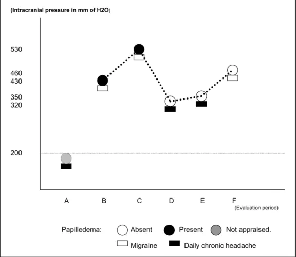
(Fig 1-A). Acetazolamide 250 mg b.i.d. was started. Fifteen days later the headache had improved without resolution of the papilledema, but a new LP revealed an intracranial pressure of 530 mmH2O (Fig 2). Forty-five days after ac-etazolamide was started the papilledema had remitted. At this time, the frequency of the throbbing headaches was twice a week. They were bilateral, associated to nausea and vomiting. Six months later the patient presented no papilledema (Fig 1-B), photopsias or headaches. A new LP revealed an intracranial pressure of 320 mmH2O (Fig 2). During the following 24 months the patient continued with-out papilledema but with episodic headaches. Thirty months after the first evaluation the daily headaches had recurred. There was no papilledema and an ophtalmological evalua-tion was normal. A new LP was carried out, disclosing an intracranial pressure of 460 mmH2O (Fig 1-B) without pa-pilledema. The patient refused a lumbo-peritoneal shunt-ing. Currently the patient persists with chronic daily head-ache, without papilledema.
Patient 2. A 40 years-old, white female, 161cm of height and 70Kg of weight, presented with a six month history of chronic daily headaches with pressing quality, bilateral lo-cation, mild to moderate intensity, increasing slowly during the day. Occasionally this headache increased in intensity, becoming throbbing and associated with nausea and/or vomiting. At the time of the appointment the patient also revealed myalgia, pulsatile tinitus in both ears and low fe-ver in the last five days. Bilateral papilledema was observed. The CAT revealed a left maxillary sinusitis. Intracranial pres-sure was 330 mmH2O (Fig 3) and the cerebrospinal fluid (CSF) revealed 26 leukocytes (95% polymorphonuclear), glucose of 64 mg/ml and 122 mg/ml of protein. Culture for fungus and virus were negative. For fifteen days she re-ceived only symptomatic medication for the treatment of viral meningitis. Thirty days after completing therapy the
patient was still presenting papilledema and pulsatile tin-nitus but her headache had improved. A new CSF examina-tion disclosed normal results but intracranial pressure was 350 mmH2O (Fig 3). A MRI of the head revealed persis-tence of the left maxillary sinusitis and a CA was normal. During ten days the patient received floxacilin. The head-ache disappeared three months later, but not the papille-dema. A new CAT of the facial sinus was normal. Another LP was carried out revealing a normal CSF but the intracra-nial pressure was 420 mmH2O (Fig 3). There was an eleva-ted serum thyroid-stimulating hormone (TSH- 12.5yU/m), and reduced triiodothyroine (T3- 30ng/dl) and tetraio-dothyronine (T4-2.3g/dl). Synthetic L-thyroxine 100 mg/day was prescribed. Fluorescein angiography (FG) revealed bi-lateral papilledema. Acetazolamide 250 mg b.i.d. was also prescribed. Six months after the treatment for the IIH and for the hypothyroisdism the papilledema and the headache had disapeared. After one year the patient stopped the medication. Fifteen days later the pulsatile tinnitus and the headache reappered without associated papilledema. The intracranial pressure was 380 mmH2O (Fig 3) and the CSF was still normal. Fluorescein angiography (FG) revealed
papillapy pallor without papilledema. Again acetazolamide 250 mg b.i.d. was started with improvement of the symp-toms after twenty days. A new LP revealed intracranial pres-sure of 250 mmH2O and a normal CSF (Fig 3). For thirty six months we followed up the patient. During this period three new episodes of headache were observed, when the in-tracranial pressure measurements were 340, 290 and 510 mm H2O (Fig 3) respectively. The patient did not agree to be submitted to a lumboperitoneal shunting.
DISCUSSION
Intracranial pressure can be increased through out
different mechanisms and several disorders. Those
dis-orders may be either primary, secundary or atypical
4.
The syndrome of IIH occurs predominantly in obese
women of childbearing age. Definitive diagnosis
can-not be made without excluiding brain tumours and
other intracranial mass lesion, infections, hypertensive
encephalopathy, pulmonary encephalopathy, and
ob-struction of the cerebral ventricles
5.
IIH occurs at a frequency of about 1 case per 100 000
456 Arq Neuropsiquiatr 2002;60(2-B)
per year in the general population and of 19.3 cases
per 100 000 per year in obese women aged 20-44
years
6. The pathophysiology of IIH is unknown, but
some postulated hypothesized mechanisms are
proposed: increase of the volume of the cerebral blood
or of the CSF
7; increase of the cerebral blood flow
8;
intracelular or extracelular edema
9; abnormalities of
the cerebral microvascular structures producing an
increase in the amount of tissue water
10; absorption
disturbances of the CSF secondary to an increase of
the pressure in the superior sagital sinus
11.
The most commom symptoms are: headache,
otal-gia, diplopia, photopsia, vomiting, mialotal-gia, dizzines
and pulsatile tinnitus
12,13. The typical characteristic of
headache in the IIH are: severe intensity (93%);
pul-sating quality (83%); bilateral location (50%);
associ-ated to nausea and/or vomiting (57%), photophobia
and phonophobia (30%) and photopsias (20%) with
a daily frequency (73%) and lasting from four days to
three years
2,14-16. The mechanisms that produce these
symptoms can be related to traction of the
nocicep-tores located in the intracranial vessels; transitory
herniation of the hipocampal uncus; traction of
cra-nial nerves (triplet and branches of C1, C2 and C3)
2.
The pulsatile tinnitus presents a duration of
sec-onds to days being unilateral in 62% of the cases.
They are reported by the patients as “falls of a ray”,
“beat of a breeze”, “heart pulsing in the ear” or
even-tually “blows in the hear”
1,16. The patients can also
present photopsias (presence of sparkles or flashes
of light of variable duration, from seconds to hours).
Less frequently, the patients refers pain in the
shoul-ders and in the arm (probably due to dilation of the
spinal roots)
17. Bilateral retro ocular pain, occuring
during movements of the head, are reported by 20%
of the pacientes
14,17.
The main abnormality disclosed by examination
is papilledema, occasionally with hemorrhagic
exsu-dates
13. However, its absence does not exclude
IIH
2,13,18-22. Paralysis of the abducent nerve, trochlear
and facial paralysis
23are also common findings. Less
frequently nystagmus, bilateral intranuclear
ophthal-moplegia, dissociation of pupilary reflex
24and
tation of the upper gaze may occur
13. Rarely the
patient may reveal ataxia
12,13and the presence of a
Babinski sign
12.
Grant
13, followin-up 79 children with IIH, showed
that papilledema disappeared in 48% of the cases
af-ter 2 months, in 54% afaf-ter 6 months, in 67% afaf-ter one
year, in 75% after two years, in 85% after 3 years and
in 86% after 10 years. The optic atrophy was only
observed respectively in two patients two and four years
after the beginning of the treatment
13. The most
frequent sequelae of IIH is the loss of visual
sharp-ness
25,26. However many patients even with
intracra-nial hypertension do not present papilledema. Why
there is no papilledema in cases of intracranial
hyper-tension is not known. Congenital or acquired optic
nerve sheath defects, IIH with resolution of
papille-dema, or early idiopathic intracranial hypertension are
alternative explanations
5. Unilateral papilledema from
IIH is an infrequent and rare situation
27. The possible
mechanisms that have been considered responsible for
lack of papilledema in IIH include: an anomaly in the
orbital optic nerve sheaths
28; an abnormality in the
venous sinuses; difference in the lamina cribosa
be-tween the two optic disc that results in reduced
trans-mission of the intracranial pressure to the optic nerve
in the scleral canal
29; axoplasmic blockage at the level
of the lamina cribosa induced by experimental elevated
intracranial pressure
30.
In both patients herein described, improvement of
the papilledema was verified even in the presence of
intracranial hypertension with daily chronic headache
and pulsatile tinnitus. As seen in the first case, the
patient presented with symptoms of daily chronic
hea-dache and the papilledema that, could had developed
during the course of the disease, remitting
after-wards.This observation has lead us to hypothesize that
the optic nerve can behave in different ways during
the evolution of this pathology, beginning without
papilledema, with further development of papilledema
that resolves later on. We believe that this pattern might
be related to the fact that the retrograde axoplasmatic
flow and anterograde axoplasmatic flow of the optic
nerve behave distinctly during the different phases of
this condition, mainly when oscillations of the
intracranial pressure occur.
We believe that the finding or not of papilledema
in IIH might reflect only the phase in which diagnosis
of IIH is done. In this condition, the presence of the
papilledema can be transitory.
REFERENCES
1. Wall M, George D. Idiopathic intracranial hypertension: a prospective study of 50 patients. Brain 1991;114:155-180.
2. Marcelis J, Silbertein SD. Idiopathic intracranial hypertension without papilledema. Arch Neurol 1991;48:392-399.
3. Classification and diagnostic criteria for headache disorders, cranial neuralgias and facial pain. Cephalalgia 1988;8(Suppl 7):51.
4. Wald SL. Disorders of cerebrospinal fluid circulation and brain edema. In Bradley WG, Daroff RB, Fenichel GM, Marsden CD (eds). Neurol-ogy in clinical practice. 2.Ed. Boston: Butterworth-Heinemann, 1996: 1431-1458.
5. Silbertein SD, Lipton RB, Goadsby PJ. Headache in clinical practice. Headache associated with non-vascular intracranial disease. Oxford: Isis Medical Media, 1998:143-164.
6. Durcan FJ, Corbett JJ, Wall M. The incidence of pseudotumor cerebri: population studies in Iowa and Louisiana. Arch Neurol 1988;45:875-877. 7. Dandy WE. Intracranial pressure without brain tumor: diagnosis and
treatment. Ann Surg 1937;106:492-513.
8. Foley J. Benign forms of intracranial hypertension “toxic” and “otitic” hydrocephalus. Brain 1955;78:1-41.
9. Sahs AL, Joynt RJ. Brain swelling of unknown cause. Neurology 1956;6:791-803.
10. Raichle ME, Grubb RL, Phelps ME, Gado MH, Caronna JJ. Cerebral hemodynamics and metabolism in pseudotumor cerebri. Ann Neurol 1978;48:740-747.
11. Johnston I, Hawke S, Halmagyim M, Teo C. The pseudotumor syn-drome: disorders of cerebrospinal fluid circulation causing intracra-nial hypertension without ventriculomegaly. Arch Neurol 1991;48:740-747.
12. Dhiravibulya K, Ouvrier R, Johnston I, Procopis P, Antony J. Benign intracranial hypertension in childhood: a review of 23 patients. J Paediatr Child Health 1991;27:304-307.
13. Grant DN. Benign intracranial hypertension: a review of 79 cases in infancy and childhood. Arch Dis Child 1971;46:651-655.
14. Wall M. The headache profile of idiopathic intracranial hypertension. Cephalalgia 1990;10:331-335.
15. Weisberg LA. Benign intracranial hypertension. Medicine 1975;54:197-207. 16. Schoenen J, Noordhout AM. Headache. In Wall PD, Melzck R. (eds)
Text-book of pain. 3.Ed. New York: Churchill Livingstone, 1994:511-512. 17. Giuseffi V, Wall M, Siegel PZ, Rojas PB. Symptoms and disease
asso-ciations in idiopathic intracranial hypertension (pseudotumor cerebri): a case-control study. Neurology 1991;41:239-244.
18. Bortoluzzi M, DiLauro L, Marini G. Benign intracranial hypertension with spinal and radicular pain. J Neurosurg 1982;57:833-836. 19. Winner P, Bello L. Idiopathic intracranial hypertension in a young child
without visual symptoms or signs. Headache 1996;36:574-576. 20. Spence JD, Amacher AL, Willis NR. Benign intracranial hypertension
without papilledema: role of 24 hour cerebrospinal fluid pressure moni-toring in diagnosis and management. Neurosurgery 1980;7:326-336. 21. Scanari M, Mingrino S, d’Avella D, DellaCort V. Benign intracranial
hypertension without papilledema: a case report. Neurosurgery 1979;5:376-377.
22. Lipton HL, Michelson PE. Pseudotumor cerebri syndrome without papilledema. JAMA 1972;220:1591-1592.
23. Lessel S. Pediatric pseudotumor cerebri (Idiopathic intracranial hyper-tension). Surv Ophthalmol 1992;37:155-166.
24. Baker RS, Baumann RJ, Bunice JR. Idiopathic intracranial hyperten-sion (pseudotumor cerebri) in pediatric patients. Pediat Neurol 1989;5:5-11.
25. Baker RS, Carter D, Hendrick BE, Buncic RJ. Visual loss in pseudotumor cere-bri of childhood: a follow-up study. Arch Ophthalmol 1985;103:1681-1686. 26. Rush JA. Pseudotumor cerebri: clinical profile and visual outcome in
63 patients. Mayo Clin Proc 1980;55:541-546.
27. Huna-Baron R, Landau K, Rosenberg M, Warren FA, Kupersmith MJ. Unilateral swollen disc due to increased intracranial pressure. Neurol-ogy 2001;56:1588-1590.
28. Moster ML. Unilateral disk edema in a young woman. Surv Ophthalmol 1995;39:409-416.
29. Monteiro MLR, Hoyt WF, Imes RK, Narahara M. Papiledema unilateral na síndrome de pseudotumor cerebral. Arq Neuropsiquiatr 1985;43:154-159. 30. Tso MOM, Hayreh SS. Optic disc edema in raised intracranial