334 Radiol Bras. 2012 Nov/Dez;45(6):334–339
Recommendations of Colégio Brasileiro de Radiologia
e Diagnóstico por Imagem, Sociedade Brasileira
de Mastologia, and Federação Brasileira das Associações
de Ginecologia e Obstetrícia for imaging screening for breast
cancer
*
Recomendações do Colégio Brasileiro de Radiologia e Diagnóstico por Imagem, da Sociedade Brasileira de Mastologia e da Federação Brasileira das Associações de Ginecologia e Obstetrícia para rastreamento do câncer de mama por métodos de imagem
Linei Augusta Brolini Dellê Urban1, Marcela Brisighelli Schaefer2, Dakir Lourenço Duarte2, Radiá
Pereira dos Santos2, Norma Medicis de Albuquerque Maranhão2, Ana Lucia Kefalas2, Ellyete
de Oliveira Canella2, Carlos Alberto Pecci Ferreira2, João Emílio Peixoto2, Luciano Fernandes Chala3,
Rodrigo Pepe Costa4, José Luís Esteves Francisco4, Simone Elias Martinelli4, Heverton Leal Ernesto
de Amorim4, Henrique Alberto Pasqualette5, Paulo Mauricio Soares Pereira5, Helio Sebastião
Amâncio de Camargo Junior5, Vania Ravizzini Sondermann5
* Document jointly prepared by Colégio Brasileiro de Radiolo-gia e Diagnóstico por Imagem (CBR), Sociedade Brasileira de Mastologia (SBM) and Federação Brasileira das Associações de Ginecologia e Obstetrícia (FEBRASGO).
1. Coordinator of the National Commission on Mammography, Colégio Brasileiro de Radiologia e Diagnóstico por Imagem (CBR). 2. Members of the National Commission on Mammography, Colégio Brasileiro de Radiologia e Diagnóstico por Imagem (CBR). 3. Invited Member of the National Commission on Mammog-raphy, Colégio Brasileiro de Radiologia e Diagnóstico por Imagem (CBR).
4. Members of the National Commission on Mammography, Sociedade Brasileira de Mastologia (SBM).
5. Members of the National Commission on Mammography, Federação Brasileira das Associações de Ginecologia e Obste-trícia (FEBRASGO).
Mailing Address: Dra. Linei A. B. D. Urban. Rua Padre Agos-tinho, 913, ap. 53, Mercês. Curitiba, PR, Brazil, 80430-050. E-mail: [email protected]
Urban LABD, Schaefer MB, Duarte DL, Santos RP, Maranhão NMA, Kefalas AL, Canella EO, Ferreira CAP, Peixoto JE, Chala LF, Costa RP, Francisco JLE, Martinelli SE, Amorim HLE, Pasqualette HA, Pereira PMS, Camargo Junior HSA, Sondermann VR. Recommenda-tions of Colégio Brasileiro de Radiologia e Diagnóstico por Imagem, Sociedade Brasileira de Mastologia, and Federação Brasileira das Associações de Ginecologia e Obstetrícia for imaging screening for breast cancer. Radiol Bras. 2012 Nov/Dez;45(6):334–339.
2010, and was responsible for 27% of new cases of cancer diagnosed in women(4). Out
of this total, about 2/3 of cases have oc-curred in women aged above 50, particu-larly in developed countries. On the other hand, in women aged under 50 (between 15 and 49 years), breast cancer incidence was two-fold higher in developing countries than in developed countries(4).
In Brazil, 52,680 new cases of breast cancer are expected to occur in 2012, with an estimated risk of 52 cases per 100,000 women. Such a risk presents a great varia-tion according to the region in the country, as follows: in the Southeastern region, it corresponds to 69/100,000; in the Southern region, 65/100,000; in the Center-Western region, 48/100,000; in the Northeastern region, 32/100,000; and in the Northern region, 19/100,000 women(5). Differences
in relation to age range are also observed, with a specific rate of four cases per 100,000 women between 40 and 49 years, and five cases per 100,000 women aged above 50(5). In a study developed in the city of Goiânia, 15% of the tumors were observed in women aged under 40, 27% between 41 and 50 years, and 57% above 50(6). That is to say,
more than 40% of cases of breast cancer occurred in patients aged under 50. period, while other countries, such as
Nor-way, demonstrated 10% decrease in mor-tality connected with only the screening(2,3).
In Brazil, there is no population screen-ing policy; only opportunistic screenscreen-ing is undertaken. Thus, it is essential to encour-age actions towards standardization of breast cancer screening, bringing informa-tion to the populainforma-tion about its relevance. With a view on this subject, Colégio Brasileiro de Radiologia e Diagnóstico por Imagem (CBR) (Brazilian College of Ra-diology and Imaging Diagnosis), Socie-dade Brasileira de Mastologia (SBM) (Bra-zilian Society of Mastology), and Federa-ção Brasileira das Associações de Gineco-logia e Obstetrícia (FEBRASGO) (Brazil-ian Federation of Gynecology and Obstet-rics Associations), through the National Commission on Mammography, present their recommendations for breast cancer imaging screening in Brazil.
Current status of breast cancer in Brazil and worldwide
The global breast cancer incidence is progressively increasing both in developed and developing countries at a yearly rate of 3.1%(4). From 641,000 cases in 1980 its
incidence has grown to 1,643.000 cases in
INTRODUCTION
The need for consensus in Brazil
Breast cancer is the most frequent type of cancer and main cause of cancer deaths among women in Brazil and worldwide. On the other hand, this is the tumor whose screening has demonstrated the greatest impact on mortality reduction. Just in United States of America, there was a 30% decrease in breast cancer mortality rates since 1990 when programs of mammo-graphic screening started being imple-mented(1). In Europe, some countries such
On the other hand, the breast cancer mortality rate is quite different among devel-oped and developing countries worldwide. In developed countries, there was a signifi-cant mortality reduction over the last years, while stability or even a continuous increase has been observed in developing countries. Such a disparity might be attributed to dif-ferences in early-detection policies, as well as to the difficulty to access appropriate treatment in poorer countries(4,5,7).
Working method and revision preview
Available scientific studies were re-viewed and data were compiled in order to present the recommendations according to age range. In the absence of evidentiary data, the recommendations reflected the consensus of the Commission comprised by specialists representing the three enti-ties. The recommendations were classified into four categories according to the degree of scientific evidence and consensus be-tween specialists, as follows:
Category 1 – Recommendation based on strong scientific evidences, with a
uni-form consensus between CBR, SBM and FEBRASGO on a vigorous support to such recommendation.
Category 2a – Recommendation based on reasonable scientific evidences, with a uniform consensus between CBR, SBM and FEBRASGO, with a vigorous support to such recommendation.
Category 2b – Recommendation based on few scientific evidences, but with a con-sensus between CBR, SBM and FE-BRASGO on a vigorous support to such recommendation.
Category 3 – Recommendation con-sensually supported by CBR, SBM and FEBRASGO specialists.
The present recommendations should be revised every three years.
RECOMMENDATIONS FOR BREAST CANCER SCREENING
Women aged under 40
MAMMOGRAPHY – Generally, at this age range mammography is not recom-mended, except on an individual basis for
women at high risk for breast cancer, as shown on Table 1.
ULTRASONOGRAPHY – At this age range, sonographic screening is not recom-mended, except on an individual basis for women at high risk for breast cancer in whom screening by magnetic resonance imaging might be appropriate but, for any reason, cannot be performed.
MAGNETIC RESONANCE IMAG-ING – At this age range, breast MRI screen-ing is not recommended, except on an in-dividual basis for women at high risk for breast cancer, as shown on Table 2.
Women aged between 40 and 69
MAMMOGRAPHY – At this age range, mammography is recommended for all women with annual periodicity.
ULTRASONOGRAPHY – Generally, at this age range, sonographic screening is not recommended, except on an individual basis for women in the situations described on Table 3.
MAGNETIC RESONANCE IMAG-ING – Generally, at this age range, MRI
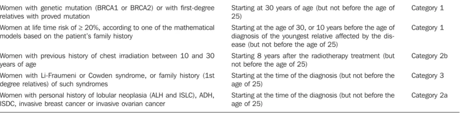
Table 1 Recommendations for mammographic screening for high-risk women aged under 40. Women with genetic mutation (BRCA1 or BRCA2) or with first-degree
relatives with proved mutation
Women at life time risk of ≥ 20%, according to one of the mathematical models based on the patient’s family history
Women with previous history of chest irradiation between 10 and 30 years of age
Women with Li-Fraumeni or Cowden syndrome, or family history (1st degree relatives) of such syndromes
Women with personal history of lobular neoplasia (ALH and ISLC), ADH, ISDC, invasive breast cancer or invasive ovarian cancer
Starting at 30 years of age (but not before the age of 25)
Starting at the age of 30, or 10 years before the age of diagnosis of the youngest relative affected by the dis-ease (but not before the age of 25)
Starting 8 years after the radiotherapy treatment (but not before the age of 25)
Starting at the time of the diagnosis (but not before the age of 25)
Starting at the time of the diagnosis (but not before the age of 25)
Category 1
Category 1
Category 2b
Category 3
Category 2a
ALH, atypical lobular hyperplasia; ISLC, in situ lobular carcinoma; ADH, atypical ductal hyperplasia; ISDC, in situductal carcinoma.
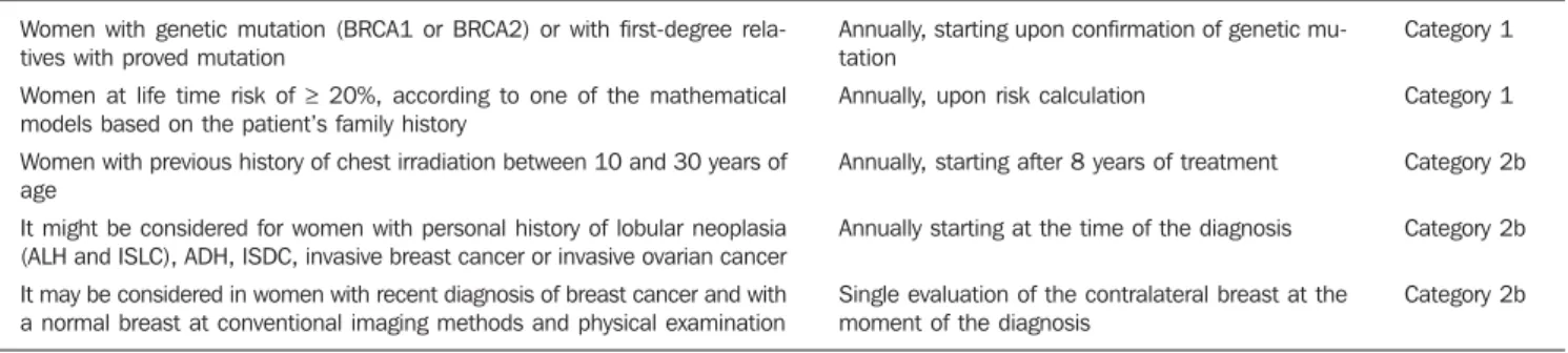
Table 2 Recommendations for screening with magnetic resonance imaging for high-risk women aged under 40. Women with genetic mutation (BRCA1 or BRCA2) or with first-degree
rela-tives with proved mutation
Women at life time risk of ≥ 20%, according to one of the mathematical models based on the patient’s family history
Women with previous history of chest irradiation between 10 and 30 years of age
Women with Li-Fraumeni or Cowden syndrome, or family history (1st de-gree relatives) of such syndromes
Women with personal history of lobular neoplasia (ALH and ISLC), ADH, ISDC, invasive breast cancer or invasive ovarian cancer
It may be considered in women with recent diagnosis of breast cancer and with a normal breast at conventional imaging methods and physical examination
Annually, starting upon confirmation of the genetic mu-tation (but not before the age of 30)
Annually upon risk calculation or 10 years before the age of diagnosis of the youngest relative (but not before the age of 30)
Annually, starting 8 years after the radiotherapy treatment (but not before the age of 30
Annually, starting at the time of the diagnosis (but not before the age of 30)
Annually, starting at the time of the diagnosis (but not before the age of 30)
Single evaluation of the contralateral breast at the mo-ment of the diagnosis
Category 1
Category 1
Category 2b
Category 3
Category 2a
Category 2a
screening is not recommended, except on an individual basis for women at high risk for breast cancer, as shown on Table 4.
Women aged above 70
MAMMOGRAPHY – At this age range, mammographic screening is recom-mended on an individual basis, as shown on Table 5.
JUSTIFICATION
Breast cancer screening is aimed at early detection of small, asymptomatic tumors with the primary objective of reducing the mortality by the disease. Secondary objec-tives of breast cancer screening include increase in patients’ survival and reduction of surgical treatment extent, allowing less mutilating surgeries and reducing the need for chemotherapy(8,9). Mammography is the
only screening method that demonstrated to be capable to promote an absolute de-crease in mortality rates(10–18).
Ultrasonog-raphy and magnetic resonance imaging have demonstrated similar capacity to de-tect early-stage breast cancer, but there is a lack of randomized, prospective studies testing their impact on the mortality reduc-tion(19–21).
The first prospective, controlled and randomized population-based study
inves-tigating the mammographic screening im-pact on breast cancer mortality was devel-oped in the 1960’s in the United States of America and was named Health Insurance Plan (HIP)(22). Such study demonstrated a
25% decrease in breast cancer mortality in a group of women submitted to mammo-graphic screening and stimulated the devel-opment of similar studies in Canada, United Kingdom and Sweden. Independent meta-analyses of such population-based studies demonstrated a reduction of 7% to 23% in breast cancer mortality in women submitted to mammographic screening, stimulating the medical societies to recom-mend the method(23,24). Population-based
mammographic screening programs were implemented in some countries and con-firmed the findings reported by population-based studies, showing reduction of 16% to 36% in mortality rates(25). Such studies
were developed with patients aged between 40 and 70, and the magnitude of the mor-tality reduction varied according to the patients’ age range.
For the group of patients aged between 50 and 69, all the medical societies in the world recommend the mammographic screening(1,26–28). Meta-analyses of the
population-based studies have shown re-duction of 20% to 35% in mortality among women at this age range(23,24). Additionally,
the adverse effects of mammographic screening are less intense in such women and lower number of them must be screened to avoid breast cancer death. The U.S. Preventive Services Task Force (USPSTF) has estimated that 1,339 women aged between 50 and 59 plus 377 women aged between 60 and 69 must be screened to avoid one breast cancer death(29). Other
more recent publication estimated a lower number of screened women to avoid one death: 351 women aged between 50 and 59 plus 233 aged between 60 and 69(30). Thus,
CBR, SBM and FEBRASGO recommend mammographic screening for such groups of women, in agreement with the other medical societies.
For women aged under 40 who are not under high risk for breast cancer, no medi-cal organization recommends mammo-graphic screening. In such group the tumor frequency is low (less than one case/1,000 women), mammography is less sensitive, and the breast parenchyma is more radi-osensitive(23,31). For patients at high risk for
breast cancer, it is recommended the screening strategy be individualized for each patient in consultation with her spe-cialist. The expected benefit should always be weighed against the involved risks, con-sidering that the youth breast is most sen-sitive to the carcinogenic effects from
ra-Table 3 Recommendations for screening with ultrasonography for women aged between 40 and 69.
It may be considered in high-risk women, particularly those where MRI screening might be appropriate but, for any reason cannot be performed
It may be considered for women with dense breast tissue, as an adjuvant to mammography
Category 2a
Category 2a Individualized
Individualized
Table 5 Recommendations for mammographic screening of women aged above 70. Women with life expectancy > 7 years, with basis on comorbidities
Women who can be submitted to invasive diagnostic investigation and treatment after abnormal result of screening
Annualy Annualy
Category 2b Category 2b Table 4 Recommendations for screening with magnetic resonance imaging for high-risk women aged between 40 and 69.
Women with genetic mutation (BRCA1 or BRCA2) or with first-degree rela-tives with proved mutation
Women at life time risk of ≥ 20%, according to one of the mathematical models based on the patient’s family history
Women with previous history of chest irradiation between 10 and 30 years of age
It might be considered for women with personal history of lobular neoplasia (ALH and ISLC), ADH, ISDC, invasive breast cancer or invasive ovarian cancer It may be considered in women with recent diagnosis of breast cancer and with a normal breast at conventional imaging methods and physical examination
Category 1
Category 1
Category 2b
Category 2b
Category 2b Annually, starting upon confirmation of genetic
mu-tation
Annually, upon risk calculation
Annually, starting after 8 years of treatment
Annually starting at the time of the diagnosis
Single evaluation of the contralateral breast at the moment of the diagnosis
diation. It is also important to note that, in dense breasts, which are most commonly found at this age range, not only the mam-mographic sensitivity is decreased, but also the radiation dose delivered by the mam-mographic apparatus is higher(32).
Major debate occurs in relation to mam-mographic screening in women aged be-tween 40 and 49. In this group, the breast cancer incidence is smaller and the fre-quency of dense breasts and fast-growing tumors is higher. Thus, according to the USPSTF estimates, the number of screened women aged between 40 and 49 (1,904) to avoid one death would be higher than women aged between 50 and 59 (1.339)(29),
although other recent publications estimate lower values (746 screened women to save one life)(30). On the other hand, several
studies and meta-analyses have shown the impact caused by mammographic screen-ing at such age range. Feigl et al. have es-timated that nearly 20% of breast cancer deaths and 34% of life expectancy years lost because of breast cancer occurred in women aged under 50(33). In a
meta-analy-ses published about the mammographic screening benefits between 40 and 49 years reported by randomized trials initiated in the period from 1963 to 1982, Smart et al. found a 23% decrease in mortality rates(34).
Such authors have suggested that the mod-ern mammography benefits must be greater, also because the screening intervals were excessively longer in those studies (18 to 28 months), utilizing only one mammo-graphic view and without utilization of the novel technologies. Such authors have also emphasized that the more delayed demon-strations of the mortality reduction could be attributed to several reasons, among them the lower number of women at this age range included in their study (less than 1/3 of the total of women included in the men-tioned eight trials(34). In other recent
pub-lication focused on this age range, Hellquist et al. have demonstrated 26% to 29% reduction in mortality as compared with the patients who did not undergo screening in Sweden(35). In Brazil, there is
Law signed in 2010 guaranteeing Access to mammography for all women aged above 40. Additionally, a Brazilian study devel-oped in Goiânia has shown that about 42% of breast cancer cases recorded in the city
occurred in patients aged under 49(6). Thus,
CBR, SBM and FEBRASGO, in agree-ment with the main medical societies, rec-ommend mammography for women at this age range. Studies estimating the potential benefit of screening suggest that, if all the women aged 40 and over were submitted to mammographic screening, the breast cancer mortality rate could drop by about 50%(33).
For women aged 70 and over, particu-larly above 75, the available data still re-main scarce. Breast cancer is one of the main causes of death among women aged above 75, but some facts suggest that the mammographic screening benefit might be smaller at this age range, namely, lower life expectancy, higher frequency of tumors with good prognosis and higher risk for death caused by other diseases(1,31). Thus,
it is suggested that the decision about the screening continuity should be individually made, taking the patient’s general health conditions and estimated life expectancy into consideration. As far as the general health conditions of the patient enable her to be submitted to a treatment for breast cancer, the mammographic screening should be continued.
Other screening techniques were also considered. Ultrasonography is not appro-priate as initial screening method for the general population, particularly because of the method limitations to evaluate microcalcifications. However, some stud-ies have demonstrated the usefulness of ultrasonography as a screening method for asymptomatic patients with negative mam-mographic results, but with dense breasts(19,20). One of the first studies was
published by Kolb et al.(20), involving
11,130 asymptomatic patients, has demon-strated that ultrasonography performed in addition to mammography increased the detection of breast cancer in 42% in pa-tients with dense breasts. Other study(36)
evaluating the role of ultrasonography in the assessment of women with dense breasts has demonstrated that the preva-lence of cancers sonographically detected corresponded to 0.41% and that the propor-tion of sonographically detected cancers in relation to the total was 22%, most of them invasive. The results of the multicenter study for screening of high-risk patients
with dense breasts (American College of Radiology Imaging Network – ACRIN) demonstrated that the addition of a single sonographic screening to mammography leads to an additional detection of 1.1 to 7.2 cancers per 1,000 women at high risk, al-though the number of false positive results is elevated(37). So, CBR, SBM and
FE-BRASGO recommend that the sonographic screening might be considered for high-risk women who do not tolerate magnetic reso-nance imaging, as well as for those at in-termediate risk and for women with dense breasts.
As compared with mammography and ultrasonography, magnetic resonance im-aging presents higher sensitivity for detect-ing breast cancer. Such data have stimu-lated the development of cohort studies focused on high-risk patients of countries in different continents: Holland(38),
Canada(39,40), United Kingdom(41),
Ger-many(42,43), Italy(44), United States of
America(45) and Norway(46). One of the first
studies was published by Kriege et al.(38) in
2004, where the accuracy of mammogra-phy, ultrasonography and magnetic reso-nance imaging was compared in 1,909 women with a remarkable family story of breast cancer or with genetic alteration (BRCA1 and/or BRCA2), demonstrating sensitivity of 33%, 60% and 100%, respec-tively. Recently, Kuhl et al. demonstrated sensitivity for breast cancer detection in high-risk patients of 33%, 37% and 92%, respectively for mammography, ultra-sonography and magnetic resonance imag-ing, with 98% specificity for all the three methods(43). In such study, no case of
inter-val carcinoma was observed, while other tumors were < 1 cm(43). A review of these
studies has confirmed that, by adding mag-netic resonance imaging in the screening of high-risk patients, there was a 44% increase in sensitivity as compared with mammog-raphy and ultrasonogmammog-raphy(47). The key
rec-ommends magnetic resonance imaging to-gether with mammography in the screen-ing of high-risk women, provided the tech-nical quality of the MRI scan is assured: the scan must be performed in a center of rec-ognized quality, relying on specifically experienced physicians, apparatuses with at least 1.5 tesla and dedicated breast coil. The center should also offer MRI-guided biopsy or being capable of indicating other service in the region that is able to do it. In the absence of access to a qualified mag-netic resonance imaging service, the present Commission recommends addi-tional screening with ultrasonography.
NOTES ABOUT SCREENING WITH OTHER TECHNOLOGIES
The mentioned studies demonstrate that the diagnostic performance of digital mam-mography in the detection of breast cancer was comparable or superior to the perfor-mance of conventional mammography for the majority of women in spite of discus-sions about the most benefited age range. In 2005, the results of the Digital Mammo-graphic Imaging Screening Trial (DIMIST) were presented(48). In such study developed
over a two-year period, 33 centers in the United States of America and Canada se-lected 49,528 women who were randomly submitted to digital and conventional mam-mography. The results demonstrated that, in terms of accuracy, digital and conven-tional mammography were similar for the general population, but digital mammogra-phy was superior in women aged under 50, in those with heterogeneously or extremely dense breasts (types 3 and 4) and in women in the pre- and perimenopausal period(48).
In 2007, Skaane et al. presented the final results of the Oslo II study(49,50). Such
ran-domized clinical trial evaluated the local population aged between 45 and 69, sub-mitted to screening with conventional mammography (n = 16,985) and digital
mammography (n = 6,944). A significant
difference was observed in the rate of early stage cancer detection between digital (0.59%) and conventional (0.38%) mam-mography, demonstrating the better perfor-mance of digital mammography in women aged up to 69. In 2009, Vinnicombe et al., in a meta-analysis involving eight large
randomized studies, observed that the rate of detection by digital mammography was higher than by conventional mammogra-phy, particularly in women aged up to 60(51). Thus, CBR, SBM and FEBRASGO
consider that digital mammography can be utilized for breast cancer screening for women aged between 40 and 69, provided it is available and accessible.
Tomosynthesis is a relatively new tech-nology which, for reducing the effects from breast tissue overlapping, may provide a better characterization of mammographic findings, reducing the necessity o addi-tional views, potentially detecting tumors previously occult at conventional mam-mography, However, data for the utilization of this method for screening the general population are not available yet(52,53). The
preliminary results of the Malmö Breast Tomosynthesis Screening Trial (MBTST) were presented in the current year at the satellite symposium of the European Con-gress of Radiology. Such study, whose fi-nal results should be presented in 2015, is intended to evaluate 15,000 women aged between 40 and 79, by means of digital mammography and tomosynthesis (with a mediolateral oblique view). Its preliminary results show an increase of approximately 15% in sensitivity, and that tomosynthesis is at least as good as digital mammography in the identification of microcalcifications, although it also presents false positive and false negative results(54). Thus, CBR, SBM
and FEBRASGO consider that it is still early to recommend tomosynthesis as a population screening method, but empha-size that such data shall be revised every three years.
REFERENCES
1. Lee CH, Dershaw DD, Kopans D. Breast cancer screening with imaging: recommendations from the Society of Breast Imaging and ACR on the use of mammography, breast MRI, breast ultrasound, and other technologies for the detection of clini-cally occult breast cancer. J Am Coll Radiol. 2010;7:18–27.
2. Jonsson H, Bordás P, Wallin H, et al. Service screening with mammography in Northern Swe-den: effects on breast cancer mortality – an up-date. J Med Screen. 2007;14:87–93.
3. Kalager M, Zelen M, Langmark F, et al. Effect of screening mammography on breast-cancer mor-tality in Norway. N Engl J Med. 2010;363:1203– 10.
4. Forouzanfar MH, Foreman KJ, Delossantos AM, et al. Breast and cervical cancer in 187 countries
between 1980 and 2010: a systematic analysis. Lancet. 2011;378:1461–84.
5. Instituto Nacional de Câncer. Perfil da morbimor-talidade brasileira do câncer de mama. Informa-tivo Vigilância do Câncer. 2012;2:1–12. 6. Martins E, Freitas-Junior R, Curado MP, et al.
Evolução temporal dos estádios do câncer de mama ao diagnóstico em um registro de base populacional no Brasil Central. Rev Bras Gine-col Obstet. 2009;31:219–23.
7. Freitas-Junior R, Gonzaga CMR, Freitas NMA, et al. Disparities in female breast cancer mortal-ity rates in Brazil between 1980 and 2009. Clin-ics (Sao Paulo) 2012;67:731–7.
8. Jacksos VP. Screening mammography: controver-sies and headlines. Radiology. 2002;225:323–6. 9. Tabar L, Yen MF, Vitak B, et al. Mammography service screening and mortality in breast cancer patients: 20-year follow-up before and after intro-duction of screening. Lancet. 2003;361:1405–10. 10. Chu KC, Smart CR, Tarone RE. Analysis of breast cancer mortality and stage distribution by age for the Health Insurance Plan clinical trial. J Natl Cancer Inst. 1998;80:1125–32.
11. Andersson I, Janzon L. Reduced breast cancer mortality in women under age 50: update results from the Malmö Mammographic Screening Pro-gram. J Natl Cancer Inst Monogr. 1997;(22):63– 7.
12. Bjurstam N, Björneld L, Duffy SW, et al. The Gothenburg breast screening trial: first results on mortality, incidence, and mode of detection for women ages 39-49 years at randomization. Can-cer. 1997;80:2091–9.
13. Brown P. UK deaths rates from breast cancer fall by a third. BMJ. 2000;321:849.
14. Frisell J, Lidbrink E, Hellström L, et al. Follow-up after 11 years – Follow-update of mortality results in the Stockholm mammographic screening trial. Breast Cancer Res Treat. 1997;45:263–70. 15. Miller AB, Baines CJ, To T, et al. Canadian
Na-tional Breast Screening Study: 1. Breast cancer detection and death rates among women aged 40 to 49 years. CMAJ. 1992;147:1459–76. 16. Miller AB, Baines CJ, To T, et al. Canadian
Na-tional Breast Screening Study: 2. Breast cancer detection and death rates among women aged 50 to 59 years. CMAJ. 1992;147:1447–88. 17. Tabar L, Fagerberg G, Chen HH, et al. Efficacy
of breast cancer screening by age. New results from the Swedish Two-County Trial. Cancer. 1995;75:2507–17.
18. Shapiro S, Venet L, Strax P, et al. Ten- to fourteen-year effect of screening on breast cancer mortal-ity. J Natl Cancer Inst. 1982;69:349–55. 19. Crystal P, Strano SD, Shcharynski S, et al. Using
sonography to screen women with mammo-graphically dense breasts. AJR Am J Roentgenol. 2003;181:177–82.
20. Kolb TM, Lichy J, Newhouse JH. Comparison of the performance of screening mammography, physical examination, and breast US and evalu-ation of factors that influence them: an analysis of 27,825 patients evaluations. Radiology. 2002; 225:165–75.
21. Liberman L. Breast cancer screening with MRI – what are the data for patients at high risk? N Engl J Med. 2004;351:497–500.
can-cer from a randomized trial. Cancan-cer. 1977;39(6 Suppl):2772–82.
23. Humphrey LL, Helfand M, Chan BK, et al. Breast cancer screening: a summary of the evidence for the U.S. Preventive Services Task Force. Ann Intern Med. 2002;137(5 Part 1):347–60. 24. Smith RA, Duffy SW, Gabe R, et al. The
random-ized trails of breast cancer screening: what have we learned? Radiol Clin North Am. 2004;42:793– 806.
25. Schopper D, de Wolf C. How effective are breast cancer screening programmes by mammography? Review of the current evidence. Eur J Cancer. 2009;45:1916–23.
26. Smith RA, Cokkinides V, Brawley OW. Cancer screening in the United States, 2009: a review of current American Cancer Society guidelines and issues in cancer screening. CA Cancer J Clin. 2009;59:27–41.
27. American College of Obstetricians-Gynecolo-gists. Practice bulletin no. 122: Breast cancer screening. Obstet Gynecol. 2011;118(2 Pt 1):372– 82.
28. National Comprehensive Cancer Network. NCCN Clinical Practice Guidelines in Oncology TM: breast cancer. Fort Washington, PA: NCCN; 2011.
29. Nelson HD, Tyne K, Naik A, et al. Screening for breast cancer: an update for the U.S. Preventive Services Task Force. Ann Intern Med. 2009;151: 727–37.
30. Hendrick RE, Helvie MA. United States Preven-tive Sservices Task Force screening mammogra-phy recommendations: science ignored. AJR Am J Roentgenol. 2011;196:W112–6.
31. Chala LF, Shimizu C, Camargo P. Rastreamento mamográfico na população em geral. In: Frasson A, Millen EC, Novita G, et al., editores. Doenças da mama: guia prático baseado em evidências. São Paulo, SP: Editora Atheneu; 2011. p. 51–7. 32. Hall FM. Mammographic screening in younger women at high risk. AJR Am J Roentgenol. 2009;193:1188.
33. Feig SA. Estimation of currently attainable ben-efit from mammographic screening of women aged 40-49 years. Cancer. 1995;75:2412–9. 34. Smart CR, Hendrick RE, Rutledgle JH 3rd, et al.
Benefit of mammography screening in women ages 40 to 49 years. Current evidence from
ran-domized controlled trials. Cancer. 1995;75:1619– 26.
35. Hellquist BN, Duffy SW, Abdsaleh S, et al. Ef-fectiveness of population-based service screening with mammography for women ages 40 to 49 years: evaluation of the Swedish Mammography Screening in Young Women (SCRY) cohort. Can-cer. 2011;117:714–22.
36. Buchberger W, Niehoff A, Obrist P, et al. Clini-cally and mammographiClini-cally occult breast le-sions: detection and classification with high-reso-lution sonography. Semin Ultrasound CT MR. 2000;21:325–36.
37. Berg WA, Blume JD, Cormack JB, et al. Com-bined screening with ultrasound and mammog-raphy vs mammogmammog-raphy alone in women at el-evated risk of breast cancer. JAMA. 2008;299: 2151–63.
38. Kriege R, Brekelmans CT, Boetes C, et al. Effi-cacy of MRI and mammography for breast-can-cer screening in women with a familial or genetic predisposition. N Engl J Med. 2004;351:425–37. 39. Warner, Plewes DB, Shumak RS, et al. Compari-son of breast magnetic reCompari-sonance imaging, mam-mography, and ultrasound for surveillance of women at high risk for hereditary breast cancer. J Clin Oncol. 2001;19:3524–31.
40. Warner E, Plewes DB, Hill KA, et al. Surveillance of BRCA1 and BRCA2 mutation carriers with magnetic resonance imaging, ultrasound, mam-mography, and clinical breast examination. JAMA. 2004;292:1317–25.
41. Leach MO, Boggis CR, Dixon AK, et al. Screen-ing with magnetic resonance imagScreen-ing and mam-mography of a UK population at high familial risk of breast cancer: a prospective multicentre cohort study (MARIBS). Lancet. 2005;365:1769–78. 42. Kuhl CK, Schrading S, Leutner CC, et al.
Mam-mography, breast ultrasound, and magnetic reso-nance imaging for surveillance of women at high familial risk for breast cancer. J Clin Oncol. 2005;23:8469–76.
43. Kuhl C, Weigel S, Schrading S, et al. Prospective multicenter cohort study to re?ne management recommendations for women at elevated famil-ial risk of breast cancer: the EVA trfamil-ial. J Clin Oncol. 2010;28:1450–7.
44. Sardanelli F, Podo F, D’Agnolo G, et al. Multi-center comparative multimodality surveillance of
women at genetic-familial high risk for breast cancer (HIBCRIT study): interim results. Radi-ology. 2007;242:698–715.
45. Lehman CD, Isaacs C, Schnall MD, et al. Cancer yield of mammography, MR, and US in high-risk women: prospective multi-institution breast can-cer screening study. Radiology. 2007;244:381–8. 46. Hagen AI, Kvistad KA, Maehle L, et al. Sensitiv-ity of MRI versus conventional screening in the diagnosis of BRCA-associated breast cancer in a national prospective series. Breast. 2007;16:367– 74.
47. Lord SJ, Lei W, Craft P, et al. A systematic review of the effectiveness of magnetic resonance imag-ing (MRI) as an addition to mammography and ultrasound in screening young women at high risk of breast cancer. Eur J Cancer. 2007;43:1905–17. 48. Pisano ED, Gatsonis C, Hendrick E, et al. Diag-nostic performance of digital versus film mam-mography for breast-cancer screening. N Engl J Med. 2005;353:1773–83.
49. Skaane P, Hofvind S, Skjennald A. Randomized trial of screen-film versus full-field digital mam-mography with soft-copy reading in population-based screening program: follow-up and final results of Oslo II study. Radiology. 2007;244: 708–17.
50. Skaane P. Studies comparing screen-film mam-mography and full-field digital mammam-mography in breast cancer screening: updated review. Acta Radiol. 2009;50:3–14.
51. Vinnicombe S, Pinto Pereira SM, McCormack VA, et al. Full-field digital versus screen-film mammography: comparison within the UK breast screening program and systematic review of pub-lished data. Radiology. 2009;251:347–58. 52. Hakim CM, Chough DM, Ganott MA, et al.
Digi-tal breast tomosynthesis in the diagnostic envi-ronment: a subjective side-by-side review. AJR Am J Roentgenol. 2010;195:W172–6. 53. Noroozian M, Hadjiiski L, Rahnama-Moghadam
S, et al. Digital breast tomosynthesis is compa-rable to mammographic spot views for mass char-acterization. Radiology. 2012;262:61–8. 54. Zackrisson S. Breast tomosynthesis: a feasible