www.jcol.org.br
Journal of
Coloproctology
Review article
Historical review of Lynch syndrome
Andrew I. Wolf
a, Adam H. Buchanan
b, Linda M. Farkas
c,*
a Division of Gastroenterology, Duke University, Durham, North Carolina
b Duke Cancer Institute, Duke University, Durham, North Carolina
c Division of Surgical Oncology, Duke University, Durham, North Carolina
a r t i c l e i n f o
Article history:
Received 22 March 2013 Accepted 15 April 2013
Keywords: Lynch syndrome
Hereditary nonpolyposis colorectal cancer
Colorectal cancer Amsterdam criteria Bethesda criteria
Microsatellite instability: MSI Immunohistochemistry: IHC Familial cancer
Muir-Torre syndrome Turcot syndrome Family X Mismatch repair
a b s t r a c t
Lynch syndrome was formerly known as Hereditary Nonpolyposis Colorectal Cancer. Cur-rently, these two nomenclatures each have their unique dei nitions and are no longer used interchangeably. The history of hereditary nonpolyposis colorectal cancer was i rst recognized formally in the literature by Henry Lynch in 1967. With advances of molecular genetics, there has been a transformation from clinical phenotype to genotype diagnos-tics. This has led to the ability to diagnose affected patients before they manifest with cancer, and therefore allow preventative surveillance strategies. Genotype diagnostics has shown a difference in penetrance of different cancer risks dependent on the gene containing the mutation. Surgery is recommended as prevention for some cancers; for others they are reserved for once cancer is noted. Various surveillance strategies are rec-ommended dependent on the relative risk of cancer and the ability to intervene with surgery to impact on survival. Risk reduction through aspirin has shown some recent promise, and continues to be studied.
© 2013 Elsevier Editora Ltda. All rights reserved.
* Corresponding author.
E-mail: [email protected] (L.M. Farkas)
2237-9363/$ - see front matter. © 2013 Elsevier Editora Ltda. All rights reserved. Palavras-chave:
Síndrome de Lynch
Câncer colorretal hereditário sem polipose
Câncer colorretal Critérios de Amsterdam Critérios de Bethesda
Instabilidade de microssatélites: MSI
r e s u m o
Revisão histórica da síndrome de Lynch
Imunohistoquímica: IHQ Câncer familial
Síndrome de Muir-Torre Síndrome de Turcot Família X
Reparação de incompatibilidade
A cirurgia é recomendada para a prevenção de alguns tipos de câncer; para outros, ela é reservada quando há o aparecimento da doença. Várias estratégias de rastreamento são re-comendadas, dependendo do risco relativo de câncer, bem como a capacidade para intervir com a cirurgia objetivando um impacto na sobrevivência. A redução do risco através do uso de aspirina recentemente mostrou ser promissor e continua a ser estudada.
© 2013 Elsevier Editora Ltda. Todos os direitos reservados.
Introduction
Lynch syndrome is a hereditary disorder with an autoso-mal dominant transmission. In addition to colorectal can-cer (CRC), those affected are at increased risk of secondary cancers such as: ovarian, uterine, renal urinary collecting system (transitional cell of renal pelvis and ureter), gastric, sebaceous gland adenomas /adenocarcinomas and brain. Since 1967, when Dr. Lynch i rst described the association of inheritance and adenocarcinoma of the colon in 1967,1 there
have been many advances, and many of these just in the past 10 years.
History
Dr. Warthin, a University of Michigan pathologist, i rst de-scribed a family affected with multiple cancers. His seam-stress would lament her inevitable death due to cancer, as had occurred with many of her family members. She did suc-cumb to endometrial cancer. Dr. Warthin drew her family tree and labeled it as Family G, as the family immigrated to Amer-ica from Germany.2 See Fig. 1.3
This information laid somewhat dormant until Dr. Henry Lynch had met with a later generation of University of Michi-gan pathologists who reintroduced this family tree to him. He found it similar to other families he had been following in Nebraska (Family N) since he was a second year medicine resident.4
Dr. Lynch met a lot of skepticism as he presented a heredi-tary link, as at that time the focus was on the environment and its relationship with cancer. The strong consensus at that time was that the familial occurrences were due to similar carcinogen exposures.
Patterns emerged as Lynch continued to follow the family. He noted in this Nebraska family that the offspring of affect-ed parents had a cumulative risk of 54.1%, comparaffect-ed to 3.6% amongst offspring of unaffected parents. He also noted a pre-dilection for the proximal colon in his families vs. the general population. Out of the 14 that were successfully treated for their colon cancer by local resection, 11 developed a second colon cancer 2-23 years later, with a mean of 8 years. There-fore, Dr. Lynch dutifully noted the autosomal transmission, the proximal location and propensity for multiple cancers over 30 years ago.5
The terminology describing this syndrome has undergone transformations throughout the years. Therefore, caution is recommended as you read earlier manuscripts, as the co-horts of patients were not always a homogenous group. The terminology Hereditary Nonpolyposis Colorectal Cancer and Lynch syndrome was i rst used in 1985.6,7,8 These two terms
were used interchangeably until Dr. Jass’ 2006 article better dei ned Lynch syndrome as a disease with a proven mismatch repair gene mutation with vertical transmission regardless of age. Prior to 2006 the terminologies were used interchange-ably, and at times studies compared apples to oranges. As we know now and will discuss later there are other families with patterns similar to Lynch syndrome but that are not proven to have a mismatch repair gene as their cause for their cancer predilection. In this review we will reserve Lynch syndrome to describe those with a proven mismatch repair gene muta-tion.9
Transforming from phenotype diagnosis to
genotype diagnosis
In the 1970’s and 1980’s, the gene mutations giving rise to Lynch syndrome were unknown. The diagnosis was made only by family history. It was not until 1990 that a collabora-tive effort was made to make consensus criteria for diagnosis. In 1990 the Amsterdam criteria were decided on by a group of scientists with special interests on hereditary disorders at the International Collaborative Group meeting in Amsterdam. It was published in 1991 (Table 1). The goal was to use this dei -nition to then place these families with common patterns in collaborative studies.10
To allow for the incorporation of many of the secondary cancers noted in these families (cancers of the endometrium,
small bowel or pelvic-ureter system), the criteria were later revised as Amsterdam II.11 See Table 2.
With each variation that followed, the goal was to increase awareness. Therefore newer criteria accepted a higher sensi-tivity for lower specii city. Newer criteria also began to incor-porate common histopathological i ndings that were noted in these colon cancers. The role of pathologists to help identify these patients emerged. As early as 1986, Mecklin and Järvin-en noted certain features in the histology of the colon cancers in these families. This included features such as poor differ-entiation, and abundant mucin secretion marked lymphocyt-ic ini ltrations. The adenomas were also noted to transform to cancer within a shorter time frame.12
It was not until 1996 that a formal evaluation of these his-topathological i ndings was reviewed. In Bethesda, The Early Detection Branch of the National Cancer Institute convened in a workshop entitled “The intersection of Pathology and Genet-ics in the Hereditary Nonpolyposis Colorectal Cancer (HNPCC) Syndrome”. From this ensued a list of guidelines to identify those who should be tested for microsatellite instability. This became known as the Bethesda Guidelines.13 See Table 3.
NCI held another workshop in 2002 that led to the Revised Bethesda Criteria14 (Table 4). In this interim the standard
pan-els for microsatellite instability testing were agreed upon. Also at this time three mismatch repair genes were found to be the cause of Lynch: MLH1 MSH2, and MSH6. The main difference in these two guidelines was that the evaluation of polyps in young patients was discarded, the age range was ex-panded to incorporate more testing, and second degree rela-tives histories were included as a risk assessment.
The University of Pittsburgh showed that the incorpora-tion of the pathologist aided in the increase of identii caincorpora-tion of high-risk patients in comparison to relying only on clinical family history information. This allowed more pathologists to then undergo further testing such as IHC and/or genetic test-ing. While 8 out of 75 CRC patients were identii ed with earlier criteria, this increased to 17/75 using the revised guidelines. In the additional 9 that were identii ed 3 had absent MSH2 on IHC, 6 had absent MLH1. This was an earlier study and IHC on
MSH6 and PMS2 were not yet incorporated in their IHC algo-rithm. Therefore this is a minimum identii cation.15
Table 1 – Amsterdam I criteria.
Amsterdam I
3 FDR with CRC, one of whom is FDR relative to the other two; and 2 generations affected; and
1 of the affected <50 years of age; and
Familial Adenomatous Polyposis has been ruled out
Table 2 – Amsterdam II criteria.
Amsterdam II
3 or more relatives with a Lynch associated cancer (colorectal, endometrial, small intestine, ureter, renal pelvis); and 2 or more successive generations affected, one is a i rst-degree
relative of the other two; and
1 or more relatives is diagnosed before the age of 50; and Familial Adenomatous Polyposis has been ruled out Tumors should be verii ed by pathologic examination.
Table 3 – Bethesda guidelines.
Individuals with cancer in families that meet the Amsterdam criteria
• Individuals with two HNPCC-related cancers, including
synchronous and metachronous colorectal cancers or associated
extracolonic cancersa
• Individuals with colorectal cancer and a i rst-degree relative with colorectal cancer and/or HNPCC-related extracolonic cancer and/ or a colorectal adenoma; one of the cancers diagnosed at age < 45 y, and the adenoma diagnosed at age < 40 y
• Individuals with colorectal cancer or endometrial cancer diagnosed at age < 45 y
• Individuals with right-sided colorectal cancer with an undifferentiated pattern (solid/cribriform) on histopathology diagnosed at age < 45 yb
• Individuals with signet ring cell type colorectal cancer diagnosed at age < 45 yc
• Individuals with adenomas diagnosed at age < 40 y
a Endometrial, ovarian, gastric, hepatobiliary, or small-bowel
cancer or transitional cell carcinoma of the renal pelvis or ureter.
b Solid/cribriform dei ned as poorly differentiated or
undifferentiated carcinoma composed of irregular, solid sheets of large eosinophilic cells and containing small gland-like spaces.
c Composed of > 50% signet ring cells.
This concept of testing tumors in an automatic sequence by pathologists had mixed implementation. Many clinicians and pathologists had concerns that PCR and IHC testing on the specimens were considered genetic testing and should not be performed without consent. Therefore, some institu-tions did incorporate this testing on their consent forms for colon resections. Other institutions felt that this was testing on the tumor and therefore no more indicative of labeling someone as Lynch short of taking a family history. It was the combination of Heather Hampel’s landmark study in 200516
which was revisited in 2008,17 and the EGAPP group,18 Dr. Jass’
dei nition of Lynch,9 and the passage of the Genetic
Informa-tion NondiscriminaInforma-tion Act (GINA)19 that led to the
ground-work for the ability to do universal screening. These will be discussed in more details later in the paper.
History of microsatellite instability (MSI) and
immunohistochemistry (IHC) testing
Microsatellites are stretches of DNA with a repetitive se-quence of nucleotides (e.g., CCCCC or CGACCACGA). These areas are susceptible to errors when a mismatch repair gene (i.e. MLH1, MSH2,MSH6, PMS2) function is impaired. The mis-match repair genes function is to repair these errors. Without repair there is an accelerated accumulation of single nucle-otide mutations and alterations in the length of simple re-petitive microsatellites. Cancers arising in cells with defective mismatch repair (MMR) gene function exhibit an inconsistent number of microsatellite nucleotide repeats when compared to normal tissue, a i nding referred to as “microsatellite in-stability”. This can be tested by polymerase chain reaction (PCR). In 1992, three groups independently published results that recognized the link between microsatellite instability and Lynch syndrome.20,21,22 Thibodeau20 noted that there was
noted enhanced survival in patients with Lynch syndrome when compared to sporadic cases. Peltomaki21 referred to it
as replication error (RER) phenotype. Alltonen22 linked the
lo-cus that would later help to identify the actual MMR genes responsible.
To facilitate communication among investigators, The Ear-ly Detection Branch sponsored a third workshop entitled the “International Workshop on Microsatellite Instability and RER Phenotypes in Cancer Detection and Familial Predisposition” on December 8-9th, 1997. Over 120 investigators attended. The
goal was to dei ne uniform criteria for MSI; to propose tech-nical guidelines for its detection; to review the literature per-taining to the implications of this phenotype; and to develop a research agenda for future research. In particular, their high priority was to identify potential areas of clinical application to cancer detection, prognosis, and therapeutic response. At this meeting, MSI was dei ned as a change of any length due to ei-ther insertion or deletion of repeating units in a microsatellite within a tumor when compared to normal tissue. It was at this meeting that the Bethesda Panel was proposed. These were to be the specii c markers for MSI assessment, including BAT25, BAT26, D5S346, D2S123 and D17S250. If two or more of the i ve microsatellites tested in the tumor were mutated it was termed MSI-high (MSI-H). If one was mutated it was termed MSI-Low (MSI-L) and if none, MS-Stable (MSS).23See Table 5.
Boland’s article23 that summarized the proceedings
stressed that MSI-H in itself was not to be diagnostic of Lynch syndrome. While MSI-H was noted in 95% of those with HNPCC cancers that met Amsterdam criteria, and in 47% of cancers in families considered high risk but not meeting Am-sterdam criteria, it was also noted in 13% of those with spo-radic cancers. In fact, in Hampel’s 2005 paper (24), only 28.1% of their patients who were MSI-H were found to carry a Lynch-associated mutation.
In 1993, the same year that the link between MSI and Lynch syndrome was reported, mutations discovered in the mis-match repair gene, MSH2, were found to be associated with the syndrome.24,25 Mutations in MLH1 and PMS2 were reported
in 199426-29 and MSH6 in 1997.30,31 The discovery of these genes
led the way for IHC testing and then to guide gene testing. IHC allows one to use antibodies to stain for the proteins produced by the MMR genes. A lack of staining is suggestive, but not indicative, of a mutation in the corresponding gene. MMR protein can be present but non-functional, and therefore can present with false positive results.32 In fact, there were
reports of MSH2 being absent on IHC testing, but no MSH2
mutation could be found. It is now known that this is due to an epimutation that leads to a silencing of MSH2. Chan33
noted this linked deletion of 3’ terminal end of epithelial cell adhesion molecule, EPCAM gene (formerly TACSTD1) to Lynch syndrome. EPCAM is located upstream from MSH2. Gross deletions that disrupt the 3’ end of EPCAM deletion leads to methylation induction of the promoter regions of MSH2. This has been reported in up to 19-30% of individuals with MSI and absence of MSH2 on IHC.34,35,36 As commercial testing for
each gene became available, the corresponding proteins were added to IHC panels.
MSI testing can be complementary to IHC testing, as false negatives can occur when the MMR protein is present but non-functional. MSI testing can be done with very little tissue and is highly reproducible.32
But because it requires microdissection and molecular analysis it is not readily available at all centers. Additionally,
Table 4 – Revised Bethesda guidelines.
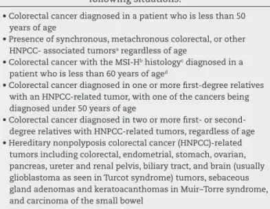
Tumors from individuals should be tested for MSI in the following situations:
• Colorectal cancer diagnosed in a patient who is less than 50 years of age
• Presence of synchronous, metachronous colorectal, or other
HNPCC- associated tumorsa regardless of age
• Colorectal cancer with the MSI-Hb histologyc diagnosed in a
patient who is less than 60 years of aged
• Colorectal cancer diagnosed in one or more i rst-degree relatives with an HNPCC-related tumor, with one of the cancers being diagnosed under 50 years of age
• Colorectal cancer diagnosed in two or more i rst- or second-degree relatives with HNPCC-related tumors, regardless of age • Hereditary nonpolyposis colorectal cancer (HNPCC)-related
tumors including colorectal, endometrial, stomach, ovarian, pancreas, ureter and renal pelvis, biliary tract, and brain (usually glioblastoma as seen in Turcot syndrome) tumors, sebaceous gland adenomas and keratoacanthomas in Muir–Torre syndrome, and carcinoma of the small bowel
a Hereditary Nonpolyposis Colorectal Cancer (HNPCC)-related
tumors include colorectal, endometrial, stomach, ovarian, pancreas, bladder, ureter and renal pelvis, biliary tract, brain (usually glioblastoma as seen in Turcot Syndrome), sebaceous gland adenomas and keratoacanthomas in Muir-Torre syndrome, and carcinoma of the small bowel.
b MSI-H, microsatellite instability–high in tumors refers to changes
in two or more of the i ve National Cancer Institute-recommended panels of microsatellite markers.
c Presence of tumor ini ltrating lymphocytes, Crohn’s-like
lymphocytic reaction, mucinous/signet-ring differentiation, or medullary growth pattern.
d There was no consensus among the Workshop participants
on whether to include the age criteria in guideline 3 above; participants voted to keep less than 60 years of age in the guidelines.
Table 5 – Recommendations for the evaluation of MSI-H and MSI-L.
The original National Cancer Institute (NCI) microsatellite panel included BAT25, BAT26, D2S123, D5S346 and D17S2507; however, the following caveats may apply:
• If only dinucleotide repeats are mutated, test a secondary panel of microsatellite markers with mononucleotide repeats (e.g., BAT40 and/or MYCL) to exclude MSI-L.
• Dinucleotide repeats are less sensitive than mononucleotide repeats for MSI-H; however, they provide an internal control for the prevention of sample mix-up
• A pentaplex panel of i ve quasimonomorphic mononucleotide repeats may be more sensitive for MSI-H tumors than other microsatellite markers and may obviate the need for normal tissue for comparison; this approach requires three or more mutant alleles to indicate MSI-H
a MSI-H, microsatellite instability–high in tumors refers to changes
in tumors with high levels of mucin, false negatives can oc-cur.16 False negatives also can occur with MSH6 germline
mu-tations, as they may have MSI-L results.37 And unlike with IHC
testing, an abnormal MSI test results does not suggest which gene to test.
Absence of MLH1 on IHC was also noted to have a low gene mutation detection rate. In time it became possible to distin-guish sporadic from a hereditary etiology in individuals with absent expression of MLH1 on IHC testing. This abnormal IHC result is frequently due to two somatic events: BRAF muta-tion Val600Glu (V600E) or MLH1 promoter hypermethylation. They both are common explanations of absent MLH1 expres-sion in patients without a germline MLH1 mutation, partic-ularly among those diagnosed with CRC after age 50. BRAF
mutations and MLH1 promoter hypermethylation are thought to be rare in Lynch syndrome-related cancers, though each has been seen in individuals with Lynch syndrome. In spite of these reported cases, the presence of a BRAF mutation or
MLH1 promoter hypermethylation essentially rules out the di-agnosis of Lynch syndrome.38-43
Remember, as abnormalities in IHC are suggestive, con-i rmatory dcon-iagnoscon-is of Lynch con-is only by poscon-itcon-ive mutatcon-ion on gene sequencing. The i rst patents for gene sequencing
of MLHH-1 and MSH2 were i led in 1997 and 1999.
Commer-cial launching then ensued in 2000.44 Commercial testing for
MSH6 soon followed. Due to technical complexity of PMS2
testing, commercial testing of the PMS2 gene did not become available until 2009.
An estimated 50% of Lynch syndrome mutations are found in MLH1,45 40% in the MSH2 gene45 7%-10% in the MSH6
gene,30,33,46 5% in the PMS2 gene47 and 1-3% EPCAM.36,48,49
Screening for Lynch syndrome
From 2000-2005 many centers were using family histories and pathological criteria by the Revised Bethesda Criteria to guide testing on tumors for MSI and or IHC. If these were abnormal, genetic counseling and then gene testing were performed. There was much controversy during this time whether MSI and IHC could be performed without patients’ consent. There was concern that these studies themselves could lead to a di-agnosis of a hereditary disorder, which could have insurance implications. With Heather Hampel’s study on all colorectal cancers,17the new dei nition of Lynch syndrome (coni rmed
by a proven MMR gene mutation),9EGAPP18and GINA19 led
the movement towards universal screening. The i nding that a family history (a standard on initial history and physicals) that met Amsterdam criteria led to a diagnosis of Lynch syn-drome with the same frequency as documenting abnormal MSI/IHC testing (about 60% of the time) also added to the defense to perform MSI and IHC without formal consent.17,50
Therefore, patients were only dei ned to have the Lynch mu-tation once mumu-tation was noted by gene sequencing. There is consensus that once germline testing is to be considered, genetic counseling is a standard.51
The United States Law began to recognize the importance and consequences of gene testing. GINA, passed in 2008 and enacted in 2009, prohibits health insurers from using genetic information (e.g., genetic test results, family history) to
de-termine insurability. It does have shortcomings that genetic counselors explain as part of their normal consenting pro-cess. If patients have a lapse of coverage, genetic diseases can be considered a pre-existing condition. It does not prevent companies from using the diagnosis of Lynch syndrome to help underwrite their disability, life insurance, and long-term policies. Since its inception, The Affordability Care Act of 2010 also allowed new coverage options to individuals who have been uninsured for at least six months because of a pre-ex-isting condition. This program will serve as a bridge to 2014, when rejecting insurance coverage due to pre-existing condi-tions will be prohibited.52
Hampel’s and De la Chappelle’s 2005 study16 on all
colorec-tal cancers resected amongst the major hospicolorec-tals in Ohio led to some very important discoveries and the feasibility of screening for Lynch syndrome on all CRC patients. It was then updated in 2008.17 In 2005, initially all colorectal
can-cers underwent MSI testing. If they were MSI-H or MSI-L they underwent IHC for MLH1, MSH2, MSH6 and PMS2-, sequenc-ing of MLH1, MSH2, and MSH6, and methylation analysis of
MLH1 promoter region if IHC for MLH1 was abnormal. If IHC revealed a lack of PMS2 and a presence of MLH1, PMS2 gene mutation was analyzed. In addition, those that were MSS but met Bethesda or Amsterdam criteria underwent IHC testing.
In 2008 an additional 500 CRC patients were screened, this time with MSI and IHC. Another 372 patients who were MSS and had normal IHC (or IHC not completed secondary to lack of tissue) underwent gene testing for two of the most com-mon MMR gene mutations in their series. One is American Founder Mutations (MSH2) and the other was another com-mon mutation in MSH2.
Combining the i ndings of the 2005 with the 2008 study patients, it was noted that the MSI-H prevalence was 12.7% in all colorectal cancers, and the prevalence of Lynch syndrome was at a minimum of 2.8%. In 2005, 10 out of the 23 identii ed Lynch syndrome patients were over 50 years of age and 5 out of 23 did not meet Bethesda or Amsterdam criteria. Testing only by MSI or IHC would each have missed two probands. MSI lacked sensitivity if there was signii cant mucin in the specimen and therefore careful dissection by pathology was recommended. For each proband, 5.79 at-risk family mem-bers were contacted; over 3 memmem-bers per proband were diag-nosed with Lynch.
Not only was prevalence now established, it also illustrat-ed the feasibility of testing all colon cancers for Lynch syn-drome. 1566 patients out 1700 patients agreed to participate. Only 2 out of 23 probands from 2005 study refused contact to be made with at-risk family members. Of the 199 mem-bers who were contacted and received counseling, only two of these refused to undergo gene testing. These i ndings led to discussion about health care policy and Lynch syndrome. The Evaluation of Genomic Applications in Practice and Pre-vention Group (EGAPP) published its position statement in 2009, concluding with moderate certainty that testing newly diagnosed CRC patients could provide moderate population benei t. It did not address the cost-effectiveness of a universal screening program.18
In 2009, Myundura et al.53 reported that universal testing
incremental cost effectiveness ratio was comparable to other preventive services. Their decision model looked at 4 main strategies:1. IHC testing for all MMR genes and utilizing BRAF
if MLH1 was abnormal 2. IHC testing for all 4 MMR genes and proceeding to gene sequencing if abnormal, or 3. MSI-H test-ing and proceedtest-ing to gene sequenctest-ing if abnormal or strat-egy 4, testing all CRC with gene sequencing.
For each of the four strategies the models calculated costs and outcomes using many of the data from Hampel’s papers, i.e. average relatives contacted, tested, calculating costs of testing, surveillance and treatment for CRC. For each strat-egy the cost-effectiveness ratios (in US dollars) were $23,206, $23,221, $28,291 and $ 79,651, respectively. See Table 6 below.
Cost-effectiveness ratios associated with Lynch syndrome testing strategies among new diagnosed colorectal cancer pa-tients and testing and surveillance for CRC among their i rst-degree relatives. See Table 7.
Analyzing the incremental cost-effectiveness ratio (ICER) using strategy 1. the incremental cost-effectiveness compar-ing to the next best strategy varies from about $18,000 to $50,000. Comparing this to colonoscopy screening (individu-als older than 50 and at every 10-year interv(individu-als) is $25,000 per LY saved. They note in these same articles that many analysts use a critical value of $50,000 or $100,000 per LY or QALY as a criterion of cost-effectiveness. They concluded that universal testing for Lynch syndrome is well within the range of acceptable ICERs for preventive services in the Unit-ed States. As with many studies looking at cost effectiveness of genetic testing, the true saving come to those which oper-ate in a hereditary center module, as there is active attempts to reach out to at-risk family members. The cost savings is truly made with the site specii c testing of the at-risk rela-tives once a mutation is known. Site specii c testing is much cheaper, frequently one tenth of the cost for diagnosing the proband’s mutation.
Current diagnostic strategies
As the preceding text shows, there are many ways to arrive at a diagnosis of Lynch syndrome, from going straight to
germline genetic testing of all Lynch genes to targeting the germline testing based on results from IHC, BRAF and MLH1
hypermethylation testing. According to their availability and policies, individual institutions follow a host of algorithms. Some hospitals only perform MSI or IHC on specimens as re-quested by clinicians on a case-by-case basis. Some institu-tions follow Bethesda Criteria and may perform MSI, IHC, or both. There is a trend after the EGAPP working group papers that more institutions are performing universal screening on ALL colorectal cancers (and some performing IHC on endo-metrial), with MSI or IHC or both. Some sites have expanded to IHC on all or a subset of endometrial cancers.
Imperative in these strategies is that the abnormal values are reported to a clinician or counselor who can appropriately interpret these results. At Duke University we currently per-form MSI and IHC on all colorectal cancers, by endoscopic biopsy or surgical resection specimen, with all results being sent to our genetic counselors. Clinicians in gastroenterology, surgery and medical oncology all agreed to allow the counsel-ors to contact their patients as an extension of their practice. This allows the treating clinician to maintain “ownership” over the follow-up of abnormal results, removes pathologists from a position of directly inl uencing patient care, and main-tains HIPAA compliance.
Many institutions begin Lynch syndrome evaluation by performing IHC testing based on the rationale that an ab-normal IHC test will lead to cheaper germline genetic ing. Cost of gene sequencing and deletion/duplication test-ing for all 4 genes varies by laboratory, but is at least $4500. Testing a single gene, as directed by an abnormal IHC result, can decrease this cost by at least $2000. Cost of site-specii c genetic testing for a known familial mutation ranges from about $150-$500. This illustrates the cost saving as hereditary centers reach out to at-risk family members.
For those found to lack MLH1 staining on IHC, centers vary whether rel exively BRAF/MLH1 hypermethylation is performed or whether genetic counseling ensues prior to performing BRAF. The presence of BRAF mutation/MLH1 hy-permethylation virtually excludes Lynch syndrome. But if the individual has a strong family history or early onset cancer, heightened surveillance may still prevail. This is because
Table 6 – Incremental cost-effectiveness ratios of the 4 testing strategies of universal to no testing, of age-targeted testing to no testing, age-targeted testing to previous strategy in dollars per life-year saved.
Strategies Description of testing strategy a
Incremental cost-effectiveness ratio of universal testing relative
to no testing and relative to previous strategy, dollars per
life-year saved
Incremental cost-effectiveness ratio of age-targeted testing
relative to no testing and relative to previous strategy,
dollars per life-year saved
Incremental cost-effectiveness ratio of universal testing relative to age-targeted testing
and relative to previous strategy, dollars per life-year
saved
1 IHC, BRAF testing
and sequencing
$22,552 and $22,552 $7,832 and $7,832 $37,010 and $37,010
2 IHC testing and
sequencing
$23,321 and $273,915 $7,944 and $60,569 $38,411 and $429,973
3 MS1 testing and
sequencing
$41,511 and $764,917 $11,680 and $168,905 $70,792 and $1,192,575
4 Genetic sequencing
for 4 genes
$142,289 and $737,025 $44,902 and $252,643 $237,278 and $1,192,575
while it is rare, MLH1 hypermethylation can be present in a Lynch syndrome patient as the second hit.36,39 On the
con-trary, MSH2 methylation has been found to be the second hit in approximately 24% of MSH2 related cancers. It has not been found in sporadic cancers.54
Because of the proteins of the MMR are frequently pres-ent as complexes/dimers, loss of one is often associated with a loss of the partner MMR protein. Loss of expression of
MLH1 is almost always associated with loss of PMS2 expres-sion. Loss of MSH2 expression is almost always accompanied by loss of MSH6 expression. On the other hand, loss of PMS2
expression or MSH6 expression is frequently seen without the accompanying loss of MLH1 or MSH2, respectively. Loss of MSH2 and MSH6 usually indicates a germline MSH2 mu-tation. Loss of MSH6, only, usually indicates a mutation in
MSH6. Non-sporadic loss of MLH1 and PMS2 (i.e., normal
BRAF and MLH1 hypermethylation testing) is typically due to an MLH1 or PMS2 mutation. Loss of PMS2, only, usually rep-resents a PMS2 mutation. Because of this relationship with paired complexes, some centers strategize by performing IHC i rst for PMS2 and MSH6. If both are present, no further testing is done. If one is absent, the other partner of the di-mer is tested. For example, absent expression of MSH6 would lead to IHC testing of MSH2.
Although there are clearly benei ts to beginning the Lynch syndrome evaluation with MSI and IHC screening on an af-fected individual’s colon or endometrial tumor, or even an adenoma with high-grade dysplasia,55,56 this is not always
possible. When another tumor within the Lynch syndrome spectrum is available, MSI and IHC testing can still guide fur-ther testing when abnormal. The same logic applies to per-forming MSI and IHC on metastases from a colorectal prima-ry. When no Lynch spectrum tumor, adenoma or metastasis is available for MSI and IHC testing, direct germline genetic test-ing of the Lynch-associated genes is the next step. However, interpretation of germline results is not always straightfor-ward. Variants of uncertain clinical signii cance can confound interpretation. And normal germline results in an individual with high prior probability of detecting a mutation (e.g., be-cause of meeting Amsterdam II criteria or having colon can-cer with histologic features noted in the revised Bethesda
cri-teria) still leave the possibility of an undetectable germline mutation. This complicates risk management for the patient and their i rst-degree relatives, as it is unclear whether they should be managed as if they have Lynch syndrome.
There are various mathematical models that have been devised using patient’s personal cancer history and family members to predict risks of gene mutations in Lynch syn-drome. Some have found these helpful to determine if gene sequencing (with its inherent short-coming without tissue availability) is worthwhile. When IHC MSI cannot be per-formed on tissue due to inavailability, these are PREMM1,2,6, MMRpredict, and MMRpro.
PREMM1,2,6
The model is based on data from 4539 individuals undergoing genetic testing of MSH2, MLH1, and MSH6 through a commer-cial laboratory. This model uses the proband’s and second-degrees relatives history of Lynch syndrome-related cancers (colon, endometrial, stomach, ovarian, small intestine, uri-nary tract/kidney, bile ducts, glioblastoma, sebaceous gland tumors, and pancreas) and age of onset of colon and endo-metrial cancers. MSI and IHC testing is not included. Based on genotype/phenotype data, this model provides specii c likeli-hood estimates for detecting a mutation in each of the MMR genes (MLH1, MSH2, MSH6). Using a 5% mutation probability as a standard for MMR testing, the model has an estimated sensitivity of 90% and a specii city of 54%.57 Using family
his-tory it can also estimate risk of MMR gene mutation in an unaffected individual.
MMRpredict
Uses a population-based cohort diagnosed with CRC before age 55 years who were tested for MLH1, MSH2, and MSH6 mu-tations. Data from MSI and IHC testing and the presence of CRC and/or endometrial cancer in i rst-degree relatives can be incorporated. This model can only be used in affected in-dividuals. Because the model is based on those diagnosed be-fore 55 years of age, it is unclear how accurate the model is for tumors diagnosed in older individuals.58
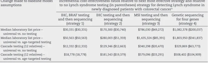
Table 7 – Interval cost-effectiveness ratios relative to next most effective strategy and relative to no Lynch syndrome testing for detecting Lynch syndrome in newly diagnosed patients with colorectal cancer.
Change made to baseline model assumptions
Incremental cost-effectiveness ratios relative to next most effective strategy and relative to no Lynch syndrome testing (in parentheses) strategy for detecting Lynch syndrome in
newly diagnosed patients with colorectal cancer a
IHC, BRAF testing and then sequencing
(strategy 1)
IHC testing and then sequencing (strategy 2)
MSI testing and then sequencing (strategy 3)
Genetic sequencing for four genes
(strategy 4)
Median laboratory list price – universal vs. no testing
$30,331 ($30,331) $170,300 ($30,740) $786,030 ($49,272) $1,082,378 ($200,037)
Median laboratory list price – universal vs. age-targeted testing
$50,563 ($50,563) $280,003 ($51,359) $1,435,324 ($85,391) $1,803,950 ($341,837)
Cascade testing (12 relatives) – universal vs. no testing
$12,332 ($12,332) $129,346 ($12,663) $340,298 ($20,470) $329,869 ($63,773)
Cascade testing (12 relatives) – universal vs. age-targeted testing
$18,778 (18,778) $181,543 ($19,379) $579,096 ($33,291) $508,402 ($104,909)
MMRpro
Uses the data obtained from clinic and population avail-able in the literature with cancer risk estimates based on pen-etrance from a meta-analysis of i ve large Lynch syndrome studies. It uses the presence of CRC and other cancers in the proband, i rst- and second-degree relatives, age of onset, and IHC and MSI testing to estimate the likelihood of identifying a germline mutation in MLH1, MSH2, or MSH6. It allows calcula-tion of a family member’s risk to inherit the germline muta-tion and the risk to develop colon or endometrial cancer.59
Risks of CRC
In the early 20th century, at the time Warthin’ i rst described
Family G, gastric cancer was a common cancer in Lynch syn-drome families. Just as sporadic cancer has seen a decline, so has gastric cancer in Lynch syndrome. The more common secondary cancers are colorectal, ovarian, gastric, and renal system (transitional cell of renal pelvis and ureter) and se-baceous cysts adenoma and adenocarcinomas. A mnemonic quite useful to remember these is COUGaRS: Colorectal, Ovar-ian, Uterine, Gastric and Renal(Urinary-transitional cell), Se-baceous tumors. Other cancers include medulloblastoma brain cancers, biliary cancers, and small bowel. More recent small increases in prostate and breast has been reported. Cur-rently, colorectal cancer is overall the most common cancer in Lynch syndrome.
Earlier in the history of Lynch syndrome, the mean age for CRC was thought to be 43 years old. Lynch describes average age of 44.6 years in his Family R.5We now know this is a false
low average due to selection bias. As we all had a higher de-gree of concern for the younger patients with CRC it falsely lowered the mean age of occurrence. When Hampel excluded the probands in her study, and with aggressive discovery of relatives with Lynch who already had CRC, the average age of cancer in the nonprobands was 61 years.60
Previously patients with Lynch syndrome were thought to have a ~80% risk of cancer by the age of 80. As our knowl-edge and diagnostic capabilities have been augmented, our numbers are tempered. Also, looking at risks based on spe-cii c mutations, more individualized risks can be predicted. These risks also vary by sex. For example, MLH1 and MSH2
mutations have CRC risks of 66-69% in men, 43-53% in women with average age of 61 years.60,61 Overall, MSH6 and PMS2 have
an attenuated risk. CRC cancer risks for patients with MSH6
mutations are 44% for males and 20% for females.62Patients
with MSH6 mutations also present with later ages of onset and a more distal distribution. They are also associated with MSI-L tumors.46,63 The risk in a patient with a PMS2 mutation
was 15%-20% by 70 years of age.47The newly noted mutation
in EPCAM has higher penetrance for CRC. Kempers estimated
from a cohort of 194 individuals with EPCAM mutations that the cumulative risk of CRC by 70 is 75%.64
Risks of non-colonic cancers
See Table 8.65
Endometrial cancer
Endometrial cancer (EC) is the second most common cancer in Lynch syndrome. Women have a 25-60% lifetime risk (Table 8).60,61,66,67 Just as studies on high-risk families found a younger
age of onset for colon cancer that was reputed in population studies, the same occurred with EC. Early studies reported av-erage age of 48 years old; the population-based studies now show average age is 62.60,67
Also similar to colon cancer, endometrial cancer has vari-ous risks based on mutation site. MLH1, MSH2, MSH632have
approximately a 44% risk for endometrial CA. Others have noted a slight increase risk of endometrial for MSH6 vs. MLH1,
and MSH2.46 Kemper64 found a 12% risk for EPCAM mutations.
Women who in their lifetime have both colon and endo-metrial cancer have an equal chance of having either can-cer i rst.69,62 For those women who are diagnosed with their
colorectal cancer i rst, their subsequent risk of later EC is 26% within 10 years of their CRC diagnosis.70 The lifetime risk (70
years) for a woman to have colon or endometrial cancer was noted to be 73% in Stoffel’s study.61
Gastric cancer
Gastric cancer in Lynch patients is usually intestinal type adeno-carcinoma, though Capelle has reported that in the Netherlands, up to 20% can present with a diffuse gastric carcinoma histol-ogy.71,72,73Microsatellite instability is noted in these tumors.74
Overall, estimates for gastric cancer risk in heterozygotes for an MLH1 or an MSH2 mutation range from 6% to 13%. Men with
MSH2 mutation have the highest risks.72,73A high incidence of H
pylori infection, or Asian populations also have increased inci-dence.75The mean age of diagnosis is 56 years old.71
Ovarian cancer
The risk for ovarian cancer is roughly twice in MSH2 (8%-11%) versus MLH1 mutation (4-6%). The mean age is 42.5 years with
Table 8 – Comparative risks of cancer types of general population and patients with MLH1 and MSH2 mutation, and mean age of onset of each cancer with patients with MLH12 and MSH2 mutations.
Cancer Type General population
risk
Lynch syndrome (MLH1 and MSH2 heterozygotes)
Risk Mean age of onset
Colon 5.5% 52-82% 44-61 years
Endometrium 2.7% 25-60% 48-62 years
Stomach < 1% 6-13% 56 years
Ovary 1.6% 4-12% 42.5 years
Hepatobiliary tract
< 1% 1.4-4%% Not reported
Urinary tract < 1% 1-4% ~55 years
Small bowel < 1% 3-6% 49 years
Brain/central nervous system
< 1% 1-3% ~50 years
Sebaceous neoplasms
30% diagnosed before the age of 35.72The histology
distribu-tion is similar to those with sporadic ovarian cancer, though borderline does not seem to be associated with Lynch syn-drome.76One metanalysis paper noted ovarian cancers with
mismatch repair dei ciency presented in earlier stages.77 The
only study that compared survival did not reveal a survival advantage for Lynch patients with ovarian cancer versus the sporadic ovarian cancer.78
Renal-urinary tract cancers
The urinary tract cancers most associated with Lynch syn-drome are transitional carcinomas of the ureter and renal pel-vis. One Dutch study suggested an increased risk with bladder cancer. Their Lynch patient with bladder cancer did show MSI and/or loss of stain on IHC that corresponded to the germline mutation. Bladder cancer, however, is not listed as one of the Amsterdam or Bethesda criteria.76,79 Watson notes a smallest
risk estimate of 1% in women with MLH1 mutation and then up to 27% in men with MSH2 mutation.72
Small bowel cancer
Lifetime risk of small bowel cancer is 3-6%, though >100 times the risk of the general population.72 50% of the small bowel
cancers are within the duodenum and jejunum, within the reach of an upper endoscopy.80 The majority in
adenocarcino-ma80 incidence is similar between MLH1 and MSH2 mutations
and rare in MSH6 and PMS2.81
Pancreatic and biliary cancer
A few studies have revealed an increased risk of pancreatic cancer, and family clustering is noted. Geary noted a seven-fold increase risk in their Lynch syndrome patients over the general population.82,83 However, other studies have not
dem-onstrated an increased risk.84
Brain tumors
The risk for brain tumors is estimated at approximately 2%.16
Risks may be underestimated as 26% of the time when the age of onset is before 25 years of age.72 The most common
histology is glioblastoma, and is rarely associated with micro-satellite instability.85 It is the third cause of cancer death for
Lynch patients in a large Dutch cohort.86
Sebaceous skin neoplasias
This includes sebaceous adenomas, sebaceous epitheliums, sebaceous carcinomas, and keratoacanthomas.87,88
Seba-ceous neoplasms associated with Lynch syndrome exhibit MSI and IHC.89,90 The data on the frequency of sebaceous
neoplasms in individuals with Lynch syndrome are limited. Studies have found that between 1% and 9% of individu-als with a germline mutation in an MMR gene have a seba-ceous neoplasm.91,92 Individuals with Lynch syndrome and
a sebaceous neoplasm have Muir-Torre syndrome, which was initially thought to be a separate entity. IHC testing of sebaceous adenomas has shown that a signii cant
propor-tion is sporadic. Among those with abnormal IHC testing in a sebaceous neoplasm, Lynch syndrome mutation carriers are more likely than those with sporadic presentation to have multiple sebaceous neoplasms and a personal or family his-tory of a Lynch spectrum cancer.93
Additional cancer risks
Hematologic cancers, laryngeal cancer and sarcomas have been suggested. Due to rarity of presentations, it is difi cult to determine the magnitude of risks.94,95 Nilbert96 did note
de-fective MMR in the histopathology of six of eight sarcomas in individuals with Lynch syndrome.
Breast cancer
The relationship between breast cancer and Lynch syndrome is unresolved.97,98,99 Studies have not consistently
demon-strated a higher than expected incidence. Walsh, however, did demonstrate that in breast cancer of patients with a mutation in a MMR gene, 51% did demonstrate a loss of immunohisto-chemical staining for the protein corresponding to the gene in which a germline mutation occurs.100
Variants of Lynch
Muir-Torre syndrome is the terminology used to describe a
Lynch syndrome patient who also has sebaceous neoplasms of the skin. The types of sebaceous skin neoplasias described include: sebaceous adenomas, sebaceous epitheliomas, seba-ceous carcinomas, and keratoacanthomas.87,88MSH2 mutation
is the most common mutation noted.92
Turcot syndrome is dei ned as CRC or colorectal adenomas
in addition to tumors of the central nervous system. This can be due to APC gene mutation as seen in FAP, or due to MMR gene mutation associated as a Lynch syndrome.101 Therefore,
the clinical colonic presentation varies from numerous colon-ic polyps to a single polyp or CRC. The brain cancer associated with APC mutation tends to be medulloblastomas; mutations of the MMR gene tend to present with glioblastomas. The brain tumors associated with mutations in a mismatch repair gene exhibit MSI.101,102
Homozygous mismatch repair mutations: rare individuals
who are homozygous for mutations in MLH1, MSH2, MSH6,
and PMS2 have been reported. Affected individuals often have onset of colon or small bowel cancer prior to the second decade of life. One third of children with biallelic mutations have been reported to have more than ten polyps. Also as-sociated is Hematologic cancer, brain tumors, and café-au-lait
macules.103,104
Survival
When matched stage for stage, colon cancers in individu-als with Lynch syndrome are associated with a better prog-nosis than sporadic colon cancer.105 This is an unexpected
poor prognosis. Due to the mutation in the MMR genes, Lynch syndrome cancers do not respond to typical chemotherapeu-tic agents like 5-l uorouracil, in fact, they may do worse.106
Surveillance
Surveillance is an important part of the management of a patient with Lynch syndrome. Optimal surveillance requires a multidisciplinary approach involving primary care physi-cians, gastroenterologists, gynecologists and colorectal sur-geons. An excellent resource for surveillance is available on the National Comprehensive Cancer Network website (www. nccn.org). There are no strong data for surveillance for many of the Lynch syndrome associated cancers and recommenda-tions outside of colon and endometrium are based on expert opinion.
CRC Surveillance
Since 1977 Dr. Lynch proposed starting colonic surveillance as early as 20 years of age.5Current surveillance
recommenda-tions also start in Lynch patients as early as age 20-25 (or 10 years prior to family member’s cancer diagnosis, whichever is earlier) with colonoscopy. Intervals are every two years, until 40, then yearly afterwards. The short interval is due to the ac-celerated progression of polyp to cancer as noted by Jass in 1992.107 Some recommend starting surveillance at the age of
30 in patients with MSH6 or PMS2 mutations since the average age of onset of colon cancer is somewhat later. Colonoscopy is repeated every 1-2 years. After diagnosis of CRC and subse-quent resection, surveillance should occur on a yearly basis. Regular surveillance is proven to reduce both incidence (11% vs 27%) and death (2% vs 12%) from CRC.108
Gynecological Surveillance
There is no clear evidence to support routine screening or surveillance for endometrial or ovarian cancer. Some rec-ommend annual transvaginal US and endometrial sam-pling at 30-35 years of age.46,47 Studies on the effectiveness
of transvaginal ultrasound examination and endometrial biopsy have had conl icting results. In most screening stud-ies, patients presented with symptoms before or during their surveillance with transvaginal ultrasound or endome-trial sampling.109,110
Ovarian cancer
No specii c ovarian cancer screening trials have been con-ducted in women with Lynch syndrome. Of note, screening for ovarian cancer using CA-125 blood tests and transvaginal ultrasound examination has not been effective in other high-risk populations such as women with a BRCA1 or BRCA2 mu-tation.111
Gastric cancer
There are no strong data for gastric cancer surveillance. Schulmann has noted that 50% of his patients with small
bowel cancer were noted proximally within the reach of an upper endoscope.80 While there are no studies on the efi cacy
of surveillance, enteroscopy is a consideration noted by the NNCN starting at age 30-35 and performing every 2-3 years. More frequent intervals can be considered if chronic inl am-mation, atrophic gastropathy and/or intestinal metaplasia is noted. Many insurance companies are now covering this procedure. Also capsule endoscopy every 2-3 years starting at 30-35 years of age can be considered for surveillance of the distal small bowel.
Urinary collecting system
A urinalysis can be performed on an annual basis starting at 30-35 years of age (NCCN 2011). There are no studies to prove efi cacy and survival. As it is a noninvasive test, recommenda-tions remain.
Other cancers
Lindor et al.46 recommend beginning annual examination at
age 21 for features of sebaceous. The National Comprehen-sive Cancer Network recommends beginning annual physical exam for such features at 25-30.112 There is no current data to
make a standard recommendation in the case of pancreati-cobiliary cancers. There are programs that are embarking as a research study to perform surveillance for Lynch and other high-risk patients for pancreatic cancer.113,114
Surgery for prophylaxis and for treatment
Gynecological cancer
Women with Lynch syndrome who are undergoing colon cancer are usually offered the choice of prophylactic hys-terectomy and bilateral salpingo-oophorectomy. Also, once the patient is past childbearing age or post-menopausal, prophylactic hysterectomy and bilateral salpingo-oopher-ectomy can be considered as a risk-reducing measure. Schmeler115 noted in their case-control study of 315 women
with Lynch syndrome, 1/3 had prophylactic hysterectomy with bilateral salpingo-oophorectomy. After 10 years of fol-low-up, there were no gynecological cancers in the women with prophylactic surgery, though there was 33% incidence of endometrial cancer and 5% ovarian cancer in the con-trol group. Chen and colleagues116 also noted the efi cacy
of prophylactic surgery. They concluded that one diagnosis of endometrial cancer was prevented for every 6 surgeries, and one ovarian cancer for every 28 surgeries. Most would recommend for the pre-menopausal women who choose prophylactic surgery to be placed on hormonal replace-ment until the age of 50.30,46,47,117
CRC
Dr. Lynch i rst described that, with good family history, rec-ommendations of colon removal may decrease the incidence of cancer.5 This was prior to signii cant use of colonoscopy in
in 1969.118 Currently prophylactic colon resection is not
rec-ommended. The extent of surgery to be performed once a CRC is diagnosed is still under investigation (NCCN 2011). There are pros and cons to consider in regards to segmental vs. total or subtotal colectomy. There is a balance of quality of life, and the risks of metachronous disease.
The initial risks of CRC as stated earlier are substantially decreased with frequent surveillance by colonoscopy. There is no reason not to believe that metachronous cancers are not also reduced with frequent surveillance. Engel noted in their Lynch patients that if a metachronous cancer was found during yearly surveillance colonoscopy it was earlier in stage, with 95% discovered in Stage I and Stage II.119
Parry120 notes in their comparison of segment versus
ex-tensive resection that the metachronous risk was reduced by 31% for each 10cm of bowel removed. While they state there was no difference in the frequency of endoscopy in the two groups, the interval was truly only estimated and not record-ed. The weakness in this study is the frequency of scoped was assumed to be distributed uniformly in the period between the i rst and last age of endoscopy. Those metachronous can-cers that were discovered, as in Engel’ study, were predomi-nantly in early stages: 27 (47%) at stage I, 20 (35%) at stage II and 10 (18%) at stage III. Of the 10 patients who developed AJCC stage III metachronous CRC (mean follow-up 12 [SD 10] years), six reported 1-2 yearly lower endoscopy, one reported no endoscopy and for three it was unknown. At 5 years after surgical resection, 49 (98%) who had extensive colectomy and 327 (98%) who had segmental colectomy were alive (p1⁄40.8). At 10 years, 49 (98%) who had extensive colectomy and 322 (97%) who had segmental colectomy were alive (p1⁄40.7). Therefore one can conclude a more substantial resection may limit second surgery but no proof in increased survival can be gathered by this study.
Maeda121 constructed a state-transition (Markov) model
based on assumptions obtained from available data sources and published literature. They compared segmental colec-tomy (SEG) to a total abdominal coleccolec-tomy (TAC) for quality adjusted life years (QALYs). They concluded that, for young (30-year-old) patients with Lynch syndrome, mean survival was slightly better with TAC than with SEG (34.8 v 35.5 years). Their QALYs were approximately equivalent, with QALYs per patient of 21.5 for SEG and 21.2 for TAC. With advancing age, SEG becomes a more favorable strategy.
There have been no studies to date that prove that a more extensive resection will translate to greater colon cancer sur-vival. Therefore, case by case management based on patients’ age of presentation, stage of initial cancer presentation, cur-rent bowel habits and continence, willingness to undergo close surveillance and patient desires need to be considered and discussed with the patient.
Risk adjustments
We are all aware that there is always an interplay between environment and genetics. We at this time cannot change our genes, but can we decrease the risks that our genes have in store for us through our environment? There are many rea-sons to stop smoking. We encourage our Lynch syndrome
pa-tients to stop smoking as it was one of the three risks factors CRC noted by Watson et al.122 The other two factors were male
sex and MLH1 mutation. The hazard ratio of being a male was 1.58, MLH1 vs. MSH2 was 2.07 and tobacco use, 1.43.
Chemoprevention
Women who take a combined oral contraceptive pill can lower their risk of endometrial and ovarian cancer. This risk seems to decrease with longer duration. While the ovaries enjoy a permanent protective effect, this affect persists for about 5 years after stopping oral contraceptives. While there is no evidence to suggest oral contraceptives in Lynch syndrome patients, its use is not contraindicated in Lynch syndrome patients. Other risks factors such as smoking and history of thromboembolism need to be considered prior to prescribing.117,122
Professor Burn and team have been looking over the years at the use of aspirin and starch to prevent CRC in patients with known mutations. His study was a two by two double blinded randomized study of starch/ASA, Starch/placebo, pla-cebo/ASA, placebo/placebo. Patients received 30 g of starch (novelose) and 600 mg of aspirin. The study was designed to note if this decreased the incidence of advanced adenomas or carcinomas. Initial results of 746 Lynch patients after 4 years did not reveal a decreased risk with the use of aspirin. In 2011, after a few patients had reached 10 years of follow-up, the results were reviewed once more in regards to their risks of CRC and other Lynch related cancers. Post-intervention review revealed 13 of 342 allocated aspirin and 27 of 329 al-located had CRC. 38 participants developed cancer at a site other than the colorectal (additionally, two participants had CRC and another Lynch syndrome cancer) of which 16 were randomly assigned to aspirin and 22 to aspirin-placebo. The study does not comment on the compliance of colonoscopy but interestingly even for non-CRC the risks decreased. From this they recommend 600 mg of ASSA per day for patients who can tolerate the regimen. CAPP3 is underway to evaluate different dosages as doses <600 have been implied in their study to be also effective. The optimum dose and duration is to be studied in CAPP3.123,124
Family X
At the beginning of the paper we discussed the importance of Lynch to be diagnosed with coni rmation of MMR gene mutation. It is now known that not all patients who meet the Amsterdam Criteria are Lynch syndrome patients. This is best illustrated with Family X. Family X was i rst described by Lindor.125 In her large database of patients that met
and small bowel, pancreas and liver. The MSS family had a CRC risk with a SIR of 2.3. There were no other cancers noted with increased risk in this group. The onset of cancer in the MSI0H group was also earlier (48.7) vs. 60.7 in the second group. The lower risk ratio for CRC and the absent risk of the secondary cancers. In retrospect this is the Lynch syndrome I patients that were discussed many years ago.
Lindor dispelled the notion that all families meeting Amsterdam I criteria are a distinct homogenous group and could now be further subdivided by their MSI status. The families that do not have an MMR defect (MSI-L/MSS in this study) have a lower risk of CRC, and a later onset. Therefore colonoscopy recommendations are to start colonoscopies for the at-risk family members at 5-10 years earlier than the earliest diagnosis of CRC in the family and occur every 5 years, if normal.
During the same year Llor et al.126 studied 1309 newly
di-agnosed CRC in Spain in which 25 probands i t Amsterdam I or II criteria. Fifteen (60%) of the tumors were MSS with the remaining MSI-H. All MSS tumors expressed MLH1, MSH2 or
MSH6. MSS probands were older at diagnosis (67.8 vs. 64.8), had more left sided colon cancers (86.7 v evenly spread), were well differentiated (33% vs. 0%), and lacked lymphocyt-ic ini ltrate (0% vs. 50%). There was no difference in synchro-nous or metachrosynchro-nous cancers. When looking at relatives, more families in the MSS group had less affected members than in the MSI-H group (18% vs. 31.5%) and were diagnosed at a later age (60.2vs 53.8). All extracolonic tumors found in both groups were endometrial and this occurred more frequently in the MSI group than in the MSS group (5.1% v3.3%). These i ndings echoed Lindor’s study that MSS fami-lies fuli lling Amsterdam criteria appear to be representing a syndrome separate from Lynch syndrome. Later studies, including a 2007 study by Valle et al.,127 continue to bolster
this important distinction.
Summary
Much has been learned since the Human Genome Proj-ect, and much more is to be discovered. As more variants of unknown signii cance are categorized as deleterious muta-tions, more patients will be properly diagnosed as Lynch syndrome. As we have better dei nitions of the mutations, and long-term follow-up on the affected patients, we will become better in tailoring patients’ risks and therefore tai-loring their management. With a better understanding of their pathophysiology we may then be able to intervene with better prevention strategies. To meet the goals of in-creasing the diagnosis of Lynch syndrome to then in turn decrease incidence of cancer in these families, a group of institutions formed the Lynch Syndrome Surveillance Network in the United States in 2011. Through universal screening and a common shared database these goals can be met.
Confl ict of interest
The authors declare no conl icts of interest.
R E F E R E N C E S
1. Lynch HT, Krush AJ. Hereditary and adenocarcinoma of the colon. Gastroenterology, 1967; 53(4), 517–527.
2. Warthin A. Heredity with reference to carcinoma as shown by the studies of cases examined in the pathological laboratory of the University of Michigan, 1895-1913. Arch Intern Med 1913; 12:546-555.
3. Lynch HT, Hitchins MP, Shaw TG, Lynch JF, Roy R. Historical Aspects of Lynch Syndrome Chapter 2 Hereditary
Colorectal Cancer MD Anderson Solid Tumor Oncology Series 5 Springer Science 2010.
4. Lynch HT, Krush AJ. Cancer family “G” revisited: 1895-1970. Cancer 1971; 27:1505-1511.
5. Lynch HT, Harris RE, Bardawil WA, Lynch PM, Guirgis HA, Swartz MJ, et al. Management of hereditary site-specii c colon cancer. Arch Surg 1977; 112(2): 170–4.
6. Lynch HT, Drouhard TJ, Schuelke GS, Biscone KA, Lynch JF, Danes BS. Hereditary nonpolyposis colorectal cancer in a Navajo Indian family. Cancer Genet Cytogenet. 1985 Feb 15; 15(3-4): 209-13.
7. Lynch HT, Schuelke GS, Kimberling WJ, Albano WA, Lynch JF, Biscone KA, et al. Hereditary nonpolyposis colorectal cancer (Lynch syndromes I and II). II. Biomarker studies. Cancer 1985; 56: 939-95.
8. Lynch HT, Kimberling W, Albano WA, Lynch JF, Biscone K, Schuelke GS, et al. Hereditary nonpolyposis colorectal cancer (Lynch syndromes I and II). I. Clinical description of resource. Cancer. 1985 Aug 15; 56(4): 934-8.
9. Jass JR. Hereditary Non-Polyposis Colorectal Cancer: the rise and fall of a confusing term. World J Gastroenterol. 2006 Aug 21; 12(31): 4943-50.
10. Vasen HF, Mecklin JP, Khan PM, Lynch HT. The International Collaborative Group on Hereditary Non-polyposis
Colorectal Cancer (ICG- HNPCC). Dis Colon Rectum 1991;34:424–5.
11. Vasen HF, Watson P, Mecklin JP, Lynch HT. New clinical criteria for hereditary nonpolyposis colorectal cancer (HNPCC, Lynch syndrome) proposed by the International Collaborative group on HNPCC. Gastroenterology 1999; 116: 1453-1456.
12. Mecklin JP, Jarvinen HJ. Clinical features of colorectal carcinoma in cancer family syndrome. Dis Colon Rectum 1986; 29 (3): 160-164.
13. Rodriguez-Bigas MA, Boland CR, Hamilton SR, Henson DE, Jass JR, Khan PM, et al. A National Cancer Institute Workshop on Hereditary Nonpolyposis Colorectal Cancer Syndrome: meeting highlights and Bethesda guidelines. . Journal of the National Cancer Institute. 1997; 89 (23): 1758-1762.
14. Umar A, Boland CR, Terdiman JP, Syngal S, Chapelle ADL, Rüschoff J, et al. Revised Bethesda Guidelines for Hereditary Nonpolyposis Colorectal Cancer (Lynch syndrome) and Microsatellite Instability. JNCI Journal of the National Cancer Institute 2004;96(4):261–268. 15. Gologan A, Krasinskas A, Hunt J, Thull DL, Farkas L,
Sepulveda AR. Performance of the revised Bethesda guidelines for identii cation of colorectal carcinomas with a high level of microsatellite instability. Archives of pathology & laboratory medicine. 2009;129(11):1390–1397. 16. Hampel H, Frankel WL, Martin E, Arnold M, Khanduja