Deorphanization of receptors: Applying screening techniques to
two orphan GPCRs
Ana Catarina Rufas da Silva Santos
Mestrado Integrado em Ciências Farmacêuticas
Deorphanization of receptors: Applying screening techniques to
two orphan GPCRs
Ana Catarina Rufas da Silva Santos
Monografia de Mestrado Integrado em Ciências Farmacêuticas apresentada à Universidade de Lisboa através da Faculdade de Farmácia
Orientadora: Ghazl Al Hamwi, PhD Student
Co-Orientadora: Professora Doutora Elsa Maria Ribeiro dos Santos Anes, Professora Associada com Agregação em Microbiologia
3 Abstract
G-Protein Coupled Receptors represent one of the largest families of cellular receptors discovered and one of the main sources of attractive drug targets. In contrast, it also has a large number of understudied or orphan receptors. Pharmacological assays such as β-Arrestin recruitment assays, are one of the possible approaches for deorphanization of receptors. In this work, I applied the assay system previously mentioned to screen compounds in two orphan receptors, GRP37 and MRGPRX3.
GPR37 has been primarily associated with a form of early onset Parkinsonism due to its’ expression patterns, and physiological role as substrate to ubiquitin E3, parkin. Although extensive literature regarding this receptor is available, the identification of a universally recognized ligand has not yet been possible. Two compounds were proposed as ligands, but both were met with controversy. These receptor association with Autosomal Recessive Juvenile Parkinson positions it as a very attractive drug target, and as such its’ deorphanization is a prime objective for investigators in this area.
Regarding MRGPRX3 information is much scarcer. Although it is part of a well-studied family, Mas Related G-Protein Receptors, this gene, found only in mammalian genome, remains elusive. Its’ expression patterns are the only indicators of a possible physiological or pathophysiological role. Similarly to other receptors of the same family, MRGPRX3 is though to be involved in the pain and/or itch pathway, but no factual evidence of said involvement has been presented yet.
Here I will focus on compounds’ screening on these receptors. The approach we used was directed for each of them and based on literature revision and on the information we had available at the time. We hoped to find a compound that produces activation of the receptors in order to allow us to withdraw some structure related clues for what the endogenous ligand of these receptors might be.
4 Resumo
Os recetores acoplados à proteína G representam uma das maiores famílias de recetores de superfície e uma das maiores fontes de alvos terapêuticos atualmente. No entanto, uma grande percentagem destes recetores não estão adequadamente caraterizados ou são classificados enquanto recetores órfãos. Ensaios farmacológicos como o ensaio de recrutamento da β-arrestina constituem uma das abordagens possíveis para desorfanizar recetores. Neste trabalho, apliquei o sistema de ensaio farmacológico que mencionei anteriormente a dois recetores: GPR37 e MRGPRX3.
O GPR37 tem sido principalmente associado a uma apresentação de início precoce de Parkinson. Esta associação deve-se aos seus padrões de expressão no organismo e ao seu papel enquanto substrato da ubiquitina E3, parkin. Não foi ainda possível determinar um ligando para este recetor que seja universalmente aceite. Dois compostos foram propostos, porém foram recebidos com controvérsia na comunidade científica, tendo surgido estudos tanto a apoiar, como a descredibilizar esta reivindicação. A sua associação com Parkinsonismo Juvenil torna este recetor um alvo farmacológico muito atrativo, pelo que a sua desorfanização se tem tornado um objetivo importante na área.
A literatura disponível relativamente ao MRGPRX3 é consideravelmente mais diminuta. Embora a família em que se insere, recetores acoplados à proteína G relacionados com o gene Mas, seja amplamente estudada, este recetor, com expressão exclusiva em mamíferos, continua a apresentar-se como um mistério. As únicas pistas relativamente a um possível papel fisiológico ou fisiopatológico são a sua expressão no organismo. É especulado que, de forma semelhante a outros recetores da sua família, o MRGPRX3 esteja envolvido em processos de sinalização da via da dor e/ou prurido.
Debruçar-me-ei sobre a triagem de compostos nestes recetores. A abordagem relativamente aos compostos a testar foi delineada com base numa revisão da literatura e com base na informação que tínhamos disponível. O objetivo deste trabalho seria a identificação de um composto que produzisse ativação do recetor e que nos permitisse retirar algumas pistas, em termos de estrutura, de qual poderá ser o ligando endógeno destes recetores.
Palavras-chave: recetor órfão, GPR37, MRGPRX3, screening, ensaio de recrutamento da β-Arrestina
5 Aknowledgements
First, I want to thank my supervisor, Professor Elsa Anes for all the support and direction she offered throughout the realization of this work. I would also like to dedicate a special acknowledgement to my Erasmus supervisor, Ghazl Al Hamwi, for teaching me how to work in the lab and guide me through my research, and all of Professor Müller’s research group in Bonn’s University. To my girls, that were by my side every step of this journey, carried me through the hard patches, and were there to celebrate all my success. To my friends of years and years, that still remain and will remain by my side. To my roommates, here and in Germany, for all the patience and late-night company. To my girlfriend, for being my biggest cheerleader and for bringing me down to earth when need be. And finally, and most importantly, to my family. My brother, for never getting mad at my long absences from home, even when he was too small to understand. To my grandparents, for all the times they made sure I had everything I needed. And finally, and most importantly, to my parents that made these five years possible and always believed in my abilities. To all of you, a heartfelt thank you.
6 Table of Contents
1. Introduction ... 12
1.1. Orphan receptors ... 12
1.2. G Protein-Coupled Receptors: Structure and physiology... 12
1.3. GPR37 ... 13
1.3.1. Discovery ... 13
1.3.2. Expression Patterns ... 14
1.3.3. Physiological role ... 14
Substrate of parkin ... 14
1.3.4. Pharmacology and Biochemistry ... 15
1.3.5. Cell Expression ... 15
1.3.6. GPR37 Neurotoxicity and ARJV’s ... 16
1.3.7. GPR37 and other pathologies ... 16
1.4. MRGPRX3 ... 17
1.4.1. Purinergic Receptors ... 17
1.4.2. MRGPR Family... 18
1.4.3. Subfamily X ... 20
1.4.4. MRGPRX3 ... 20
2. Material and Methods ... 22
2.1. Experimental part ... 22
2.1.1. PCR ... 22
2.1.2. Restriction ... 22
2.1.3. Ligation ... 22
2.1.4. Transformation ... 23
2.1.5. Plasmid preparation and sequencing ... 23
2.1.6. LipofectamineTM 2000 transfection ... 23
2.1.7. β-Arrestin recruitment assays ... 24
3. Results ... 25
3.1. Molecular cloning of GPR37 into a plasmid suitable for mammalian transfection .. 25
a. Pharmacological Assays: effects of the agonist Prosaptide on GPR37 ... 27
b. Screening of approved drug library for β-arrestin recruitment assays in GPR37 ... 28
3.2. The MRGPRX3 : Molecular cloning of MRGPRX3 into a plasmid suitable for mammalian transfection ... 29
7
a. Pharmacological Assays: Screening of Xanthine Library ... 30
b. Nucleotide/Nucleoside Screening for MRGPRX3 ... 31
c. In-house Purinergic Library screening for MRGPRX3 ... 35
4. Discussion ... 36
5. Conclusion ... 37
6. References ... 38
8 Figure Index
Figure 1. G-Protein Coupled Receptors. ... 13
Figure 2. Phylogeny of Mas-related G protein-coupled receptors ... 19
Figure 3. Purine, pyrimidine and xanthine base chemical structure ... 21
Figure 4. The PCR products GPR37 ... 25
Figure 5. Minipreparation of the ligated plasmids GPR37 ... 26
Figure 6. Prosaptide TX14(A) response in the β-arrestin recruitment assays ... 27
Figure 7. The β-arrestin blind screening assay: the approved drug library ... 28
Figure 8. Minipreparations of the ligated plasmids MRGPRX3 ... 29
Figure 9. The β-arrestin recruitment screening assays: the xanthine library ... 30
Figure 10. Nucleoids and Nucleosides tested at the MRGPRX3. ... 31
9 Table Index
Table 2.1.6. Media Composition………. 24 Table 4.1.2. Nucleotides and Nucleosides tested in MRGPRX3……… 32
10 List of Abbreviations
7TM Seven Transmembrane
AR-JV Autosomal Recessive Juvenile Parkinson ADP Adenosine Diphosphate
ATP Adenosine Triphosphate BAM Bovine Adrenal Medulla Ca2+ Calcium Ion
cAdeR Chinese Hamster Adenine Receptor cAMP Cyclic Adenosine Monophosphate cDNA Complementary Deoxyribonucleic Acid cGMP Cyclic Guanosine Monophosphate
CHIP Carboxyl Terminus Of Hsc70-Interacting Protein CHO Chinese Hamster Ovary Cells
D2R D2 Dopamine Receptor
DA Dopamine
DMSO Dimethyl Sulfoxide
dNTP Deoxynucleotide
DRG Dorsal Root Ganglia
ERK Extracellular Signal-Regulated Kinase ET(B)R-LP-2 Endothelin B Receptor-Like Protein 2 ETB Endothelin B Receptor
FCS Fetal Bovine Serum
G418 Geneticin
GAP Gtpase-Accelerating Proteins GBA Gα-Binding And Activating GDP Guanosine Diphosphate GPCR G Protein-Coupled Receptor GPR G-Protein Regulator
GPR37 G- Protein Coupled Receptor 37 GPR37L1 G Protein-Coupled Receptor 37 Like 1 GTP Guanosine Triphosphate
HA Head Activator
HCC Hepatocellular Carcinoma
hET(B)R-LP Human Endothelin B Receptor-Like Protein
IB4+ Isolectin B4+
LB Lysogeny Broth
mAde1R Mouse Adenine Receptor 1 mAde2R Mouse Adenine Receptor 2
MRGPR MAS-Related G Protein-Coupled Receptors pCMV Citomegalovirus Plasmid
PD Parkinson’s Disease
PSPA Prosaposin
PCR Polymerase Chain Reaction
qPCR Polymerase Chain Reaction Quantitative Real Time rAdeR Rat Adenine Receptor
RAS Reninangiotensin System
RGS Regulators Of G-Protein Signalling
11 SOC Super Optimal Broth With Catabolites Repression
TX14A Prosaptide Synthetic Analog UDP Uridine Diphosphate
12
1. Introduction
1.1.Orphan receptors
An orphan receptor is a receptor for which no ligand has yet been identified (1). The lack of such knowledge presents as a striking obstacle when trying to understand the physiological or pathological role of said receptor.
Even without the identification of their endogenous ligand, orphan receptors become attractive pharmaceutical research targets when, for example, their augmented or diminished expression correlates with a disease.
Pharmacological assays are one of the approaches used to try and deorphanize receptors. During these assays, the screening of many compounds is essential in order to try to produce a hit (a compound that leads to the activation of the receptor). When hits are discovered and validated, they can give us clues to what the endogenous ligand of a receptor may be, mainly through structure similarities.
In this work I’ll explore two receptors that fall into this classification: GPR37 and MRGPRX3. The objective of the work was to apply screening techniques to try to deorphanize the receptors.
1.2.G Protein-Coupled Receptors: Structure and physiology
G protein-coupled receptors (GPCRs) are the largest class of cell surface receptors in humans, composing nearly all the signalling pathways for regulation of physiological processes. That type of widespread representation, along with their expression at cell surface level and sometimes specific distribution in the organism means they are preferential targets for drug development. Their malfunction could be related to the onset of several diseases.
An important characteristic of GPCRs, regarding structure, is the fact they possess seven transmembrane alpha helices, warranting them the designation of 7TM receptors as well.
The distinguishing feature of G-proteins is their ability to bind guanosine diphosphate (GDP) when it is inactive and guanosine triphosphate (GTP) when it’s active. This generic definition comprises small G-proteins, made up of only one subunit, as well as heterotrimeric G-proteins. These last G-proteins are the ones that associate with GPRCs, and therefore the ones I will focus on following forward.
Structurally, heterotrimeric G proteins are composed of three subunits, Gα, Gβ and Gγ. However in terms of function, the subunits are divided in two, Gα and Gβ/γ, Gβ and Gγ binding together. While in its’ basal state, the conformation of the G-protein allows the subunit Gβ/γ to block Gα interactions with other proteins (2).
The subunit Gα is responsible for the binding specificity of each G-protein. Gα proteins are grouped into four families: Gαs, Gαi, Gαq, and Gα12. Each of this families is divided in several members, and the expression varies family to family.
Although the α subunit is the main responsible for the specificity, the β/γ subunit is also divided in subfamilies that contribute to a specific coupling. There are 5 subfamilies of the Gβ and 12 of the Gγ (3).
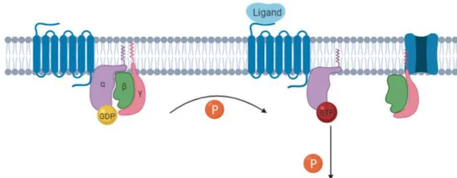
The stimulation of GPCRs is responsible for the activation of the attached G-protein. When the ligand binds to the GPCR, a conformational change occurs in the G-protein. The Gα exchanges the previously bound GDP for GTP, becoming activated. Simultaneously the Gα dissociates from the dimer Gβ/γ. The now dissociated subunits can bind to another
13 Figure 1. G-Protein Coupled Receptors. Generalized schematic for GPCR activation. On ligand binding to the
GPCR, the receptor undergoes a conformational change whereby the α subunit of its associated G protein is activated by exchanging bound GDP for GTP. The α and and βγ subunits of the G protein subsequently dissociate to activate their respective downstream signaling cascades. Adapted from Belmonte et al. 2011.
membrane protein and regulate its’ function, this type of regulation being more common with the Gα protein.
While it remains active, the G-protein will keep relaying signal unless this signal is suppressed by the inactivation of said protein. This inactivation occurs when GTP is hydrolysed to GDP, which can occur over time on its’ own because G-proteins are designed with a GTPase activity that allows for that biochemical process. The GTPase activity works as a regulation mechanism, being activated when the GPCR is no longer in contact to its’ ligand (2).
There exist other methods of G-protein regulation, besides the GTPase pathway, that are not mediated by the G-protein itself. Ric-8 proteins, originally identified in C. elegans, is one example, regulating protein at different levels. Another cellular mechanism of G-protein regulation that can accelerate their deactivation through inhibition are GPR (G-G-protein regulator) proteins, domains of approximately 25 amino acid segments. However, some studies show that GPR can function as a promoter for G-protein signalling by sequestering the Gα subunit and allowing the Gβ/γ signalling, or by allowing Ric-8 to prolong said signalling. GBA motif-containing proteins are capable of regulation the duration and extent of the signal. Finally, the family of RGS (Regulators of G-protein Signalling) are important in the regulation of GTP to GDP dephosphorilation and re-association of G-protein subunits, and are theorised to function as GAP for Gα and promoting their conformational change back to the form that is connected to the other subunits (4).
1.3.GPR37
GPR37, as indicated by the name, is a G-protein coupled receptor, that has gained attention by the scientific community due to its’ correlation with Parkinson’s disease.
When speaking of GPR37, we should not neglect to mention GPR37L1, a receptor that shares a high homology with GPR37.
As stated before, both these receptors remain orphan receptors.
1.3.1. Discovery
GPR37 belongs to the rhodopsin-type GPCR family, also known as Class A. It was first discovered in 1997 through a screening of human hippocampus tissue cDNA library. The
main identifying characteristics were its’ high homology with endothelin B receptor (ETB), respectively 52% similarity and 26.7% identity, and an unusually long N-terminus, containing 234 amino acids and four potential glycosylation sites. A high homology (approximately 40%) to bombesin receptors 1 and 2 was also reported (5,6).
The receptor was named endothelin B receptor-like protein (hET(B)R-LP) due to its’ ETB homology (6).
GPR37 was mapped to chromosome 7q31, and it includes two exons with an intron in between (5).
Furthermore, GPR37 was shown to undergo post-translational processing, the N-terminal being susceptible to metalloproteins proteolytic cleavage, leading to shedding of the
14 N-terminus afterwards and to be predominantly expressed at the plasma membrane in its’ cleaved form (7).
GPR37L1 was discovered one year later. A screening for ETB receptor homologs confirmed the discovery of GPR37 and also yielded a new finding. A transcript highly related to GPR37 was identified and named ET(B)R-LP-2. This receptor showed a 68% similarity and 48% identity to GPR37, but only had possible glycosylation site in the N-terminus.
The existence of both these receptors was also shown in rats, showing and homology of 42% between them and high similarity to the human receptors (2,8).
1.3.2. Expression Patterns
GPR37 theorised physiological role is based on its’ distinct expression. It is almost exclusively expressed in the brain, and only some residual expression has been found in other tissues by some studies, for example in the thyroid gland, oesophagus, liver, tesits, prostate, placenta and smooth muscle (2,3).
Expression has been identified predominantly in neurons, Purkinje cells of the cerebellum, pyramidal cells of the hippocampus and granule cells of the dentate gyrus, but also glial cells, even if not as substantially.
The analysis of GPR37 expression was done through Northern Blot technique in most of the studies I consulted.
In contrast, GPR37L1 expression has only been reported in glial cells and has shown no relevant expression in peripheral tissues until recent studies highlighted some level of expression in human heart explants and the gastrointestinal system, through qPCR or Western Blot (2).
1.3.3. Physiological role Substrate of parkin
Parkin is an E3 ubiquitin-protein ligase playing a critical role in ubiquitination of proteins on the outer membrane of the mitochondria and clearing damage mitochondria through autophagy (9). In 1998 this protein was connected to the onset of Autosomal Recessive Juvenile Parkinson, presenting high expression in the substantia nigra, and was named Parkin according to this connection (10,11). A mutation that leads to the malfunction of parkin has been linked to the accumulation of misfolded substrate proteins in the endoplasmic reticulum, causing a cellular stress and resulting in subsequent cell death in the substantia nigra that is responsible for the onset of disease (12). This highlights parkin’s role in suppressing unfolded protein stress-induced cell death via its’ ubiquitin activity and its’ importance in the physiopathology of early onset Parkinson.
GPR37 alternative name, Pael-R, was given when it was identified has a substrate of parkin. Pael-R showed a distribution concordant with AR-JV dopaminergic neuron destruction, respectively in the corpus callosum and the substantia nigra (11).
Since then, and due to the now established link between the receptor and AR-JV, the interest in GPR37 increased significantly.
Similarly to other parkin substrates in the absence of its’ ubiquitination activity, GPR37 accumulates in a misfolded form. Under normal circunstances it would be bound to its’ chaperone Hsc/Hsp70, that helps with refolding. However, during cellular stress, a protein named CHIP (carboxyl terminus of Hsc70-interacting protein) is upregulated promoting the
15 dissociation of GPR37 and its’ chaperone. Therefore misfolded GPR37 accumulates, triggering neuronal death (2).
1.3.4. Pharmacology and Biochemistry
Possible ligands: Receptor of prosaposin and prosaptide?
Several efforts have been made to try and deorphanize these receptors.
A decapeptide firstly identified in the organism Hydra, Head Activator (HA) is a neuropeptide that exhibits mitogen and neuroprotective activity and that was shown to mediated receptor internalization and signal transduction through ERK stimulation, increasing Ca2+ mobilisation in cells containing GPR37 (4). Further studies supported these findings and also showed cAMP response to HA stimulation (13).
The proposition of an mammalian ortholog of HA as endogenous ligand for GPR37 has been mainly discredit in following years (2).
Some reports of a homolog mammalian neuropeptide of HA were presented (14,15), but the claim was never proved and, years latter, other studies amounted as evidence to the inexistence of HA or an homolog on the human genome (16). Besides this, other studies also disputed the effect of HA on GPR37 internalization, ERK1/2 phosphorylation or cAMP stimulation (17).
Two other theorised ligands have been proposed and met with controversy (2). Prosaposin (PSPA) is a multifunctional protein, first identified as precursor of saposins, proteins activators of lysosomes, that facilitate lysosomal hydrolysation of sphingolipids (18). PSPA also acts as a neurotrophic factor, stimulating neural growth and differentiation (19) and protection against cellular stress. Prosaptide is prosaposin’s active peptide fragment, TX14A as its’ synthetic analogue.
Both these peptides bind to the receptors and mediate receptor internalization and signalling through ERK phosphorylation. It was theorised that the neuroprotection offered by both prosaposin and prosaptide was mediated by their binding to GPR37 and GPR37L1, an idea supported by the overlapping distribution of these receptors and the patterns of putative prosaposin receptors (20). In these studies, it was shown that GPR37 and GPR37L1 were Gαi-coupled receptors (2).
This claim was again met with some controversy. The concentration of agonist needed to evoke a response being considerably higher than usual concentrations for GPCR activation (21); Gαi/o-mediated agonism led to a augment of signal not considerably higher when compared to baseline, even more discredited when compared with positive controls (22); TX14A not surging as a hit for our receptors in other GPCR screenings, were points against the claim that prosaposin and prosaptide are GPR37 and GPR37L1 endogenous ligands. It remains under debate whether there’s validity to the claim.
1.3.5. Cell Expression
The major obstacles in studying GPR37 function is its’ poor cell surface expression and cytotoxicity (6), which difficulties assays’ performance as most receptors must reach the plasma membrane to have proper activity.
Based on the example of other GPCRs with deficient surface expression, it was discovered that a truncation of the receptor N-terminal could improve trafficking to the plasma membrane and so increase surface expression while decreasing cell death. Likewise,
16 co-expression with other receptors such as Dopamine D2 receptor and adenosine A2A receptor, and interaction with PDZ scaffold syntenin-1 increase GPR37 surface expression.
It should be mentioned that the apparent interaction between GPR37 and the dopamine receptor could be of pharmaceutical interest due to the possibility of targeted therapy of D2R antagonists (used to treat schizophrenia) to neurons that express this dimer instead of D2R alone (23).
Recent studies showed that the deficient cell surface expression was due to a 6-Cysteine motif in the C-terminus that caused intracellular retention (13).
1.3.6. GPR37 Neurotoxicity and ARJV’s
Autosomal Recessive Juvenile Parkinson (ARJV’s) is a form of parkinsonism, distinguished by an onset before age 40, slow progression, occasional dystonia and long-lasting response to low doses of L-dopa. It has a hereditary character, and has been found to be mainly related to mutations in the gene that encodes parkin (24,25).
The connection between GPR37 and ARJV’s is established through parkin, as explained previously. In cases of parkin mutation derivate ARJV, an accumulation of misfolded GPR37 is observed, along with other proteins, leading to neuronal cell death. GPR37, specifically has been linked to dopamine (DA) neuron loss (26), although it’s also associated to non-dopamine neuron death. Some studies have revealed that GPR37 knockout in mice will alter dopamine signalling, and significantly decreased striatal DA and elevate dopamine transporter (DAT) activity due to increased membrane expression and trafficking which was shown to be due to loss of intracellular retention by GPR37 binding (27). However, GPR37 knockout mice were also shown to be resistant to DA neurodegeneration and a PD-like phenotype in a model of PD neurotoxicity (28). Furthermore, activation of GPR37 with prosaposin was shown to protect DA neurons (29).
It is unknown how overexpression and activation of the same gene have opposite effects, but it is believed to be related to GPR37 folding. Also, it is not yet explained how the deletion of this gene also leads to neuroprotection.
The several complex mechanisms of neurotoxicity and neuroprotection that surround GPR37 make it very alluring for neuronal disease studies, mainly ARJV’s. The relation of GPR37 to this disease appears to be mainly due to its’ accumulation in the misfolded form, when there is a disfunction in parkin.
It is possible that GPR37 role is not exclusive to AR-JV. Studies have found GPR37 to be part of Lewy Bodies’ core structure, indicating they may be involved in their formation. Lewy Bodies are a hallmark cytologic evidence of idiopathic Parkinson’s Disease, although their exact role in the pathology has yet to be determined (30).
This is where the interest in GPR37 peeks for it presents as an appealing target for anti-parkinsonism therapy.
1.3.7. GPR37 and other pathologies
Some studies have reported GPR37 involvement in other neurological pathologies, such as depression major and bipolar disorder (31). However, these findings have been controversial, with more recent studies presenting contradictory results (32). Similarly, GPR37 role in non-motor symptoms in PD, such as gastrointestinal involvement, memory, sleep, etc, has been widely debated. The inconsistency of the results indicates that further investigation is still necessary to reach a definite conclusion. However, it is possible GPR37 is related to anxiety symptoms (2).
Two mutations in the gene for GPR37 were related to Autism Spectrum Disorder in one study (33), but further investigation is necessary to confirm this connection.
17 Other associations that have been made with this receptor were, for example, with cancer. Specifically, a study indicated that under-expression of GPR37 had a potential relation with the severity of malignancy of Hepatocellular Carcinoma (HCC), and that under-expression of GPR37 was closely related to proliferation of HCC. As a result, low expression of GPR37 was correlated with a poor prognosis (34). This presents a possible new biomarker for this tumour.
1.4.MRGPRX3
1.4.1. Purinergic Receptors
Purinergic receptors are, as the name indicates, receptors that are activated by purines or pyrimidines. Two categories were originally described: receptors that bind to adenosine, P1; and receptors that bind to ATP, P2. P2 receptors are divided in P2X, ionotropic receptors, and P2Y, G-protein coupled receptors. Structure wise, they are not related, sharing only a ligand.
P2Y receptors
Purinergic P2Y receptors are a family of G-protein coupled receptors whose ligands are extracellular nucleotides, such as adenine and uridine nucleotides, dinucleotide polyphosphates and nucleotide sugars. The family is composed of eight mammalian subtypes: P2Y1, P2Y2, PY4, PY11, PY12, PY13 and PY14. Their expression is not, however, exclusive to humans, they are also found in other animal species, which shows evidence of an ancient origin (35).
Due to the considerable extent of this family of receptors, it is not surprising they play a role in several important signalling pathways. Neural signal transmission through P21 receptors (36) and control migration of microglia by P2Y12 receptors modulated by adenine nucleotide; UDP mediated microglial phagocytosis by P2Y6 receptors are both evidence of the strong importance of these receptors in the nervous system (35,37) Other physiological pathways mediated by purinergic P2Y receptors are: modulation of cardiomyocyte glucose transport, vascular tone and vascular smooth muscle proliferation, platelet aggregation, immunocyte differentiation, etc. The vast array of signal pathways they mediate makes them very good targets for pharmaceuticals, as is the case with drugs like clopidogrel, that block platelet aggregation through the blockage of P2Y12 receptors, and are used in cardiovascular events therapeutics and profilaxy (35). Their pharmacological interest has, in recent studies, extended for the Alzheimer’s disease, a possible role of P2Y2 in its’ pathology having been revealed (38).
Similarities between some of the receptors allow them to be group in two smaller subfamilies. P2Y1, 2, 4, 6 and 11 receptors form the first, a subfamily of receptors with Gq-protein coupling responding to the stimulation of phospholipase C and the subsequent increase in inositol phosphates and mobilisation of Ca2+. The receptors P212, 13 and 14 have Gi-protein coupling and lead to inhibition of adenylate cyclase activity or ion channel activity (35).
18 1.4.2. MRGPR Family
The MAS-related G protein-coupled receptors (MRGPR) were discovered approximately 20 years ago through the screening of cDNA libraries, using probes for Mas gene itself, and revealing a gene that shared a 35% homology with the Mas gene. A few years later the same gene was identified through a second screening, that was able to restrict the expression of these genes to the Dorsal Root Sensory ganglia. They were called sensory neuron–specific G protein-coupled receptors (SNSR) because they showed expression only in sensory neurons. Sensory neurons are speculated to be involved in nociceptive transmission in both acute and chronic pain. The localization of these genes therefore indicates that they might have a role in the pain pathway.
Curiously, most MRGPRs are unrelated to the Mas gene, that is part of the reninangiotensin system (RAS).
The nomenclature of Mas-related G protein receptors was unanimously adopted later due to their high homology to the MAS1 oncogene. Currently MRGPR family is composed by approximately 50 members (figure 2) in mouse, rat, human, macaque, and rhesus monkey. There are several subsets of this family: MRGPRA, MRGPRB, MRGPRC, MRGPRD, MRGPRE, MRGPRF, MRGPRH and MRGPRX. Subfamilies A, B, C, and H exist only in rodents, whereas subfamily X’s expression is exclusive to primates. Subfamilies D to G are expressed in different mammalian species, including rodents and primates.
All genes of the family other than Mas, Mas1L, and MRGPRH are located on chromosome 11 in human, chromosome 1 and rat, and chromosome 7 in mouse. In rats, the genes for Mas and MRGPRH are also on chromosome 1, but in mouse these two genes are on chromosome 17, and in humans they are situated on chromosome 6 (39).
19 Figure 2. Phylogeny of Mas-related G protein-coupled receptors consisting of nine subfamilies of MRGPRs.
Rodent-specific subfamilies (A, B, C, and H), primate-Rodent-specific subfamily (X), conserved subfamilies in mammalian species (D, E, F, and G). Adapted from Solinski et al. 2011.
No MRGPR family has met all the necessary criteria to be considered deorphanized, even if some ligands have been identified for each of the subfamilies.
In the subfamily MRGPRA, comprised of 19 genes, four receptors have been deorphanized and characterized as purinergic receptors with adenine as their endogenous ligand. It was suggested to call this family P0, to differentiate them from the two purinergic families existing (P1 and P2). In rat, the corresponding receptor was named Rat Adenine Receptor (rAdeR), in mouse two receptors were identified (mAde1R and mAde2R) and one in chinese hamster (cAdeR) (40–42).
Ligands for every member of MRGPRB family remain elusive.
The two functional MRGPRC genes, one in rat and one in mice, rMrgC and mMrgC11 have been renamed rMrgprX1 and mMrgprX1, respectively, even though they bare very low homology with MRGPRX. Several peptides were shown to activate the receptors, suggesting involvement in more than one signalling pathway. Activation of these receptors was shown to enhance pain, with signals such as thermal hyperalgesia and mechanical allodynia (43); induce itch (44); and to have analgesic effects (45). The rest of the subfamily is mostly comprised of pseudogenes.
MRGPRD has one receptor per species, with several ligands having been identified. Physiologically it is implied in itch stimuli, and, due to its’ expression in arteries, is also thought to produce relaxation of aortic rings (46).
20 MRGPRH exists only in rodents, whereas the families MRGPRE-G exist in rodents and primates. No member of these families as yet been deorphanized (39).
1.4.3. Subfamily X
The MRGPRX subfamily includes four receptors and another three that are not listed because they share a very high similarity to the ones listed, approximately 98%, most likely constituting polymorphisms and not distinct subtypes. All these genes are located on chromosome 11p15.1 in humans.
MRGPRX are highly expressed in the dorsal root ganglia, co-localized mainly to lectin IB4+ sensory neurons, which reinforces the suggestion they may be related to the pain transmission pathway.
Recently, genes from the subfamily X have also had expression reported in human Mast Cells, mimicking MRGPRA and B expression in rodents. This fact suggests that the X subfamily inherited promoter elements from ancestral MRGPRB and -A/C genes.
Furthermore, human MRGPRX2 has shown expression in adrenal glands and in several brain areas (47).
Both MRGPRX1 and MRGPRX2 have been deorphanized, their better characterized ligands being BAM peptides and cortistatin-14 peptide, respectively. The MRGPRX3 and MRGPRX4, however, remain orphan receptors, without any identified ligands (39). Our investigated receptor, the MRGPRX3, is the most enigmatic one in the MRGPRX subfamily.
1.4.4. MRGPRX3
It is expressed in the DRG, the cerebellum, the lungs and the corneal endothelial cells. Additionally, MRGPRX3 as well as MRGPRX4 were found to be expressed in the keratinocytes and the angiogenic peptide AG-30/5C (MLKLIFLHRLKRMRKRLKRKLRLWHRKRYK) was suggested to activate both receptors. However, the latter suggestion was never confirmed (48).
Additional to human expression, MRGPRX3 was identified in the rhesus monkey (rhMRGPRX), with a very high percentage (90%) of homology to its’ human counterpart. A Gq coupling was determined for rhMRGPRX3. However, due to null constitutive activity being identified for hMRGPRX3, no conclusions were drawn for the human receptor (49).
Further studies have offered correlation between sites of this gene expression and a possible physiological or pathophysiological role. Lung expression of MRGPRX3 reducing after exposure to oxidative stress suggested a possible role in pulmonary disease (50). A study conducted in transgenic rats showed that overexpression of this gene lead to the development cataracts and an abnormal skin symptoms, both related to cell hyper proliferation (48,51). Unfortunately, these isolated clues have not made possible to conclude any information relating to the role of MRGPRX3 in the human organism yet.
In the research group I was inserted in one agonist of MRGPRX3 was identified for the first time. Due to confidentiality reasons, I will refer to it as Standard Agonist X from now
21 on. This compound shared some structure similarities with nucleotides, what lead us to believe that the endogenous ligand for our receptor could be either a nucleotide or a nucleoside, similarly to what was verified in MRGPRX1 case, and possibly, it could be classified as a purinergic receptor. Therefore, the next logical step, when trying to identify the endogenous ligand of our receptor would be to test the libraries of compounds that contained purine or pyrimidine bases. We screened the xanthine library, a library of nucleosides and nucleotides we assembled and an in-house purinergic library.
22 2. Material and Methods
2.1.Experimental part
2.1.1. PCR
PCR mixture was composed of DNA Template (30ng), Pyrobest™ DNA Polymerase, Pyrobest™ Buffer II 10X, dNTP Mixture 2.5 mM, forward primer (f-NheI-pCMV :aagtcagctagcaccaccatgcagagtgtgaagacagtcccc), reverse primer (r-HindIII-pCMV : aacacgaagcttgagcaatgagttccgacagaagcaaaagtggac), DMSO, PCR-H2O. The reaction was optimized for each receptor, but it consisted of the same phases: initial denaturation at 98ºC, for 45s, afterwards denaturation, followed by annealing and finishing in extension, this sequence was repeated 25-30 times, and the reaction finishes with a final extension cycle at 72ºC for 4 minutes.
The PCR products were visualized using a 1% agarose gel in Tris buffer with GelRed® (Biotium, Fremont, USA) with the exception of mutagenesis PCR products which were used directly for transformation. Then the desired bands were excised and purified using Zymoclean™ Gel DNA Recovery Kit (ZymoResearch, California, USA).
DMSO was added to optimize the PCR (52).
2.1.2. Restriction
The PCR products and vectors (four PathhunterTM plasmid variants namely pCMVProlink1, pCMV Prolink2, pCMV Prolink2 ARMS1, and pCMV Prolink2 ARMS2) were double digested using the two restriction enzymes NheI-FD and Hind III-FD (Thermo Scientific, Waltham, MA, USA) according to standardized procedures described by the enzymes manufacturer. The amount of DNA used was 650ng.
After an incubation of 15 minutes at 37 ℃ enzymes were heat inactivated at 60℃ for 5 minutes. The DNA was cleaned using the DNA Clean & Concentrator™ Kit (ZymoResearch, California, USA).
2.1.3. Ligation
Ligation of the restricted inserts and plasmids (sticky ends) was performed with T4 DNA Ligase. DNA fragments were added in a molar ratio of vector to insert of 1:3. The ligation reaction was incubated at room temperature for one hour or at 16 ℃ overnight.
23
2.1.4. Transformation
The chemically competent Top 10 (Escherichia coli) cells were thawed on ice for 30 minutes. The ligation product of each ligation reaction was directly added to the competent cells (100 μl), then incubated for 30 minutes on ice followed by a heat-shock for 30 seconds at 42°C and returned to the ice. After 2 minutes 300 μl of SOC medium was added. The bacteria were incubated at 37 °C for 1 hour while shaking at ~300 rpm. Then each bacterial mixture was spread on LB agar plates with Kanamycin and the plates were incubated at 37°C overnight. On the next day, six clones from each plate were incubated separately overnight in 4 ml of LB medium with 50 μg ml-1 Kanamycin.
2.1.5. Plasmid preparation and sequencing
The ZR Plasmid Miniprep kit (ZymoResearch, California, USA) was used to isolate the plasmids from the overnight cultures. The yielded DNA amounts were measured using a Nanodrop spectrophotometer and then sent for sequencing to GATC (Konstanz, Germany). Then, the clone with the plasmid of the desired sequence was pre-incubated for 6 hours in 4 ml LB medium with 50 μg ml-1 Kanamycin, which was subsequently transferred to 100 ml LB medium/Kanamycin to yield highly purified plasmid DNA in a big scale using the PureLink® HiPure Plasmid Midiprep Kit (Invitrogen, California, USA).
The sequencing results of both receptors are presented in attachment.
2.1.6. LipofectamineTM 2000 transfection
The β-arrestin CHO cells were split one day before transfection (1.5x10⁶ in 25 cm2 flask) in F12 medium (10% FCS. 100 U ml-1 penicillin G, 100 μg ml-1 streptomycin, and 0.3 g L-1 Hygromycin B). On the day of transfection the medium was exchanged for the same medium without antibiotics. Then, 10 μg of DNA was incubated in a final volume of 625 μl of F12 (without supplement). Subsequently, 25 μl of LipofectamineTM 2000 was incubated in 600 μl F12 (without supplement) for 5 minutes. The lipofectamine in the medium was mixed with the medium containing the DNA and incubated for 20 minutes. The DNA lipid mixture was added to the β-arrestin CHO cells and incubated for 24 hours. Then the selection medium was added, and the cells were transferred into a 175 cm2 flask with selection medium containing 800 μg ml-1 G418. The selection medium was changed every two days for two weeks.
24
Table 2.1.6. Media Composition
Media Composition
Cell-line Medium Additives Transfected cells PathHunterTM CHO β-Arrestin GIBCO TM F-12 Nutrient Mixture (Thermofisher Scientific) 10 % FCS Fetal bovine serum (Sigma-Aldrich, F0804 100 U ml-1 Penicillin(Invivogen ) 100 μg ml-1 Streptomycin(Invivo gen) 200 μg ml-1 Hygromycin B Gold™ (Invivogen) 800 μg ml-1 G418(Invivogen)
2.1.7. β-Arrestin recruitment assays
The assay system is based in Enzyme Fragment Complementation technology. The G-protein is produced with a part of a β-galactosidase enzyme, called the Pro-Link. The investigated receptor is co-expressed in CHO cells, which express β-arrestin and the larger subunit of the β-galactosidase. Upon ligand binding and receptor activation, the β-arrestin subunit will be recruited to prevent the receptor further activation and hence the enzyme β-galactosidase is completed. After the addition of the substrate, the enhanced enzyme activity breaks it, emitting light that can be measured through luminescence reader. The higher the measurement the more efficient the binding of the β-arrestin to the G-protein.
The system for MRGPRX3 and for GPR37 was established and optimized previously. The day before the assay, 20.000 of the transfected β-arrestin CHO cells were seeded in each well of a 96-well plate in 90 μl of medium (2% Opti-MEM™ for cells expressing truncated GPR37 and for cells expressing MRGPRX3). Two hours before the assay the medium was exchanged for medium without FCS and selection antibiotics. For agonist assays 10 μl of agonist was added and incubated for 90 minutes at 37 ℃ (95% humidity, 10 % CO2) then 50 μl of the PathHunter detection reagent (DiscoverX, Birmingham, UK) was added to each well and then incubated for 60 minutes in the dark at room temperature. Then the data were analysed, and concentration-response curves were determined using GraphPad PrismTM program.
25
3. Results
3.1.
Molecular cloning of GPR37 into a plasmid suitable for mammalian
transfection
In order to establish the pharmacological assays, GPR37 was cloned natively in a plasmid that is suitable for mammalian transfection. This involved a molecular biology process, starting with PCR expansion, then restriction, ligation, transformation, and lastly, transfection. The methods were previously described.
As stated in the introduction, GPR37 surface expression is problematic, and therefore, we used previously truncated GPR37, since it was shown to enhance its’ surface expression.
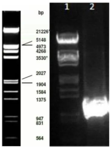
Figure 3 depicts the truncated GPR37 PCR product and the controls utilized indicating that the PCR was successful, with an accumulation of product around the 1340bp, the correspondent size of tGPR37.
Figure 4. The PCR products: The PCR product was visualized using 1% agarose gel and GelRed. 6x loading dye was added
to the PCR mixture. Amplification of the truncated GPR37 (1340 bp). The desired bands were excised and purified. Lane 1 is Phage Lambda DNA EcoRI / Hind III and lane 2 is Pyrobest product GPR37t +DMSO.
26 Figure 5. Minipreparation of the ligated plasmids. Lane 1 is the Phage Lambda DNA EcoRI / Hind III. Lane 2 is the empty
plasmid pCMV used as a control for negative clones. Lane 3 till 8 are the clones picked. Only lane 7 (clone 5) is positive with the GPR37 insert. The plasmids are visualized using 1% agarose gel with GelRedTM.
After transformation six picked clones from different parts of the plate (figure 4) were analysed by agarose gel electrophoresis. An empty plasmid was used as negative control while a plasmid recovered with an inserted was used as a positive control. Figure 4 shows the empty plasmid in lane 2, smaller, and therefore presenting a longer migration, than the plasmid with the insert would. In lanes 3 to 8 are the clones, from which only 7 is positive.
It would be preferable to have more positive clones in case some problem arouse with clone 7. For example, if it was heavily mutated during preparation. The conditions of stress the DNA is exposed to during PCR and the other methods could induce mutations. If the receptor used in the pharmacological assays is heavily mutated the results might not be viable, as the mutations presents a bias of variation.
The sequencing results are present in attachment. Our clone presented no mutations and was therefore suitable for use.
27 a. Pharmacological Assays: effects of the agonist Prosaptide on GPR37
Prosaptide was identified as a ligand for GPR37 in several published studies, although not unanimously, since it sparked some controversy when some studies found no activation of the receptor when treated with this compound. Based off the published literature, we tested prosaptide (figure 5).
The compound was tested at 1 µM in triplicates of two independent experiments. However, no increase in activity was seen compared to cells treated only with buffer. Therefore, we did not consider prosaptide as standard agonist for following experiments.
Testing of Prosaptide
prosaptide TX14(A) Buffer
0 2000 4000 6000 8000 10000 12000 L u n m in e s c e n c e (R L U )
Figure 6. Prosaptide TX14(A) response in the β-arrestin recruitment assays: The data represent the mean ± SEM from
triplicates of two independent experiments. 1 µM of prosaptide TX14(A) was tested at the tGPR37. No significant effect was observed.
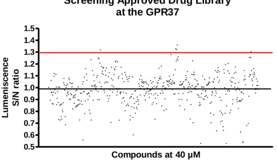
28 b. Screening of approved drug library for β-arrestin recruitment assays in GPR37 The research group possesses an approved drug library composed of several hundred compounds. We tested the entire library at 40 µM at the truncated GPR37 CHO β-arrestin cell-line in duplicates in two independent experiments resorting to β-arrestin recruitment assays (figure 6).
The S/N ratio represents the value of luminescence obtained for each compound divided by the background (value obtained in wells treated only with buffer). 1,3 was established as the cutoff for the consideration of a potential hit.
5 potential hits were identified namely, fenbendazole, carbamazepine, betaxolol-HCl, chinidine base and naphazolinde. Next step would be validating these potential hits further establishing concentrations-responses curves.
Screening Approved Drug Library
at the GPR37
0.5 0.6 0.7 0.8 0.9 1.0 1.1 1.2 1.3 1.4 1.5 Compounds at 40 µM L u m e n is c e n c e S /N ra ti oFigure 7. The β-arrestin blind screening assay: the approved drug library was screened for agonist at 40 µM at the truncated
GPR37 CHO β-arrestin cell-line. Data represents the means of duplicates from two independent expirements, The res line represents the cutoff of considering the compound as potential hit.
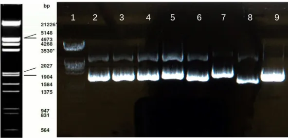
29 Figure 8. Minipreparations of the ligated plasmids. Lane 1 is the Phage Lambda DNA EcoRI / Hind III.
Lane 2 till 7 are the clones picked. Lane 8 is the empty plasmid and lane 9 contains the plasmid with and insert. Only lane 7 (clone 6) is positive with the MRGPRX3 insert. The plasmids are visualized using 1% agarose gel with GelRedTM.
3.2. Molecular cloning of MRGPRX3 into a plasmid suitable for mammalian transfection
After transformation six picked clones from different parts of the plate (figure 8) were analysed by agarose gel electrophoresis. An empty plasmid was used as negative control while a plasmid recovered with an inserted was used as a positive control. Figure 8 shows the empty plasmid in lane 8, smaller, and therefore presenting a longer migration, than the plasmid with the insert would. In lanes 2 to 7 are the clones, from which only 7 is positive.
Clone 5 was sent for sequencing and the results are present in attachment.
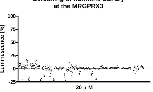
30 a. Pharmacological Assays: Screening of Xanthine Library
The compounds were tested at a concentration of 100 µM in two or three independent experiments in duplicates.
In this experiment, we considered 100% luminescence to be correspondent to the value obtained by Standard Agonist X and 0% the value of the buffer. For every compound their percentage of luminescence was calculated taking in account the value of standard agonist and buffer in their testing plate.
The testing of the Xanthine Library yielded no potential results. No compound presented a % luminescence high enough to be considered a potential hit. The highest record was around 25% of the standard agonist activation (figure 8).
Screening of Xanthine Library
at the MRGPRX3
-25 0 25 50 75 100 20 M Lumi ne s c e nc e (% )Figure 9. The β-arrestin recruitment screening assays: more than 800 Compounds were tested at final concentrations of 20
µM at β-arrestin CHO cells expressing MRGPRX3 with the Prolink2 ARMS1 variant. The data represent the means of duplicates and normalized to 100% standard agonist X (300 µM) and 0% buffer with final DMSO concentration of 1%.
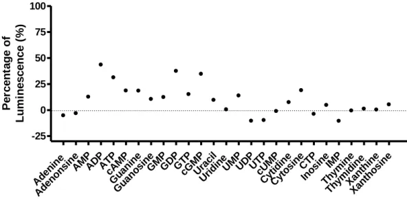
31 b. Nucleotide/Nucleoside Screening for MRGPRX3
We constructed a library containing several nucleotide and nucleoside compounds in 10mM stock solutions.
The compounds were tested at a concentration of 100 µM in two or three independent experiments in duplicates. Although none of the compounds showed >50 % of standard agonist-induced activation, some nucleosides showed an effect of 25-40 % at the MRGPRX3 (figure 9). Moreover, adenosine 5′-diphosphate (ADP) and adenosine 5'-triphosphate (ATP) showed a moderate effect at the MRGPRX3 with 44 and 31% of maximal activation, respectively. However, adenine and adenosine did not induce any signal at the receptor in the β-arrestin recruitment assays. Guanine and its nucleoside derivatives were slightly active in the MRGPRX3, with guanosine 5′-diphosphate (GDP) and cyclic guanosine monophosphate (cGMP) showing the highest percentage of activation with 37 and 35% effect, respectively. Thus, MRGPRX3 could be a purinergic receptor, however with apparently weak potency.
Screening of Nucleotides/Nucleosides Aden ine Aden onsi ne AM P AD P ATP cAM P Guani ne Gu ano sineGM P GD P GT P cGM P Urac il Urid ine UMPUDPUTP cUM P Cyt idin e Cyt osi ne CTP Inosi neIMP Thymi ne Thymi dine Xant hine Xant hosi ne -25 0 25 50 75 100 P e rc e n ta g e o f L u m in e s c e n c e ( % )
Figure 10. Nucleoids and Nucleosides tested at the MRGPRX3: The results were generated using the β-arrestin recruitment
assays. The data represent the means of duplicates generated in three independent experiments. Each compound was tested at a concentration of 1100% standard agonist X (300µM) and 0% buffer. The final DMSO concentration was 1%.
32
Table 2.2. Nucleotides and Nucleosides tested in MRGPRX3
Compounds Structure Percentage of activation
±SEM (%) at 100 µM Adenine -5 ±6 Adenosine -3 ±3 AMP 13 ±10 ADP 44 ±12 ATP 31 ±1 cAMP 19 ±6 Guanine 19 ±10 Guanosine 10 ±6 GMP 12 ±2 GDP 37 ±5
33
Table 2.2. Nucleotides and Nucleosides tested in MRGPRX3
Compounds Structure Percentage of activation
±SEM (%) at 100 µM GTP 15 ±0 cGMP 35 ±13 Uracil 10 ±5 Uridine 1 ±10 UMP 14 ±16 UDP -10 ±0 UTP -10 ±9 cUMP -1 ±5 Cytosine 19 ±10 Cytidine 7 ±10
34
Table 2.2. Nucleotides and Nucleosides tested in MRGPRX3
Compounds Structure Percentage of activation
±SEM (%) at 100 µM CTP -4 ±7 Inosine 5 ±12 IMP -11 ±9 Thymine -1 ±15 Thymidine 1 ±14 Xanthine 0 ±17 Xanthosine 5 ±5
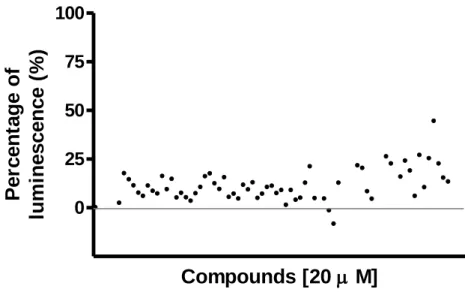
35 c. In-house Purinergic Library screening for MRGPRX3
An In-house purinergic library consisting of 70 compounds was also screened in the MRGPRX3. This library included known agonists and antagonists of different purinergic receptors, as well as compounds inhibiting important targets related to the purinergic signaling. These compounds were tested at a concentration of 20 µM. The standard agonist X was in the purinergic library and appeared to have the highest signal among all other compounds (figure 10). Compounds and detailed results are included as supplement material (attachment 2). This library is recommended to be screened for other MRGPRX receptors. ADP is also included in this library and it showed a moderate effect of 21%. Additionally, ATP is in this library too. However, it had no effect in this screening. One explanation could be that we recently established the nucleoside library and compounds were collected in solid form and stock solutions were freshly prepared before the assays. In contrast to the purinergic library, where the compounds were already stored in plate as 2 mM stock solution in DMSO hence unstable compounds like ATP were compromised.
0 25 50 75 100
Compounds [20
M]
P
e
rc
e
n
ta
g
e
o
f
lu
m
in
e
s
c
e
n
c
e
(
%
)
Figure 11. The β-arrestin recruitment assays: the purinergic library the compounds were tested at final concentrations of
100 µM using β-arrestin CHO cells expressing MRGPRX3 with the Prolink2 ARMS1 variant. The data represent the means of duplicates from two or three independent experiments and normalized to 100% standard agonist X (100 µM) and 0% buffer with final DMSO concentration of 1%.
36
4. Discussion
The challenges presented along the process of trying to deorphanize a receptor are several. Even in cases were a ligand has been identified they are, most often than not, poorly characterized pharmacologically and biochemically. Information regarding their G-protein coupling or the pathway through which they exert their function poorly understood or not known at all. Use of unspecific assays, such as β-arrestin recruitment assay, is a way of getting around this problem. But not without presenting some disadvantages: possible high backgrounds and rapid substrate catalysis resulting in very brief windows in which measurements are detectable and viable following compound addition. Furthermore, coloured compounds will interfere with the results, since they quench the light (53).
A decline in rates of receptor deorphanization resorting to traditional reverse pharmacology studies has been observed over the last few years, which also suggests a need for new approaches (54).
Regardless of the challenges, investigation surrounding the deorphanization of GPCR is essential for the therapeutic potential they present, and reverse pharmacology studies that presuppose the identification of a ligand and then screening of further compounds to determine structure-activity relations, remains the most used technique.
In my work I was faced with two distinct cases: one receptor for which I had a standard agonist, MRGPRX3, and one receptor for which I had not, GPR37. Although I used a similar experimental approach for both, the analysis of results became easier for the receptor that had an agonist, for it was possible to establish a positive control that served, in addition, to verify appropriate receptor expression in the cells I was using. In GPR37’s case, since the possible agonist suggested in literature did not produce signal in our experiments, we had no positive control. My tutor had previously resorted to immunofluorescence assays to verify receptor expression in the cells we were using, so the results could be validated. I do not present the image in my results because I did not have access to it.
For MRGPRX3 we faced some problems during the molecular biology part of the experiment. The cloning did not yield positive clones in one of our attempts and in the other, the positive clone we had was too mutated to be viable. In the third attempt we were able to produce a viable clone, the sequencing of which is presented in the attachment. The first screening we performed, we used standard agonist X at 100 µM. However, the readings got progressively lower, which lead us to believe a shift in the response curve might have occurred. In following screenings the standard agonist was used at 300 µM.
The results we obtained for MRGPRX3 were promising, but inconclusive. Screening of purinergic and nucleotide/nucleoside libraries suggested that the receptor could be purinergic. ADP, ATP, GDP and cGMP showed promising results when tested in the nucleotides/nucleosides library. These results were not reproducible in the purinergic library screening, since only ADP showed any activation, even if in smaller percentage, whereas ATP showed no increase in signal. Thus, MRGPRX3 could be a purinergic receptor, however with apparently weak potency. Realization of dosage response curves would’ve been the next step in order to verify this activation and determine minimal active concentration.
For GPR37 5 potential hits were identified. Namely, fenbendazole, carbamazepine, betaxolol-HCl, chinidine base and naphazolinde. Next step would be validating these
37 potential hits and further establishing concentrations-responses curves. Imidazole compounds were previously found to interfere with the assay system, therefore signal produced by fenbendazole could derive only from such interference. In this case conformation with another assay should be performed.
The brief amount of time I had to work in the lab did not allow me to develop further this project. Mainly for MRGPRX3, extra time to try and confirm its’ connection to purinergic receptors would be useful.
It would’ve also been interesting to try and determine the coupling of the receptors, for it could give us further information regarding their function.
5. Conclusion
Although I was not able to identify a ligand for either receptor, I do believe it shed some lights on the path that should be followed moving forward.
The importance of continuing to search for ligands and clues regarding the function of both these receptors is pronounced. GPR37 could present a very interesting therapeutic target for Parkinson’s disease, perhaps bringing a breakthrough in this area. There is, however, a big road ahead of making this viable. Regarding MRGPRX3, it remains completely enigmatic, the full possibilities of its’ deorphanization not having been uncovered yet.
38
6. References
1. Orphan receptor | definition of orphan receptor by Medical dictionary [Internet]. [citado 7 de Novembro de 2019]. Disponível em:
https://medical-dictionary.thefreedictionary.com/orphan+receptor
2. Smith NJ. Drug Discovery Opportunities at the Endothelin B Receptor-Related Orphan G Protein-Coupled Receptors, GPR37 and GPR37L1. Front Pharmacol [Internet]. 17 de Novembro de 2015 [citado 5 de Novembro de 2019];6.
Disponível em:
http://journal.frontiersin.org/Article/10.3389/fphar.2015.00275/abstract
3. Tissue expression of GPR37 - Summary - The Human Protein Atlas [Internet]. [citado 5 de Novembro de 2019]. Disponível em:
https://www.proteinatlas.org/ENSG00000170775-GPR37/tissue
4. Rezgaoui M. The neuropeptide head activator is a high-affinity ligand for the orphan G-protein-coupled receptor GPR37. Journal of Cell Science. 1 de Fevereiro de 2006;119(3):542–9.
5. Marazziti D, Golini E, Gallo A, Lombardi MS, Matteoni R, Tocchini-Valentini GP. Cloning of GPR37, a Gene Located on Chromosome 7 Encoding a Putative G-Protein-Coupled Peptide Receptor, from a Human Frontal Brain EST Library. Genomics. Outubro de 1997;45(1):68–77.
6. Zeng Z, Su K, Kyaw H, Li Y. A Novel Endothelin Receptor Type-B-like Gene Enriched in the Brain. Biochemical and Biophysical Research Communications. Abril de 1997;233(2):559–67.
7. Mattila SO, Tuusa JT, Petäjä-Repo UE. The Parkinson’s-disease-associated receptor GPR37 undergoes metalloproteinase-mediated N-terminal cleavage and ectodomain shedding. J Cell Sci. 1 de Abril de 2016;129(7):1366–77.
8. Valdenaire O, Giller T, Breu V, Ardati A, Schweizer A, Richards JG. A new family of orphan G protein-coupled receptors predominantly expressed in the brain 1. FEBS Letters. 1998;424(3):193–6.
9. Seirafi M, Kozlov G, Gehring K. Parkin structure and function. FEBS J. Junho de 2015;282(11):2076–88.
10. Kitada T, Asakawa S, Hattori N, Matsumine H, Yamamura Y, Minoshima S, et al. Mutations in the parkin gene cause autosomal recessive juvenile
parkinsonism. Nature. Abril de 1998;392(6676):605–8.
11. Imai Y, Soda M, Inoue H, Hattori N, Mizuno Y, Takahashi R. An Unfolded Putative Transmembrane Polypeptide, which Can Lead to Endoplasmic
Reticulum Stress, Is a Substrate of Parkin. Cell. Junho de 2001;105(7):891–902.
12. Imai Y, Soda M, Takahashi R. Parkin Suppresses Unfolded Protein Stress-induced Cell Death through Its E3 Ubiquitin-protein Ligase Activity. J Biol Chem. 17 de Novembro de 2000;275(46):35661–4.
39 13. Gandía J, Fernández-Dueñas V, Morató X, Caltabiano G, González-Muñiz R,
Pardo L, et al. The Parkinson’s disease-associated GPR37 receptor-mediated cytotoxicity is controlled by its intracellular cysteine-rich domain. J Neurochem. Maio de 2013;125(3):362–72.
14. Bodenmüller H, Schaller HC, Darai G. Human hypothalamus and intestine contain a hydra-neuropeptide. Neuroscience Letters. 1 de Janeiro de
1980;16(1):71–4.
15. Bodenmüller H, Schaller HC. Conserved amino acid sequence of a
neuropeptide, the head activator, from coelenterates to humans. Nature. 15 de Outubro de 1981;293(5833):579–80.
16. Davenport AP, Alexander SPH, Sharman JL, Pawson AJ, Benson HE, Monaghan AE, et al. International Union of Basic and Clinical Pharmacology. LXXXVIII. G protein-coupled receptor list: recommendations for new pairings with cognate ligands. Pharmacol Rev. Julho de 2013;65(3):967–86.
17. Southern C, Cook JM, Neetoo-Isseljee Z, Taylor DL, Kettleborough CA, Merritt A, et al. Screening β-arrestin recruitment for the identification of natural ligands for orphan G-protein-coupled receptors. J Biomol Screen. Junho de
2013;18(5):599–609.
18. Kishimoto Y, Hiraiwa M, O’Brien JS. Saposins: structure, function, distribution, and molecular genetics. J Lipid Res. Setembro de 1992;33(9):1255–67.
19. O’Brien JS, Carson GS, Seo HC, Hiraiwa M, Kishimoto Y. Identification of prosaposin as a neurotrophic factor. Proceedings of the National Academy of Sciences. 27 de Setembro de 1994;91(20):9593–6.
20. Meyer RC, Giddens MM, Schaefer SA, Hall RA. GPR37 and GPR37L1 are receptors for the neuroprotective and glioprotective factors prosaptide and prosaposin. Proc Natl Acad Sci USA. 4 de Junho de 2013;110(23):9529–34. 21. Alexander SPH, Benson HE, Faccenda E, Pawson AJ, Sharman JL, Spedding
M, et al. The Concise Guide to PHARMACOLOGY 2013/14: G protein-coupled receptors. Br J Pharmacol. Dezembro de 2013;170(8):1459–581.
22. Mistry R, Dowling MR, Challiss RAJ. [35S]GTPγS binding as an index of total G-protein and Gα-subtype-specific activation by GPCRs. Methods Mol Biol. 2011;746:263–75.
23. Dunham JH, Meyer RC, Garcia EL, Hall RA. GPR37 Surface Expression Enhancement via N-Terminal Truncation or Protein−Protein Interactions. Biochemistry. 3 de Novembro de 2009;48(43):10286–97.
24. Safadi SS, Barber KR, Shaw GS. Impact of Autosomal Recessive Juvenile Parkinson’s Disease Mutations on the Structure and Interactions of the Parkin Ubiquitin-like Domain. Biochemistry. 5 de Abril de 2011;50(13):2603–10.