The e ffe ct o f CagA status o n re spo nse to
Helicob acter p ylori
e radicatio n the rapy
in We ste rn Turke y
Departments of 1Gastroenterology and 2Infectious Disease,
Faculty of Medicine, Celal Bayar University, Manisa, Turkey M. Saruç1, G. Goksel1,
S. O zkaya1, F. Guclu1,
B. O zbakkaloglu2
and H. Yuceyar1
Abstract
If cytotoxin-associated gene A (CagA) status affects the response rates of therapy, then it may be possible to predict Helicobacter pylori
eradication rates. We aimed to evaluate the response to eradication treatment of H. pylori infection in CagA-positive and CagA-negative patients. A total of 184 patients (93 males, 91 females, mean age 42.6 ± 12.8 years) with H. pylori-positive chronic gastritis were studied. Subjects underwent a gastroscopy and biopsy specimens were taken from the gastric antrum, body, and fundus. Before the eradication therapy was given all patients were tested for CagA, TNF-a and gastrin levels. They were then prescribed lansoprazole (30 mg bid), clarithromycin (500 mg bid), and amoxicillin (1.0 mg bid) for one week. On the 8th week a second endoscopy was performed and further biopsy specimens were obtained from the same sites as in the initial endoscopy. One hundred and twenty-seven patients (69.1%) were found to be CagA positive and 57 patients (30.9%) were CagA negative. The total eradication rate was 82.6%. In the CagA-positive group this rate was 87.4%, and in the CagA-negative group it was 71.9% (P = 0.019). TNF-a levels were higher in the CagA-positive than in the CagA-negative group (P = 0.001). However, gastrin levels were not different between groups (P = 0.421). Our findings revealed that CagA-negative status might be a risk factor for failure of H. pylori
triple therapies. The CagA pathogenicity island gives a growth advan-tage to H. pylori strains and has been associated with an increase in the inflammatory response at the gastric mucosal level. These properties could make CagA-positive H. pylori strains more susceptible to antibiotics.
Co rre spo nde nce
M. Saruç Merkez Efendi Mah.
Altin Sok. Kirazlik-ince Apartment 8/5 Manisa Turkey
E-mail: msaruc@ hotmail.com
Received January 10, 2001 Accepted September 3, 2001
Ke y words
·Helicobacter pylori ·Eradication
·Cytotoxin-associated gene A ·CagA
Intro ductio n
Infection with Helicobacter pylori is a causal factor in the development of peptic ulcers (1). There is a strong association be-tween gastritis induced by H. pylori and ulcer development, and peptic ulcer disease
has been reported that CagA-bearing H. py-lori strains are more virulent and cause more severe gastroduodenal diseases (4,5) than CagA-negative strains.
Despite a multitude of clinical trials dur-ing recent years, no therapeutic regimen has clearly emerged as the optimal treatment for
H. pylori infection. The success rates of the same eradication regimen reported by differ-ent cdiffer-enters show wide variability (6). One explanation for this variability may be dif-ferent prevalence of infection with CagA-bearing strains according to geographic gion. If CagA status affects the rates of re-sponse to treatment, then it may be possible to predict eradication rates previously and choose appropriate regimens for each pa-tient according to whether the H. pylori strain is CagA positive or negative.
Thus, in the present study our objective was to evaluate the response to treatment for the eradication of H. pylori infection in CagA-positive and CagA-negative patients.
Mate rial and Me thods
The study was conducted according to good clinical practice and the Declaration of Helsinki. The study was approved by the local Ethics Committees before the enroll-ment of patients. Written informed consent was obtained from all patients.
Patients attended to routine diagnostic gastroscopy and likely to need H. pylori
eradication therapy were recruited for this study. All patients belonged to the same mid-socioeconomic class with an annual in-come of US$ 2900 ± 120. All of them were from Manisa, a city in Western Turkey. The patients were also evaluated in terms of other epidemiological features like the number of rooms in the house, living place (city or country), educational level, gender, tooth brushing, smoking and alcohol use, but there was no significant difference between CagA-positive and -negative patients (P>0.05). One hundred and eighty-four patients (93 males,
91 females, mean age 42.6 ± 12.8 years) with chronic gastritis were studied. All of the 184 patients had H. pylori infection. Anti-H. py-lori IgG (Pyloriset EIA-G, Orion Diagnostica, Espoo, Finland) was positive in all patients.
H. pylori positivity, a requirement to take part in the study, was determined by both the rapid urease test and histological examina-tion. If one of these tests yielded negative results then this particular patient was not included in the study. Patients with previous gastric surgery, known bleeding diathesis, taking oral anticoagulants, or who had been treated with bismuth compounds, proton pump inhibitors, or antibiotics known to be active against H. pylori within the previous two months, were excluded from the study. Subjects underwent an upper gastrointes-tinal endoscopy and biopsy specimens were taken from the gastric antrum, body, and fundus. Before the eradication therapy was given all patients were tested for CagA, se-rum tumor necrosis factor a (TNF-a) and gastrin levels. They were then prescribed lansoprazole (30 mg bid), clarithromycin (500 mg bid), and amoxicillin (1 g bid) for one week. During the 8th week a second endoscopy was performed and further bi-opsy specimens were obtained from the same sites as in the initial endoscopy. Compliance with medication was assessed by tablet count-ing and by direct questioncount-ing at the second endoscopy. Possible side effects of treat-ment were also assessed at these times by open and direct questioning, by the same gastroenterologist, using a standardized ques-tionnaire.
in formalin were sent to the pathology labo-ratory for histological examination. Forma-lin-fixed tissues were processed routinely, embedded in paraffin and cut into 5-µm sections. Along with the usual hematoxylin-eosin stain, all sections were also stained with toluidine blue in order to better reveal the bacteria. Slides were examined inde-pendently by two pathologists. Endoscopy was performed under sedation with 0 to 5 mg intravenous midazolam. An Olympus GIF1T-30 gastroscope was used and was thoroughly cleaned and disinfected between endo-scopies. This involved internal and external brushing using a neutral detergent, washing for 7 min in an endoscope washer (model EW-20, Olympus Optical Company Lim-ited, Tokyo, Japan) with neutral detergent, and 4-min disinfection with 2.2% glutaral-dehyde.
Anti-CagA IgG antibodies (VIVA Dia-gnostika, Cologne, Germany, by ELISA) were used to determine the CagA status of H. pylori before treatment. Serum gastrin levels were studied by radioimmunoassay (double antibody gastrin, KGAD1, Diagnostic Prod-ucts Corporation, Los Angeles, CA, USA). TNF-a levels were determined with an Amicyte kit by ELISA.
Patients with eradicated and non-eradi-cated H. pylori were compared according to their pretreatment CagA status. Fisher’s ex-act probability test and the c2 test were used
to determine the statistical significance. P<0.05 was considered to be significant.
Re sults
One hundred and twenty-seven patients (69.1%) were CagA positive and 57 (30.9%) were CagA negative. After the end of treat-ment patients were divided into two groups according to CagA status. The distribution according to age and gender was the same in both groups: 51 and 48.9% males, mean age 41.3 ± 11.2 and 43.3 ± 13.3 years in the CagA-positive and -negative groups,
respec-tively (P1 = 0.280, P2 = 0.390).
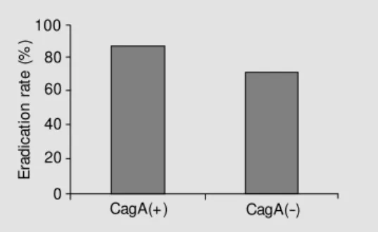
After eradication therapy, 152 patients had no infection, but 32 still continued to be infected with H. pylori. The total eradication rate was 82.6%. H. pylori was eradicated in 111 of 127 (87.4%) CagA-positive patients and in 41 of 57 (71.9%) CagA-negative pa-tients. CagA positivity seems to increase the eradication rates of H. pylori when one-week triple therapy is used. This difference was statistically significant (P = 0.019) (Fig-ure 1).
H. pylori density was greater in the positive group (2.1 ± 1.0) than in the CagA-negative group (1.1 ± 0.6) (P = 0.01). H. pylori activity and chronic inflammation also were significantly higher in the CagA-posi-tive group (1.6 ± 0.6 and 2.2 ± 0.9) than in the CagA-negative group (0.6 ± 0.3 and 1.2 ± 0.6, respectively) (P = 0.001, P = 0.002). The presence of atrophy and lymphoid aggre-gates was not different between the two groups (P>0.05). However, intestinal meta-plasia was found to be significantly more frequent in patients infected with CagA-posi-tive H. pylori strains (P<0.001).
TNF-a levels were also significantly dif-ferent between CagA-positive and -negative groups (18.33 ± 4.7 vs 9.84 ± 2.3 pg/ml, respectively; P = 0.001) before treatment (Figure 2). Although gastrin levels seemed to be higher in the CagA-positive group than in the CagA-negative group, there was no statistical significance between these values, probably because of the wide range of stan-dard deviation (56.7 ± 21.4 vs 47.5 ± 18.2 pg/ml; P = 0.421).
Figure 1. Eradication rates of the patients infected w ith cytotoxin-associated gene A (CagA)-posi-tive and -nega(CagA)-posi-tive Helicobacter pylori strains. P = 0.019 (Fisher’s exact probability test).
E
ra
d
ic
a
ti
o
n
r
a
te
(
%
)100
CagA(+) CagA(-) 80
60
40
20
D iscussio n
The gastroduodenal response to chronic
H. pylori infection is characterized by the infiltration of plasma cells, lymphocytes, neutrophils and monocytes into the mucosa (7). Eradication studies have shown that this inflammatory response represents a specific reaction to the presence of H. pylori (6,7). In addition to stimulating local T and B cell responses and a systemic antibody response,
H. pylori infection also induces a local pro-inflammatory cytokine response (8). There is now increasing evidence from both in vivo
and in vitro studies that CagA-positive strains induce an enhanced inflammatory response and mucosal damage (6-8). The presence of CagA-positive H. pylori strains also signifi-cantly delays healing of ulcers (5). However, little information is available about whether this strain variability has any effect on the eradication rates of H. pylori.
In a study to evaluate the response to eradication treatment of H. pylori infection in CagA-positive and CagA-negative patients, the efficacy of the eradication treatment with triple therapy was found not to be dependent on the presence of the CagA gene (9).
In another study on the same subject from France (10), the authors tested whether the absence of CagA, which is more com-mon in strains isolated from non-ulcer dys-peptic patients, was a risk factor for the failure of H. pylori eradication. Their results showed that the absence of H. pylori eradi-cation was positively correlated with CagA-negative status (10). This result can explain
the low efficacy of the current regimen when used in non-ulcer patients (10,11). Some recent data also indicate that eradication rates are higher for patients with CagA-positive
H. pylori strains (12,13).
Strains of H. pylori expressing the CagA protein induce gastric epithelial cells to se-crete inflammatory cytokines (14-16). Cyto-toxins like bacterial cytoCyto-toxins, TNF-a, in-terleukin-1 and interleukin-6 were released from epithelial cells (16,17). In our study we used TNF-a and gastrin levels to determine the degree of chronic inflammation, as done in previous studies (18,19). TNF-a levels correlated with CagA positivity and showed enhanced inflammation by more virulent strains. There was no such correlation be-tween gastrin levels and CagA status. The major limitation of our study was the meas-urement of antibody levels against CagA protein without the determination of the CagA protein itself. This is why we may have obtained some negative results due to some host factors that inhibit synthesis of an anti-body against this specific protein. However, some data from the literature suggest that patients infected with CagA-negative H. py-lori strains can produce anti-CagA antibody when infection with CagA-positive strains occurs (6,7).
We may speculate that treatments of longer duration may be necessary to increase the success rates of eradication treatment in patients infected with CagA-negative H. py-lori strains (12,13). Another approach may be using some different combinations like three antibiotics plus a proton pump inhibi-tor or two antibiotics and a proton pump inhibitor plus bismuth citrate. Randomized, multicenter trials are needed to evaluate and determine the most effective and safe regi-men and duration of treatregi-ment for these par-ticular patients with CagA-negative strains. One may ask if CagA-negative strains, which are less virulent and more resistant to cation therapy, should be necessarily eradi-cated, and if the indication of eradication
T
N
F
-a
(
p
g
/m
l)
30
25
20
15
10
5
0
CagA(+) CagA(-) Figure 2. Serum TNF-a levels of
treatment should be different for CagA-nega-tive and -posiCagA-nega-tive strains.
Our findings revealed that CagA-nega-tive status might be a risk factor for failure of
H. pylori triple therapies. The CagA patho-genicity island gives a growth advantage to
H. pylori strains and has been associated
with an increase in the inflammatory re-sponse at the gastric mucosal level. These properties could make CagA-positive H. py-lori strains more susceptible to antibiotics. Recommendations should be elaborated to manage these CagA-negative patients differ-ently.
Re fe re nce s
1. Blum AL (1996). Helicobacter pylori and peptic ulcer disease. Scandinavian Jour-nal of Gastroenterology, 214 (Suppl): 24-27.
2. Buckley JM & Deltenre M (1997). Therapy of Helicobacter pylori infection. Current Opinion in Gastroenterology, 13: 56-62. 3. O’M orain C, Dettmer A, Rambow A, von
Fritsch E & Fraser AG (1996). Double blind, m ult icent er, placebo-cont rolled evaluation of clarithromycin and omepra-zole for Helicobacter pylori-associated duodenal ulcer. Helicobacter, 3: 130-137. 4. M aeda S, Ogura K, Yoshida H, Kanai F, Ikenoue T, Kato N, Shiratori Y & Omata M (1998). M ajor virulence factors, VacA and CagA, are commonly positive in Helico-bacter pylori isolates in Japan. Gut, 42: 338-343.
5. Brzozow ski T, Konturek PC, Kw iecien S, Sliw ow ski Z, Pajdo R, Konturek SJ & Hahn EG (1998). Helicobacter pylori expressing CagA and VacA delays the healing of is-chemia-reperfusion injury, a new model of studying Hp-pathogenesis. Digestion, 59 (Suppl 3): 376 (Abstract).
6. Lind T, Veldhuyzen van Zanten S, Unge P, Spiller R, Bayerdorffer E, O’M orain C, Bardhan KD, Bradet t e M , Chiba N, Wrangstadh M , Cederberg C & Idstrom JP (1996). Eradication of Helicobacter py-lori using one-w eek triple therapies com-bining omeprazole w ith tw o antimicrobi-als: The M ACH I study. Helicobacter, 3: 138-144.
7. Crabtree JE (1996). Immune and inflam-matory response to Helicobacter pylori in-fection. Scandinavian Journal of Gastro-enterology, 215 (Suppl): 3-10.
8. Crabtree JE (1996). Gastric mucosal in-flammatory response to Helicobacter py-lori. Alimentary Pharmacology and Thera-peutics, 10 (Suppl 1): 29-37.
9. Laudanno O, Berli D, Ferrer J, Fay M , Linari M , Fay F & Banchio J (1998). Eradi-cation treatment of Helicobacter pylori in-fection in CagA positive and CagA nega-tive patients. Digestion, 59 (Suppl 3): 400 (Abstract).
10. Broutet N, M arais A, Liu D, Lamouliatte H, Samoyeau R & M egraud F (1998). CAg negative status: a risk factor for failure of
Helicobacter pylori triple therapies in non-ulcer dyspepsia. Gut, 43 (Suppl 2): 79 (Ab-stract).
11. van der Hulst RW, van der Ende A, Dekker FW, Ten Kate FJ, Weel JF, Keller JJ, Kruizinga SP, Dankert J & Tytgat GN (1997). Effect of Helicobacter pylori eradi-cation on gastritis in relation to CagA: a prospective 1-year follow -up study. Gas-troenterology, 113: 25-30.
12. van Doorn LJ, Schneeberger PM , Nouhan N, Plaisier AP, Quint WG & de Boer WA (2000). Importance of Helicobacter pylori
CagA and VacA status for the efficacy of antibiotic treatment. Gut, 46: 321-326. 13. Broutet N, M arais A, Lamouliatte H, de
M ascarel A, Samoyeau R, Salamon R & M egraud F (2001). CagA status and
eradi-cation treatment outcome of anti- Helico-bacter pylori triple therapies in patients w ith nonulcer dyspepsia. Journal of Clini-cal M icrobiology, 39: 1319-1322. 14. Crabtree JE & Spencer J (1996).
Immuno-logic aspects of Helicobacter pylori infec-tion and malignant transformainfec-tion of B cells. Seminars in Gastrointestinal Dis-ease, 7: 30-40.
15. Kutukculer N, Aydogdu S, Goksen D, Caglayan S & Yagevi RV (1997). Increased mucosal inflammatory cytokines in chil-dren w ith Helicobacter pylori-associated gastritis. Acta Paediatrica, 86: 928-931. 16. Pober S (1988). Cytokine-mediated
activa-tion of vascular endothelium. American Journal of Pathology, 133: 426-433. 17. Rasi V, Ikkala E & Valtonen V (1982).
Plasma beta-thromboglobulin in severe infections. Thrombosis Research, 26: 267-274.
18. Patel P, Gasbarrini G, Pretolani S, Gasbar-rini A & Franceschi F (1997). Extradiges-tive diseases and Helicobacter pylori in-fection. Current Opinion in Gastroenterol-ogy, 13: 52-55.