Ciências da Saúde
Xanthine analogues and adipose tissue
metabolism
David José Fadista Carrageta
Dissertação para obtenção do Grau de Mestre em
Ciências Biomédicas
(2º ciclo de estudos)
Orientador: Prof.ª Doutora Branca Silva (CICS-UBI)
Co-orientadores: Prof.ª Doutora Mariana Monteiro (UMIB-ICBAS-UP)
e Mestre Tânia Dias (CICS-UBI)
iii
Acknowledgments
The conclusion of this master's dissertation symbolizes not only the end of another academic stage, but also a trophy of personal fulfillment in the course that I have been drawing throughout life. Despite its individuality, I cannot but emphasize that its concretization is due to a collective. For this purpose, I wish the sincerest thanks to all those who, directly or indirectly, contributed to the development of this project and to my personal and professional growth.
To my supervisor, Professor Branca Silva, for the opportunity and for accepting me as her student. For the scientific competence, monitoring, availability, criticisms, corrections, and suggestions provided during the whole realization of this project.
To my co-supervisor, Professor Mariana Monteiro, for the wisdom, example, availability and sympathy, as well as for all the corrections and suggestions.
To Professor Pedro Oliveira for the reception in his laboratory at ICBAS-UP and financial support. For all the wisdom, support, accompaniment, availability, and good advice.
To Dr. Mietha Van der Walt and Dr. Gisella Terre'Blanche from the School of Pharmacy, North-West University, South Africa, for the collaboration and for synthesizing and providing the compound, which in its absence, this project would not be realized.
To Professor Marco Alves for all the wisdom, availability, good advice, and suggestions given throughout the project.
To Tânia, my co-supervisor and "mother" in the laboratory, for all the sympathy, friendship, good disposition, motivation, concern, and patience. For all the knowledge transmitted, good advice, support and for being always present.
To all my laboratory colleagues: Raquel, Susana, Ana, and Maria João for receiving me so well; to Bernardo, Hugo, Luís, Tatiana, and Rute for the good disposition and moments that we shared in our room down the hall. To all for the scientific and laboratory support, for the friendship and good work environment. When we work on what we like with who we like, we do not work a single day of our lives.
To Dr. Sofia, Madalena, and the entire Anatomy Department of ICBAS-UP for all the support and availability. To Dr. Ivana for the acquisition of the NMR spectra and for all the scientific support. To Fernanda for the support with the coloring techniques and photographs. To all the great friends I made in Covilhã. Even with distance, friendship still prevails. To Carlos and Pedro, my best friends since childhood, for being always present. For all the good memories, support, and advice.
To Taís for all the love, affection, support, and patience. For all the good memories that we have and will have, without you my life would not be the same.
To my parents, Luísa and Fernando, and my sister, Daniela, who are always present, encourage me to fight for my goals and support me in all my decisions. For good advice and for helping me to be the person I am today.
v
Resumo
A obesidade tem aumentado nas últimas décadas e é um dos problemas de saúde mais preocupantes a nível mundial. As metilxantinas, como é o caso da cafeína (1,3,7-trimetilxantina), teofilina (1,3-dimetilxantina) e teobromina (3,7-dimetilxantina), demonstraram propriedades anti-obesogénicas bastante promissoras. Estes compostos naturais encontram-se bastante difundidos na dieta humana, nomeadamente no café, no chá e no chocolate. De entre estes fitoquímicos, a cafeína é descrita como moduladora do metabolismo da glucose e ácidos gordos, assim como o seu consumo aparenta ser inversamente proporcional ao aumento de peso corporal. Com base na estrutura química das metilxantinas, vários compostos análogos têm sido sintetizados. Foi colocada a hipótese de que um desses compostos, a 8-(3-fenilpropil)-1,3,7-trietilxantina, poderia apresentar um potencial anti-obesogénico bastante elevado. Deste modo, o nosso projeto visou avaliar os efeitos desta nova molécula no perfil metabólico e oxidativo de adipócitos, com o objetivo de avaliar o seu potencial como opção farmacológica para o tratamento da obesidade e suas complicações. Para este fim, foi utilizada como modelo in vitro a linha celular de pré-adipócitos de rato 3T3-L1 e a cafeína, de origem sintética, para fins comparativos. As células foram incubadas na presença de concentrações crescentes de cafeína e 8-(3-fenilpropil)-1,3,7-trietilxantina (0.1, 1, 10 e 100 µM) e o perfil citotóxico de ambos os compostos foi avaliado espectrofotometricamente pela redução do sal de tetrazólio (MTT) e quantificação de lactato desidrogenase (LDH) libertada. Os metabolitos presentes no meio de cultura foram identificados e quantificados recorrendo à ressonância magnética nuclear de protão (1H-NMR)
e as células foram recolhidas para a caracterização do perfil oxidativo. Os nossos resultados demonstram que a 8-(3-fenilpropil)-1,3,7-trietilxantina não induziu citotoxicidade em nenhuma das concentrações estudadas. Comparativamente à cafeína, este composto aumentou significativamente o consumo de glucose, piruvato e glutamina, assim como a produção de lactato, alanina e acetato. Estes resultados ilustram o elevado potencial da 8-(3-fenilpropil)-1,3,7-trietilxantina como modulador metabólico, mesmo quando comparado com a cafeína. Adicionalmente, a 8-(3-fenilpropil)-1,3,7-trietilxantina promoveu um efeito antioxidante, diminuindo os níveis de oxidação proteica e protegendo contra os danos causados pelo stress oxidativo. Em suma, a 8-(3-fenilpropil)-1,3,7-trietilxantina apresenta-se como um ótimo candidato para o design de fármacos anti-obesidade seguros e inovadores.
Palavras-chave
8-(3-fenilpropil)-1,3,7-trietilxantina; Cafeína; Fármacos anti-obesidade; Metabolismo de adipócitos; Obesidade; Xantina.
vii
Resumo Alargado
O tecido adiposo é um órgão metabolicamente ativo que atua como o principal repositório de energia do corpo humano, sob o formato de gordura (triacilgliceróis). Este órgão complexo e dinâmico atua como um tampão energético, armazenando e libertando energia consoante as necessidades do organismo. Sempre que existir um excesso energético, o tecido adiposo irá armazenar esta energia sob a forma de moléculas de triacilgliceróis, dentro das gotas lipídicas da sua célula predominante, o adipócito. Este processo é denominado lipogénese. Em contrapartida, sempre que existir um défice de energia, o adipócito irá proceder à lipólise, ou seja, à hidrólise de moléculas de triacilgliceróis em glicerol e ácidos gordos livres, que são transportados e oxidados de forma a produzir grandes quantidades do nucleótido adenosina trifosfato (ATP).
A obesidade tem aumentado nas últimas décadas e é um dos problemas de saúde mais preocupantes a nível mundial. Estima-se que em 2015 mais de 2.2 biliões de indivíduos apresentavam peso excessivo (índice de massa corporal (IMC) > 25 kg/m2), dos quais 710
milhões eram obesos (IMC > 30 kg/m2). Esta condição é definida pela acumulação excessiva de
gordura no tecido adiposo e é geralmente atribuída a maus hábitos alimentares e a sedentarismo, contudo pode ser igualmente atribuída a fatores genéticos, epigenéticos, fisiológicos e ambientais. A acumulação de massa adiposa é acompanhada pela hipertrofia e hiperplasia de adipócitos, ou seja, um crescimento em tamanho e número. Este constitui um mecanismo adaptativo ao excesso de gordura, protegendo o organismo de lipotoxicidade. No entanto, a quantidade de gordura que o tecido adiposo consegue armazenar é limitada e, quando ultrapassada, poderá levar a complicações metabólicas, como a resistência à insulina e, consequentemente, diabetes mellitus tipo 2, assim como a disfunções cardiovasculares e infertilidade. Deste modo, é crucial prevenir e tratar a obesidade. Atualmente, os pilares do tratamento da obesidade assentam na restrição calórica da dieta associada ao aumento da atividade físico. Contudo, este é um processo lento e difícil, pelo que existe uma crescente procura por opções farmacológicas seguras e eficazes que possam agilizar e simplificar o processo.
O estudo de compostos naturais com potencial anti-obesogénico tem suscitado interesse entre a comunidade científica. De entre estes, as metilxantinas apresentam um potencial anti-obesogénico particularmente elevado. As metilxantinas mais estudadas e conhecidas são a cafeína (1,3,7-trimetilxantina), a teofilina (1,3-dimetilxantina) e a teobromina (3,7-dimetilxantina), muito presentes na dieta através do café, chá e chocolate. Estes compostos têm a capacidade de modular o metabolismo da glucose e ácidos gordos, promovendo a lipólise e inibindo a diferenciação de adipócitos, ou adipogénese, assim como o seu consumo aparenta ser inversamente proporcional ao ganho de peso. Existem vários mecanismos sugeridos para explicar estes efeitos, sendo que o predominante é o antagonismo
viii
de recetores de adenosina. Ao bloquear a cascata de sinalização da adenosina nos adipócitos, as metilxantinas induzem um sinal lipolítico, levando à hidrólise de triacilgliceróis em glicerol e ácidos gordos livres.
Devido às suas propriedades e efeitos benéficos para a saúde, a estrutura química das xantinas naturais tem sido utilizada como modelo para a síntese de compostos inovadores com novas características. Um desses compostos, a 8-(3-fenilpropil)-1,3,7-trietilxantina, foi sintetizado pela equipa de Van der Walt e Terre’Blanche (School of Pharmacy, North-West University, South Africa). Este composto apresenta uma alta afinidade por recetores de adenosina, característica esta que poderá estar associada a um potencial anti-obesogénico elevado. Deste modo, este projeto visou caracterizar a modulação conferida por esta nova molécula no perfil metabólico e oxidativo dos adipócitos, com o objetivo de avaliar o seu potencial como opção farmacológica para o tratamento da obesidade e suas complicações. Para este fim, utilizou-se como modelo in vitro a linha celular de pré-adipócitos de rato 3T3-L1 e a cafeína, de origem sintética, para fins comparativos. As células foram cultivadas na presença de doses crescentes de cafeína ou 8-(3-fenilpropil)-1,3,7-trietilxantina (0.1, 1, 10 e 100 µM) e o perfil citotóxico de ambos os compostos foi avaliado espectrofotometricamente pelos ensaios da redução do sal de tetrazólio (MTT) e quantificação da lactato desidrogenase (LDH) libertada. Os metabolitos presentes no meio de cultura (lactato, alanina, acetato, piruvato, glutamato e glucose) foram identificados e quantificados recorrendo à ressonância magnética nuclear de protão (1H-NMR) e as células foram recolhidas para a caracterização do
perfil oxidativo, através da análise da oxidação e nitração proteica, assim como da peroxidação lipídica.
Os nossos resultados demonstraram que a 8-(3-fenilpropil)-1,3,7-trietilxantina não induz citotoxicidade nas concentrações estudadas, ao passo que a cafeína induziu na concentração mais elevada (100 µM). Adicionalmente, a 8-(3-fenilpropil)-1,3,7-trietilxantina aumentou significativamente o consumo de glucose, piruvato e glutamina, assim como a produção de lactato, alanina e acetato comparativamente aos grupos expostos à mesma concentração de cafeína. Estes resultados sugerem que a 8-(3-fenilpropil)-1,3,7-trietilxantina tem uma capacidade superior à da cafeína para modular o metabolismo celular para produção de energia. Adicionalmente, a 8-(3-fenilpropil)-1,3,7-trietilxantina reduziu a oxidação proteica, revelando propriedades antioxidantes e um possível papel protetor contra o stress oxidativo. Por outro lado, a cafeína exibiu comportamentos pro-oxidantes acompanhados pelo aumento da oxidação proteica.
Em suma, o composto 8-(3-fenilpropil)-1,3,7-trietilxantina aparenta ser um ótimo candidato para o design de fármacos anti-obesidade seguros e inovadores.
x
Abstract
Obesity has been increasing in the last decades and is one of the most prolific health concern worldwide. Methylxanthines, such as caffeine (1,3,7-trimethylxanthine), theophylline (1,3-dimethylxanthine), and theobromine (3,7-dimethylxanthine), have demonstrated potential anti-obesity properties. These natural compounds are widely distributed in the human diet, especially food products such as coffee, tea, and chocolate. In fact, caffeine is known to modulate glucose and fatty acid metabolism and its consumption seems to be inversely associated with body weight increase. Based on methylxanthines chemical structure, several xanthine analogues have been synthetized. We hypothesized that one of those compounds, 8-(3-phenylpropyl)-1,3,7-triethylxanthine, may have a promising anti-obesity potential. Our study aims to characterize the modulation conferred by 8-(3-phenylpropyl)-1,3,7-triethylxanthine in the metabolic and oxidative profile of adipocytes in order to evaluate its potential as a pharmacological option to address obesity and its complications. For this purpose, the anti-obesogenic potential of 8-(3-phenylpropyl)-1,3,7-triethylxanthine was evaluated in mouse preadipocyte cell line 3T3-L1, using synthetic caffeine for comparative purposes. Cells were cultured in the presence of increasing concentrations of caffeine or 8-(3-phenylpropyl)-1,3,7-triethylxanthine (0.1, 1, 10 and 100 µM) and the cytotoxic profile was accessed spectrophotometrically by reduction of tetrazolium salt (MTT) and quantification of released lactate dehydrogenase (LDH). The metabolites in culture medium were identified and quantified by proton nuclear magnetic resonance (1H-NMR) and
cells were collected for analysis of the oxidative profile. Our results show that 8-(3-phenylpropyl)-1,3,7-triethylxanthine presented no cytotoxicity at all studied concentrations. When compared with caffeine, 8-(3-phenylpropyl)-1,3,7-triethylxanthine significantly increased glucose, pyruvate, and glutamine consumption, and lactate, alanine, and acetate production. These findings illustrate that 8-(3-phenylpropyl)-1,3,7-triethylxanthine has a high potential to act as a metabolic modulator, even when compared with caffeine. Additionally, 8-(3-phenylpropyl)-1,3,7-triethylxanthine promoted an antioxidant environment, decreasing protein oxidation, and protecting against oxidative stress-induced damage. Thus, 8-(3-phenylpropyl)-1,3,7-triethylxanthine appears as a promising candidate for new and safe anti-obesity drug design.
Keywords
8-(3-phenylpropyl)-1,3,7-triethylxanthine; Adipocyte metabolism; Anti-obesity drug; Caffeine; Obesity; Xanthine.
xii
Table of contents
I. Introduction ... 1
1.1. The human adipose tissue ... 2
1.2. Obesity... 4
1.3. Methylxanthines ... 7
1.3.1. Chemical structure, biosynthesis, properties, and natural sources... 7
1.3.2. Anti-obesity potential ... 9
1.3.3. Lipolytic activity ... 11
1.3.3.1. Adenosine receptors antagonism ... 11
1.3.3.2. Modulation of catecholamines’ release ... 13
1.3.4. Anti-adipogenic activity ... 15
II. Objectives ... 17
III. Materials and Methods ... 19
3.1. Xanthine analogues synthesis ... 20
3.2. Mouse preadipocyte cell line 3T3-L1 culture ... 20
3.2.1. Adipocyte differentiation... 21
3.2.2. Oil Red O lipid staining ... 22
3.3. Experimental groups ... 22
3.4. Characterization of the cytotoxic profile ... 23
3.4.1. Characterization of the cytotoxic profile through the MTT assay ... 23
3.4.2. Characterization of the cytotoxic profile through the LDH assay ... 24
3.5. Characterization of the metabolic profile ... 24
xiii
3.6.1. Total protein extraction and quantification ... 25
3.6.2. Protein oxidation analysis ... 25
3.6.3. Lipid peroxidation analysis ... 26
3.6.4. Tyrosine residues nitration analysis ... 27
3.7. Statistical analysis ... 27
IV. Results ... 28
4.1. 3T3-L1 preadipocytes differentiation ... 29
4.2. ETX presents no cytotoxic effects in 3T3-L1 preadipocytes ... 31
4.3. ETX enhances metabolism towards energy expenditure in 3T3-L1 preadipocytes ... 33
4.3.1. ETX increases glucose, pyruvate, and glutamine consumption ... 33
4.3.2. ETX increases lactate, acetate, and alanine production ... 35
4.5. ETX decreases protein oxidation in 3T3-L1 preadipocytes ... 36
V. Discussion ... 39
VI. Conclusions ... 43
xv
List of figures
Figure 1 – Anatomy of the adipose tissue and its composition in humans ... 2
Figure 2 – White adipose tissue as an energetic buffer ... 4
Figure 3 – White adipose tissue expansion during obesity scenarios ... 5
Figure 4 – Evolution from non-obese to severe obesity with ectopic lipid deposition ... 6
Figure 5 - Chemical structures of the three predominant methylxanthines (caffeine, theobromine and theophylline) and paraxanthine, the major dimethylated metabolite of caffeine ... 8
Figure 6 - Schematic illustration of methylxanthines’ activity in adipocytes ... 13
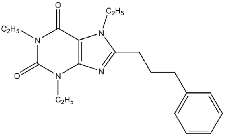
Figure 7 – Chemical structure of the new synthetic xanthine (8-(3-phenylpropyl)-1,3,7-triethylxanthine) ... 20
Figure 8 - Schematic illustration of the 3T3-L1 preadipocytes differentiation into adipocytes. ... 21
Figure 9 – 3T3-L1 preadipocytes differentiation attempt ... 30
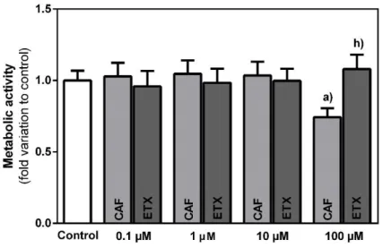
Figure 10 – Evaluation of the metabolic activity of 3T3-L1 preadipocytes after 24 h incubation with increasing concentrations of caffeine (CAF) or 8-(3-phenylpropyl)-1,3,7-triethylxanthine (ETX) as determined by the MTT assay ... 31
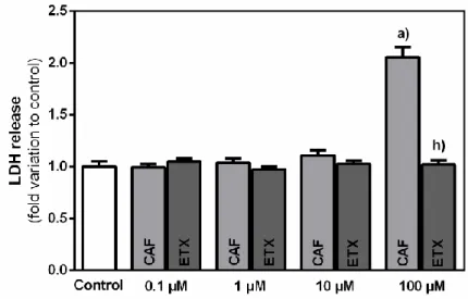
Figure 11 – Released lactate dehydrogenase (LDH) by 3T3-L1 preadipocytes after 24 h incubation with increasing concentrations of caffeine (CAF) or 8-(3-phenylpropyl)-1,3,7-triethylxanthine (ETX) ... 32
Figure 12 – Consumption of glucose (A), pyruvate (B), and glutamine (C) in 3T3-L1 preadipocytes after 24 h incubation with increasing concentrations of caffeine (CAF) or 8-(3-phenylpropyl)-1,3,7-triethylxanthine (ETX) ... 34
Figure 13 – Production of lactate (A), alanine (B), and acetate (C) in 3T3-L1 preadipocytes after 24 h incubation with increasing concentrations of caffeine (CAF) or 8-(3-phenylpropyl)-1,3,7-triethylxanthine (ETX) ... 35
Figure 14 – Characterization of the oxidative profile in 3T3-L1 cells after 24 h incubation with increasing concentrations of caffeine (CAF) or 8-(3-phenylpropyl)-1,3,7-triethylxanthine (ETX) ... 37
xvii
List of abbreviations
13C-NMR Carbon-13 nuclear magnetic resonance 1H-NMR Proton nuclear magnetic resonance
4-HNE 4-hydroxynonenal
5’AMP 5’ adenosine monophosphate
AC Adenylyl cyclase
ACC Acetyl-CoA carboxylase
ACTH Adrenocorticotropic hormone
ADSCs Adipose-derived stem cells
Akt Protein kinase B
AMPK 5’AMP-activated protein kinase
aP2 Adipocyte protein 2
AR Adenosine receptor
ATGL Adipose triglyceride lipase
ATP Adenosine triphosphate
BCA Bicinchoninic acid
BMI Body mass index
BSA Bovine serum albumin
C/EBP ccaat enhancer binding protein
C/EBPα ccaat enhancer binding protein alpha
C/EBPβ ccaat enhancer binding protein beta
C/EBPδ ccaat enhancer binding protein delta
CAF Caffeine
cAMP Cyclic adenosine monophosphate
DMSO Dimethyl sulfoxide
DNP 2,4-dinitrophenyl
DNPH 2,4-dinitrophenylhydrazine
EDAC N-(3-dimethylaminopropyl)-N’-ethylcarbodiimide
ERK Extracellular signal-regulated kinase
ETX Synthetic xanthine
FDA Food and Drug Administration
GLUT4 Glucose transporter type 4
GPDH Glycerolphosphate dehydrogenase
GSK3β Glycogen synthase kinase 3 beta
HSL Hormone-sensitive lipase
IBMX 3-Isobutyl-1-methylxanthine
xviii IR Insulin receptor Ki Inhibitory constant LDH Lactate dehydrogenase MGL Monoacylglycerol lipase MTT 3-(4,5-dimethylthiazol-2-yl)-2,5-diphenyltetrazolium bromide MTX Methylxanthine
NCS Newborn calf serum
OS Oxidative stress
PBS Phosphate buffered saline
PDE3B Phosphodiesterase 3B
PDE4 Phosphodiesterase 4
PFK-2 Phosphofructokinase-2
PI3K Phosphoinositide 3-kinase
PKA Protein kinase A
PPARγ Peroxisome-proliferator activated receptor gamma
PVDF Polyvinylidene difluoride
RNS Reactive nitrogen species
ROS Reactive oxygen species
SDS Sodium dodecyl sulfate
TAG Triacylglycerol
TFA Trifluoracetic acid
UV Ultra violet
1
2
I. Introduction
1.1. The human adipose tissue
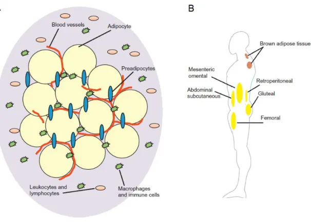
The adipose tissue is a metabolically active organ that acts as the main energy repository in the human body and as an endocrine organ able to synthesize several biologically active molecules that regulate metabolic homeostasis (Coelho et al., 2013). Different cell types compose this dynamic and complex organ (Figure 1A), including adipocytes, which are the dominant cells, preadipocytes, vascular endothelial cells, pericytes, macrophages and fibroblasts (Cedikova et al., 2016; Geloen et al., 1989).
Figure 1 – Anatomy of the adipose tissue and its composition in humans. (A) The adipose tissue is highly vascularized and composed by different cell types, such as adipocytes, preadipocytes and immune cells. (B) Brown and white adipose tissue are distributed through the human body. The brown adipose tissue is mainly located in the interscapular region, while the white adipose tissue is mainly located subcutaneously. The main depots of white adipose tissue are located in the abdominal and gluteal-femoral region, or near the kidney and digestive organs, constituting the retroperitoneal or mesenteric and omental adipose tissue, respectively (adapted from Tsiloulis & Watt, 2015).
The adipose tissue fulfils several functions, which may vary among fat depots due to its size, distribution, and heterogeneity according to their molecular, morphological, and metabolic profiles (Ibrahim, 2010). In humans, there are two main types of adipose tissue, white and brown, with several differences in adipocytes’ morphology and function
3
(Figure 1B). The brown adipose tissue is mainly located in the interscapular region and its size is reduced from birth to adulthood (Coelho et al., 2013). While this tissue also stores energy in form of fat, its main activity is heat production, or thermogenesis. Brown adipocytes are described as being smaller in comparison to white adipocytes, with relatively abundant cytoplasm, lipid droplets of different sizes and numerous mitochondria that produce heat by fatty acids oxidation (Saely et al., 2012). Although its participation in thermogenesis is nearly irrelevant, white adipose tissue presents much broader functions. White adipose tissue is widely distributed in the human body. Most of this tissue is located subcutaneously, storing 80% of total body fat, with the main depots found in the abdominal and gluteal-femoral region. The remaining 20% are located around the digestive organs, constituting the visceral adipose tissue (mesenteric and omental), and around the kidney (retroperitoneal) (Tsiloulis & Watt, 2015). By involving organs and infiltrating tissues, the white adipose tissue not only offers mechanical protection, but also plays an important role in the regulation of the body temperature, acting as a thermal insulator (Fonseca-Alaniz et al., 2007; Saely et al., 2012). Furthermore, white adipose tissue accomplishes multiple other functions, such as immune, endocrine and regenerative (Coelho et al., 2013; Thomou et al., 2010).
Although the white adipose tissue presents several functions, its main function is storing and releasing energy in response to the energetic needs, acting as a buffer for energy imbalance not only in cells, but also in whole-organisms. White adipose tissue stores energy whenever there is a surplus and releases energy for other organs whenever needed, such as in fasting conditions. This energy storage takes place in the form of triacylglycerol within adipocyte lipid droplets, which occupies most of the adipocyte volume. On the other hand, energy is released in the form of free fatty acids (or non-esterified fatty acids) and glycerol (Tsiloulis & Watt, 2015). These are highly efficient energy storage species, since they can be readily oxidized to produce large quantities of adenosine triphosphate (ATP) (Kiess et al., 2008).
Fat accumulation and the size of the white adipose tissue is determined by the balance between triacylglycerols synthesis and its breakdown into free fatty acids and glycerol (Figure 2). Both these pathways are controlled by a complex protein-protein interactions, intracellular signalling and neurohumoral regulators (Lass et al., 2011). Lipogenesis is the process that results in the production and storage of energy in the form of triacylglycerol within adipocyte lipid droplets. This process is stimulated when there is a surplus of energy, with high free fatty acids and glucose concentration in bloodstream (Kersten, 2001). With high glucose concentration, insulin’s release is stimulated and insulin-dependent glucose transporter 4 (GLUT4) transports glucose into the adipocytes. Glycolysis occurs and glycerol-3-phosphate is produced. Together with fatty acids coming from the liver and intestines, both substrates are esterified to form lipid droplets of triacylglycerol (Bernlohr et al., 2002). On the other hand, in fasting state or when there is a lack of energy, lipolysis occurs. Triacylglycerol is hydrolysed to glycerol and fatty acids for energy production (Tsiloulis & Watt, 2015). Glycerol is transported out of adipocytes via aquaporin-type
4
molecules and is used in the liver for oxidation or gluconeogenesis. Released fatty acids are carried by albumin to the liver, muscles and other tissues for oxidation, being converted into acetyl coenzyme A, in a process designated β-oxidation (Bernlohr et al., 2002; Laclaustra et al., 2007).
Figure 2 – White adipose tissue as an energetic buffer. When there is an energy surplus, there is an uptake of glucose by adipocytes, through GLUT4. Glycolysis occurs and glycerol-3-phosphate is produced. Glycerol-3-phosphate and fatty acids, that are carried by VLDL from the liver and chylomicrons from the intestine, are esterified to form TAGs, which is stored in lipid droplets. However, when energy is needed, TAGs are hydrolysed into glycerol and fatty acids, which are released and transported to be oxidized and produce energy. In the bloodstream, the fatty acids are bounded and transported by albumin. Both the lipogenic and lipolytic pathways are regulated by several molecules, activating or inhibiting key molecules in the process, such as one of the most important enzymes in the TAG hydrolysis, the hormone-sensitive lipase. For instance, insulin is known to inhibit its activity, stimulating the accumulation of lipids in the adipocyte. On the other hand, several other molecules may stimulate lipogenesis, such as glucagon, catecholamines, ACTH or serotonin (adapted from Coelho et al., 2013). Abbreviations: ACTH – Adrenocorticotropic hormone; Glycerol-3-P – glycerol-3-phosphate; GLUT4 – glucose transporter 4; TAGs – triacylglycerols; VLDL – very low-density lipoprotein.
1.2. Obesity
Obesity is clinically defined by the excessive accumulation of body fat. Obesity usually leads to an impaired health status, evidencing the deleterious impact of this chronic condition. The increasing prevalence of obesity makes it a global concern among modern societies (do Carmo et al., 2008; Ng et al., 2014). It is estimated that in 2015 more than 2.2 billion individuals were overweight (body mass index (BMI) from > 25 kg/m2), where over 710
million of these were obese (BMI > 30 kg/m2) (Afshin et al., 2017). This condition has
multifactorial causes that include genetic, epigenetic, physiological, sociocultural, and environmental factors. Overall, obesity is a consequence of a positive energy balance that occurs when the energy intake is higher than the expenditure. This scenario is generated by
5
an excessive consumption of food, while having a lack of physical activity, thus leading to the accumulation of the extra energy as fat (Bray et al., 2016; Pi-Sunyer, 2002). Genetic predisposition is also an important factor to consider in the onset of obesity.
White adipose tissue mass accumulation is associated with a bigger size and a higher number of adipocytes. When the organism has an energy surplus it is accumulated as lipids in adipocytes, which then suffer hypertrophy. This white adipose tissue buffering activity is an adaptive response to energy excess, which protects other tissues from lipotoxicity (Chavey et al., 2013). The maintenance of white adipose tissue homeostasis also includes pre-adipocyte hyperplasia, where these cells proliferate and differentiate into mature adipocytes (adipogenesis) (Nishimura et al., 2007). Simultaneously, there is an excessive extracellular matrix deposition in the form of fibrosis (Figure 3), which seems to limit cell hypertrophy and thus promote adipocyte hyperplasia. In fact, it has been suggested that hyperplasia and hypertrophy may be reciprocally regulated, although the mechanisms underlying these processes are still poorly understood (Muir et al., 2016). Altogether, obesity depends on the hypertrophy of pre-existing adipocytes, but also on hyperplasia, since there is a formation of new adipocytes from precursor cells through adipogenesis. However, in severe obesity conditions, a hypertrophic threshold may be reached, exceeding adipocyte buffering capacity and leading to decreased pre-adipocyte proliferative capacity, impaired metabolic functions, ectopic lipid deposition, inflammation, and dysregulated leptin and adipokines secretion (Cotillard et al., 2014; Landgraf et al., 2015; Ryden et al., 2014).
Figure 3 – White adipose tissue expansion during obesity scenarios. This mass accumulation is associated with an increase in size and number of adipocytes, fibrosis, impaired metabolic and endocrine functions, inflammation and hypoxia (adapted from Alligier et al., 2013).(Alligier et al., 2013)
6
A decreased capacity for lipid clearance accompanies the increased lipid storage due to the dysregulation of lipolysis, which leads to increasing lipid accumulation. The white adipose tissue expandability hypothesis supports the argument that if an individual has the capacity to store fat in this tissue, the process is harmless and there is no ectopic deposition of lipids with resulting metabolic complications (Langin, 2011; Langin & Mouisel, 2013; Virtue & Vidal-Puig, 2008). However, when this storage capacity is overridden, there is an ectopic deposition of lipids in the skeletal muscle and in the liver (Figure 4). This ectopic lipid deposition may favour several complications through lipotoxic mechanisms, including insulin resistance and ultimately diabetes mellitus (type 2) (Castro et al., 2014; Zimmet et al., 2001), cardiovascular dysfunctions (Luna-Luna et al., 2015) and infertility (Esmaeilzadeh et al., 2014; Mission et al., 2015; Palmer et al., 2012).
Figure 4 – Evolution from non-obese to severe obesity with ectopic lipid deposition. Increased fat storage and reduced lipolysis increases the fat accumulation in obesity. If the white adipose tissue capacity of storage is overcome, there is ectopic lipid deposition in the skeletal muscle and liver, a metabolic complication that may lead to diabetes and cardiovascular dysfunctions (adapted from Langin & Mousiel, 2013). Abbreviations: FA – Fatty acids; TG – triacylglycerols.
Thus, it is crucial to prevent and treat obesity. Currently, low-calorie diets and increased physical activity are the ground stones in obesity treatment. The pharmacological approaches to address obesity are scarce and the existing drugs with anti-obesity potential are prone to tolerance and only provide short-term weight loss (Bray, 2008; Snow et al., 2005). Although several possible targets for obesity treatment have been recently identified (Monteiro, 2014), there is still a need to find new, safe and cost-effective therapeutic approaches.
The study of natural products along the years has evidenced their preventive and therapeutic potential in the control of obesity. A variety of medicinal plants, fruits, and
7
vegetables have been used in different anti-obesity products as food supplements to promote weight loss (de Resende et al., 2015; Sun et al., 2016). Methylxanthines constitute a group of natural occurring molecules with growing interest for researches in this field. These compounds are naturally produced by both animals and plants, being caffeine, theobromine and theophylline the most well-studied (Monteiro et al., 2016). These compounds can be found in tea leaves (Camellia sinensis L.), cocoa (Theobroma cacao L.), and coffee beans (Coffea sp.), food products that are consumed worldwide on a daily basis. Moreover, methylxanthines have proven to have biological effects that could contribute not only for obesity management (Liu et al., 2015), but also for neurological (Fredholm et al., 1999; Nehlig et al., 1992; Roll, 1980), respiratory (Barnes, 2013; Dent et al., 1994), cardiac (Moffat, 1986; Roll, 1980) and renal disorders (Osswald & Schnermann, 2011), as well as in male reproductive function (Brokaw, 1987; Dias et al., 2015; Dias et al., 2014; Yamaguchi et al., 2009).
1.3. Methylxanthines
1.3.1. Chemical structure, biosynthesis, properties, and natural sources
Methylxanthines are methylated xanthine-derivatives that may be produced in plants and animals and can be found in most human fluids and tissues (Zrenner et al., 2006). These purine bases are heterocyclic complexes that result from the coupling of pyrimidinedione with imidazole rings (Talik et al., 2012). Among the most well-known methylxanthines (Figure 5) are 1,3,7-trimethylxanthine, 1,3-dimethylxanthine, and 3,7-dimethylxanthine, also commonly recognized as caffeine, theophylline, and theobromine respectively.
In plants, xanthosine is the primary substrate for methylxanthines’ biosynthesis. Xanthosine can be produced through the degradation of adenine nucleotides, adenosine and guanosine nucleosides, and/or de novo purine biosynthesis (Ashihara et al., 2011). The pathway that leads to caffeine production starts with xanthosine methylation to 7-methylxanthosine, then this ribose is hydrolysed to form 7-methylxanthine and subsequent methylations yield theobromine and, finally, caffeine (Koshiishi et al., 2001; Misako & Kouichi, 2004). While theobromine may be a precursor of caffeine, theophylline can be considered as a catabolite. In almost all caffeine-producing plants, caffeine is degraded by three consecutive demethylations, with the formation of theophylline in the process (Ashihara et al., 2011). On the other hand, the major demethylated metabolite of caffeine in animals is paraxanthine (1,7-dimethylxanthine), an isomer of theobromine and theophylline, but it is not naturally produced by plants (Monteiro et al., 2016). Additionally, caffeine (Zajac et al., 2003), theobromine (McKeague et al., 2016), theophylline (Nanjundaiah et al., 2016) and paraxanthine (Müller et al., 1998) can also be obtained by chemical synthesis.
8 Figure 5 - Chemical structures of the three predominant methylxanthines (caffeine, theobromine and theophylline) and paraxanthine, the major dimethylated metabolite of caffeine.
Methylxanthines are considered weak Brønsted bases due to the imino nitrogen at position 9. Caffeine possesses methyl groups at position 1, 3 and 7, which confers electrophilic properties. Although theophylline shares the same electrophilic sites at position 1 and 3, it has a proton at position 7 instead of a methyl group, becoming a Brønsted acid site (Monteiro et al., 2016). For that reason, only theophylline may become a proton donor. In fact, this molecule acts as a proton donor in most pharmaceutical systems (Beale & Jr John, 2011). Theobromine differs from both compounds due to the absence of the methyl group at position 1. The presence of this methyl group was reported to be the responsible of several physicochemical properties of caffeine and its physiological effects (Pavia, 1973). Additionally, caffeine structure comprises lipophilic properties (Salihović et al., 2014), which should improve its permeability through cell membranes and cross biological barriers, such as the blood-brain barrier (McCall et al., 1982). Several synthetic modifications of these natural compounds have been produced to pursue pharmacological purposes, resulting in drugs like dyphylline, proxyphylline and enprofylline. Moreover, novel modifications are being investigated for different therapeutic applications (Baraldi et al., 2007; Moro et al., 2006), thus enhancing the interesting properties of these compounds in health promotion.
Plants containing methylxanthines are common in food products daily ingested worldwide, such as coffee, tea, cocoa, and chocolate. Coffee and tea are globally consumed and are the principal sources of caffeine intake through diet (Frary et al., 2005). Although
9
coffee is usually expected to contain more caffeine than tea, it can be found in considerable amounts in certain types of tea (Camellia Sinensis species). One study (Gilbert et al., 1976) showed that the median caffeine concentration in tea is 27 mg per cup (median size of 225 mL), while in coffee is 74 mg per cup (median size of 200 mL). Theobromine is the predominant methylxanthine found in cocoa beans, where it represents about 2% of its dry weight. Furthermore, some Camellia species contain more theobromine than caffeine (Ashihara et al., 2008). Despite caffeine and theobromine are prominent in dietary sources, the same does not happen with theophylline. This methylxanthine is naturally present in tea leaves and in vestigial amounts in cocoa and coffee beans (Barnes, 2013). For that reason, only few amounts of this methylxanthine are thought to be obtained through diet (Stavric, 1988). Although coffee, tea and cocoa are the most well-known plant-derived products containing methylxanthines, there are other plants reported as methylxanthines producers, such as Paullinia sp. (guarana), Cola sp. (cola beverages), Ilex paraguariensis A. St.-Hil. (mate), and Citrus sp. (Atawodi et al., 2007; Baumann et al., 1995; Kretschmar & Baumann, 1999; Weckerle et al., 2003).
Methylxanthines have been described to exert multiple physiological effects in the human body, including in the nervous (Fredholm et al., 1999; Nehlig et al., 1992; Roll, 1980), respiratory (Barnes, 2013; Dent et al., 1994) and cardiac systems (Moffat, 1986; Roll, 1980). They stimulate the skeletal muscle and promote diuresis (Tarka, 1982). Male fertility is another field in which these compounds may have positive outcomes. For instance, methylxanthines have been described to be beneficial for sperm Ca2+ transport (Tash &
Means, 1982). Moreover, methylxanthine-rich beverages, such as tea, and caffeine were also reported to be effective additives for sperm storage and in vitro fertilization (Brokaw, 1987; Dias et al., 2014; Yamaguchi et al., 2009). Caffeine by itself has also improved the nutritional support of spermatogenesis by Sertoli cells (Dias et al., 2015). Some of these beneficial effects were associated with methylxanthines antioxidant properties (Azam et al., 2003; Grucka-Mamczar et al., 2009; Leon-Carmona & Galano, 2011; Ofluoglu et al., 2009). More recently, the anti-obesity potential of methylxanthines has also been reported. These compounds may interact with the adipose tissue and its effects could comprise a promising therapeutic approach for obesity management.
1.3.2. Anti-obesity potential
In the past couple of decades, the search for new pharmacological tools to address obesity has increased. Phytochemicals, such as methylxanthines, have been reported to promote weight loss in obese individuals, thus attracting the interest of several researchers. For instance, it has been reported that trained and untrained young (20-40 years) males consuming 4 mg/kg of caffeine after an overnight fast demonstrated increased resting metabolic rate values, although the magnitude of this effect was greater in trained subjects
10
(LeBlanc et al., 1985). Higher levels of free fatty acids were also observed in the plasma, as well as increased lipid oxidation, due to enhanced lipolysis. Furthermore, when caffeine consumption is associated with physical exercise, the effects are exacerbated (Schubert et al., 2014). A controlled trial was performed with lean individuals (18-45 years) taking 3 mg/kg of caffeine 90 min before, and 30 min after doing physical exercise. After performing physical activity (1 h of cycling, with periods of rest 1 h before and 2 h after), the participants had a test meal in ad libitum conditions. It was shown that the combination of physical exercise and caffeine supplementation led to an increase in energy expenditure and fat loss relative to placebo. In addition, when exposed to caffeine the participants reduced the energy and fat intake and the exercise was perceived as less difficult and more enjoyable. Another study (Gavrieli et al., 2013) showed that obese and overweight individuals consumed less food in an ad libitum meal after an intake of 2-4 cups of coffee than those with lower or no coffee consumption, which suggest that caffeine may have appetite suppressing effects. Besides, caffeine intake has been linked with an increased energy expenditure per day in lean subjects, but also in post-obese participants during weight loss maintenance (Dulloo et al., 1989). Caffeine also demonstrated to improve weight loss, when following a diet with caloric restriction, and prevent weight regain (Davoodi et al., 2014). Overweight females having a calorie shifting diet for 6 weeks and then a follow-up diet for 4 weeks with caffeine treatment (5 mg/kg/day) or placebo, demonstrated that after the calorie shifting diet period, the females consuming caffeine exhibited more weight and fat loss in comparison with the placebo group. Moreover, after the follow-up diet period, the weight loss on the caffeine group persisted, while the placebo group weight was regained.
Methylxanthines’ consumption can be also combined with other natural compounds for superior results (Zheng et al., 2004). Female mice were fed with a standard powder diet containing several combinations of caffeine, green tea catechins and L-theanine for 16 weeks. The combination of 0.05% caffeine and 0.3% green tea’s catechins was the most effective for preventing weight gain, in comparison with the normal diet group from the 4th to the 16th
week. Furthermore, it was also shown that a diet with a theobromine-rich cocoa powder promoted weight loss in Wistar rats, compared to control (Eteng et al., 2006).
Altogether, the molecular activity of methylxanthines seems to lead to the stimulation of lipolysis and inhibition of adipogenesis, decreasing the accumulation of the adipose tissue. By this means, methylxanthines can promote weight loss in overweight and obese subjects. Several mechanisms were proposed to explain their actions, which will be addressed on the following topics.
11
1.3.3. Lipolytic activity
1.3.3.1. Adenosine receptors antagonism
Lipolysis is the pathway that leads to free fatty acids and glycerol release from triacylglycerols, being unique to adipocytes. One known regulator of lipolysis is the adenosine receptor (AR), which can be divided in four types: A1, A2A, A2B and A3. These are present in
every cell, with the four types exhibiting a differential cell and tissue expression (Olah & Stiles, 1995). Adenosine is an endogenous nucleoside released from adipose tissue during sympathetic nerve activation. In 1972, it was shown for the first time that adenosine and its analogues could inhibit lipolysis in adipocytes (Fain et al., 1972). Later, it was reported that this lipolysis inhibition by adenosine was mainly mediated by activation of the A1R. Moreover,
it has been demonstrated that these receptors are important regulators of lipolysis, fatty acid storage and tissue partitioning of fat (Johansson et al., 2007). Adipose tissue has a high expression of A1R, which are functionally active in differentiated adipocytes (Trost &
Schwabe, 1981; Vassaux et al., 1993). However, accumulating evidence indicates that the number of receptors may differ among fat repositories. Data from animal studies reported much higher affinity and binding capacity of white adipose tissue for one specific A1Ragonist
in comparison to brown adipose tissue, suggesting that white adipose tissue might have a higher expression of A1R than brown adipose tissue (Saggerson & Jamal, 1990). The human
subcutaneous adipose tissue showed a higher number of receptors than the visceral adipose tissue, supporting the hypothesis that the A1R may exert a major role in the regulation of
subcutaneous fat storage (Barakat et al., 2006). Furthermore, it has been proposed that these receptors are efficiently coupled with a Gi protein in adipocytes and cannot be affected
independently (Liang et al., 2002). This feature suggests that G proteins inactivation cannot be overcome by the activation of a higher number of A1R (Dhalla et al., 2009). Besides,
functional uncoupling of A1R from these proteins leads to the synthesis of more receptors
(Jajoo et al., 2006). Each receptor appears to activate more than one G protein and signal amplification seems to be independent of the total number of receptors (Baker et al., 2000). Overall, in adipocytes, A1R activation by its agonists (e.g. adenosine) inhibits of the adenylyl
cyclase activity, which consequently reduces cyclic adenosine monophosphate (cAMP) formation, inhibits protein kinase A (PKA) and, ultimately, blocks phosphorylation of the lipases involved in lipolysis, leading to its inhibition (Dhalla et al., 2009). Additionally, adenosine and its analogues might modulate insulin action and sensitivity in adipose tissue through A1R, by increasing insulin sensitivity and potentiating insulin-induced activation of
phosphoinositide 3-kinase (PI3K), resulting in the decrease of cAMP concentration in adipocytes (Budohoski et al., 1984; Rolband et al., 1990; Takasuga et al., 1999). On the other hand, while A1Rand A3R stimulation leads to lower concentrations of cAMP via Gi proteins, the
stimulation of A2AR and A2BR increases cAMP concentration via Gs proteins, translating into
12
adipocyte differentiation is low and, consequently, its role in lipolysis is reduced (Borglum et al., 1996).
A vital mechanism of methylxanthines’ anti-obesity activity is the non-specific antagonism of AR, as these molecules are able to block and inhibit, in a competitive manner, the role of adenosine in the cells, thus stimulating lipolysis (Figure 6A) (Chen & Chern, 2011). Methylxanthines have the capacity to inhibit the four subtypes of AR at physiological doses (<100 µM), but most of their actions seem to be mediated through the inhibition of A1 and A2A
subtypes (Sattin & Rall, 1970). The AR inhibitory effects are mostly mediated by the 1-methyl group of methylxanthines structure (Green & Stanberry, 1977). In fact, caffeine and theophylline were shown to be potent inhibitors of AR (Boulenger et al., 1982). Still, in other studies (Daly et al., 1983; Fredholm, Irenius, et al., 2001; Klotz et al., 1997) theophylline and paraxanthine were reported to have slightly higher affinities than caffeine for the A1R (in
similar concentrations). On the other hand, theobromine does not possess the 1-methyl group and was described to have lower affinity for A1R and A2AR subtypes (Carney et al., 1985;
Schwabe et al., 1985; Shi & Daly, 1999).
Additionally, methylxanthines are also nonselective competitive inhibitors of phosphodiesterases at pharmacological doses (> 1 mM) (Beavo et al., 1970; Butcher & Sutherland, 1962; Cardinali, 1980; Nicholson et al., 1989). Phosphodiesterases are responsible for hydrolysing cAMP and have a central role in regulating cAMP signalling, most of which is accountable to phosphodiesterase-4 (Wu & Rajagopalan, 2016). Reversible inhibition of phosphodiesterases by methylxanthines impairs the hydrolysis process, preventing the degradation of cAMP and consequently increasing its concentration (Sassone-Corsi, 2012). Therefore, phosphodiesterase-4 inhibition increases lipolysis via activation of hormone-sensitive lipase that is induced by the increased cAMP concentration (Figure 6A) (Wu & Rajagopalan, 2016). The three natural-occurring methylxanthines are described as competitive inhibitors of phosphodiesterases, where theophylline is reported to be a more potent inhibitor than caffeine (Daly, 2007; Stavric, 1988).
A possible drawback of methylxanthines use is the increased release of free fatty acids into circulation as an end product of lipolysis that is observed after A1R antagonism.
Ultimately, this scenario could potentially lead to the worsening of insulin resistance (Dhalla et al., 2009). Interestingly, A1R antagonists were reported to improve glucose tolerance,
which could be attributed to a selective increase in the adipose tissue receptors (Xu et al., 1998).
While natural occurring methylxanthines have affinity for the AR, synthetic compounds based on the xanthine chemical structure have been developed, evidencing more potent and selective antagonism activity for all four receptor subtypes (Moro et al., 2006). In general, substitutions at position 8 with aryl or cycloalkyl groups were reported as promising potential for the identification of novel adenosine A1R and A2AR antagonists. Conversely, it
was concluded that ethyl substitution at the positions 1, 3 and 7 may enhance adenosine A1R
13
Terre'Blanche, 2015). Moreover, several ring-extended xanthines have been developed to increase phosphodiesterase-4 inhibition (in the nM range) (Arnold et al., 2002; Pissarnitski et al., 2004).
Figure 6 - Schematic illustration of methylxanthines’ activity in adipocytes. Methylxanthines can stimulate lipolysis (A) due to the inhibition of A1 receptors, increased concentration of catecholamines
and inhibition of PDE4. This results in an increased cAMP concentration, which activates PKA and the lipases involved in lipolysis. Methylxanthines were also reported to inhibit adipogenesis (B), by disturbing the Akt and ERK axis and by activating AMPK. Altogether, its action leads to the decrease expression of C/EBPα and PPARγ, the two main adipogenic transcription factors. Abbreviations: 5’AMP – 5’ adenosine monophosphate; AC – Adenylyl cyclase; ACC – Acetyl-coA Carboxylase; Akt – Protein kinase B; AMPK – 5’AMP-activated protein kinase; aP2 – Adipocyte protein 2; ATGL – Adipose triglyceride lipase; ATP – Adenosine triphosphate; C/EBPα – ccaat enhancer binding protein alpha; C/EBPβ – ccaat enhancer binding protein beta; C/EBPδ – ccaat enhancer binding protein delta; cAMP – Cyclic adenosine monophosphate; ERK – Extracellular signal-regulated kinase; FFA – Free fatty acids; FAS – Fatty acid synthase; GLUT4 – Glucose transporter type 4; GPDH – Glycerolphosphate dehydrogenase; GSK3β – Glycogen synthase kinase 3 beta; HSL – Hormone-sensitive lipase; IR – Insulin receptor; MGL – Monoacylglycerol lipase; MTX - Methylxanthine; PDE3B – Phosphodiesterase 3B; PDE4 – Phosphodiesterase 4; PI3K – Phosphoinositide 3-kinase; PKA – Protein kinase A; PPARγ – Peroxisome-proliferator activated receptor gamma; TAG – Triacylglycerols.
1.3.3.2. Modulation of catecholamines’ release
Catecholamines, such as adrenaline and noradrenaline, are major regulators of lipolysis in humans (Morigny et al., 2016). These compounds activate or inhibit lipolysis by bounding/unbounding to adrenergic receptors. Adrenergic receptors can be divided in two major types, α and β, each with several subtypes (Langin, 2006). In general, when β-adrenergic receptors are stimulated, a lipolytic response is triggered by its action on the stimulatory G alpha (Gs) subunit of heterotrimeric G proteins. Subsequently, adenylyl cyclase
14
other hand, the activation of α-adrenergic receptors induces an antilipolytic signal, since they are coupled with inhibitory G alpha (Gi)subunit of heterotrimeric G proteins, thus inhibiting
adenylyl cyclase activity and cAMP production (Stich et al., 1999). Therefore, lipolysis regulation relies on the relative affinity of catecholamines for the distinct adrenergic receptors and on the expression of each receptor on the adipocytes’ membrane (Morigny et al., 2016).
Methylxanthines can stimulate the sympathetic nervous system, leading to the release of noradrenaline and activating the β-adrenergic receptors (Acheson et al., 2004). It has been observed that caffeine increases catecholamine levels (Chen et al., 1994). Moreover, catecholamines and caffeine can act synergistically, increasing cAMP concentration in a superior extent than induced by the hormones alone, thus further promoting lipolytic activity (Figure 6A) (Butcher et al., 1968). In addition, methylxanthines have been shown to potentiate the effects of ephedrine (the most active alkaloid of Ephedra sp.), an α- and β-adrenergic receptor agonist (Greenway, 2001). This sympathomimetic agent, originally used as a bronchodilator, was found to stimulate lipolysis and induce weight loss in asthmatic patients. Ephedrine also stimulates the release of noradrenaline, which then binds to β-adrenergic receptors on adipocytes (Diepvens et al., 2007). A combined administration of methylxanthine (caffeine or theophylline) and ephedrine to obese mice for 6 weeks led to a decrease in body weight and fat, as well as an increase in energy expenditure (Dulloo & Miller, 1986a). Later, administration of the same combination of compounds (22 mg of ephedrine, 30 mg of caffeine and 50 mg of theophylline) to human subjects demonstrated that ephedrine/methylxanthine combination was more effective in raising resting metabolic rate in post-obese and lean subjects than ephedrine alone (Dulloo & Miller, 1986b). There are several other studies that corroborate these findings (Liu et al., 2013). Obese subjects that received 200 mg caffeine/20 mg ephedrine for over 24 weeks, lost 6.5% of total body weight and 12.4% of whole body fat mass. However, in 2004 the Food and Drug Administration (FDA) banned from the market the ephedra-containing supplements due to the potential adverse effects, and efforts to find an optimized replacement are ongoing (Bray & Greenway, 2007; Haller & Benowitz, 2000). Ann Liu et al. (2015) studied albuterol for that purpose. Albuterol is another selective β2-adrenergic agonist previously reported to stimulate lipolysis when used in a dose four-fold higher than the inhalatory bronchodilation dose used for asthma treatment (200 µg) (Amoroso et al., 1993; Goldberg et al., 1975). The combination of albuterol (7-17 ng/mL) and caffeine (3-10 µg/mL) resulted in a 30-40% increase in lipolysis over buffer-treated human adipocytes. Furthermore, in human subjects, the combination of caffeine (100-200 mg) with albuterol (2-4 mg) also resulted in significant increase of energy expenditure, irrespective of treatment-dose combination. Besides, an increase in lean mass along with a decrease on fat mass also occurred in caffeine/albuterol treated rats.
15
1.3.4. Anti-adipogenic activity
Mild obesity results mainly from adipocytes hypertrophy, whereas more severe cases of obesity also involve cell hyperplasia. To maintain the metabolic homeostasis upon energy overload, preadipocytes are recruited to differentiate into mature adipocytes, in a process called adipogenesis. This process is important for healthy adipose tissue growth, remodelling, and expansion, which may prevent the adverse metabolic dysregulation associated with obesity (Eisenstein & Ravid, 2014; Rosen & MacDougald, 2006). Certain transcription factors have been shown to be pivotal for adipogenesis, such as the ccaat enhancer binding proteins (C/EBP) family, and peroxisome-proliferator activated receptor-gamma (PPARγ) (Rosen et al., 1999; Tontonoz et al., 1994; Wu et al., 1999). The expression of two C/EBP proteins, C/EBPβ and C/EBPδ, promotes the expression of PPARγ, possibly via binding sites on its promoter (Fajas et al., 1997; Zhu et al., 1995). Then, PPARγ stimulates the expression of another C/EBP protein, C/EBPα, which induces the differentiation process (Freytag et al., 1994; Lin & Lane, 1992, 1994). For that reason, these latter transcription factors are considered the main regulators of adipogenesis. PPARγ and C/EBPα trigger differentiation through the regulation of adipocyte-specific genes that are necessary for adipocyte function, including fatty acid binding protein/adipose protein 2 (FABP/aP2), insulin receptor, GLUT4, acetyl-coA carboxylase, fatty acid synthase, and glycerolphosphate dehydrogenase (GPDH) (Kubota et al., 1999; Spiegelman et al., 1993; Tontonoz, Hu, Devine, et al., 1995; Tontonoz, Hu, & Spiegelman, 1995). On the other hand, there is evidence that C/EBPβ and C/EBPδ are not essential for adipocyte differentiation, suggesting the existence of adipogenic transcriptional cascades that do not involve these proteins (Tanaka et al., 1997).
More recently, it was shown that methylxanthines may inhibit adipogenesis. Primary rat adipose-derived stem cells (ADSCs) and mouse bone marrow stromal cell line (M2-10B4) were used to evaluate the in vitro effects of caffeine on adipogenesis (Su et al., 2013). The continuous exposure of cells to caffeine (0.1-1 mM) during differentiation showed that caffeine dose-dependently reduced lipid droplet and adipocyte differentiation in both cell types, and that it also decreased the expression of C/EBPα and PPARγ, the two main adipogenic transcription factors. Similar results were obtained in 3T3-L1 cell line when cells were incubated with a 5% coffee solution (Aoyagi et al., 2014). Later, it was described that caffeine effects in adipocytes were mediated by reducing the activation of the protein kinase B (Akt) and disturbing the Akt/glycogen synthase kinase 3 beta (GSK3β) axis. This axis is known for its role in multiple cellular processes such as metabolism, proliferation, or transcription. In fact, it was shown that reduced activation of this axis leads to the inhibition of the mitotic clonal expansion and inhibition of the C/EBPβ in 3T3-L1 cells.
Theobromine was also shown to inhibit differentiation of 3T3-L1 cells (Jang et al., 2015). When preadipocytes were exposed to theobromine (50, 100 and 150 µg/mL) for 7 days during their differentiation period, a decreased accumulation of lipid droplets and a decreased expression of C/EBPα and PPARγ were reported, in a concentration-dependent
16
manner. The authors also reported an increased phosphorylation of the AMP-activated protein kinase (AMPK) and a decreased phosphorylation of extracellular signal-regulated kinase (ERK), also in a concentration-dependent manner. Both protein pathways intervene in the regulation of PPARγ, whose inhibition leads to the inhibition of adipogenesis (Burns & Vanden Heuvel, 2007; Farmer, 2005).
Overall, methylxanthines seem to inhibit adipogenesis by disturbing the adipocyte signalling and inhibiting the main adipogenic transcription factors (Figure 6B). However, adrenergic and adenosine receptors also appear to have a role in adipogenesis. In fact, the expression of β-adrenergic receptors increases during the differentiation process, whereas upon stimulation by agonists, there is an inhibition of adipocyte differentiation due to PKA activation (Li et al., 2010). Moreover, it has been reported that the activation of A1 and A2
receptors stimulates adipocyte differentiation, by promoting lipogenesis and lipid accumulation, which are accompanied by an increased PPARγ and C/EBPα expression, respectively (Gharibi et al., 2012). However, in other studies it appears that AR coupled to Gs
proteins inhibit adipocyte differentiation, while receptors coupled to Gi proteins promote
differentiation (Wang et al., 1996; Wang & Malbon, 1999). Thus, although their actions and adipogenesis mechanism are not completely understood, it has been suggested that they may play a role in cAMP-independent adipogenesis regulation (Eisenstein & Ravid, 2014).
17
18
II. Objectives
Methylxanthines are widely distributed in the human diet and its consumption seems to be inversely associated with body weight increase. As previously described, caffeine (the most well-known and studied natural methylxanthine) is reported to modulate glucose and fatty acid metabolism. Recently, Van der Walt and Terre’Blanche (2015) synthetized several xanthine analogues. One of those synthetic xanthine analogues, 8-(3-phenylpropyl)-1,3,7-triethylxanthine (ETX), was reported to be a very potent A1 receptor antagonist. Thus, ETX
may be a promising drug candidate for obesity treatment.
This project aims to study the modulation conferred by new this molecule in the metabolic and oxidative profile of adipocytes in order to evaluate its pharmacological potential for obesity treatment. For this purpose, the anti-obesogenic potential of ETX was evaluated in mouse adipocytes (derived from differentiation of the cell line 3T3-L1), using synthetic caffeine as comparator.
Therefore, the following specific objectives were established:
1. Culture and differentiation of 3T3-L1 preadipocytes into adipocytes; 2. Characterization of the cytotoxic profile of ETX in 3T3-L1 adipocytes;
3. Evaluation of the metabolic profile in 3T3-L1 adipocytes after treatment with ETX as compared to caffeine;
4. Evaluation of the oxidative profile in 3T3-L1 adipocytes after treatment with ETX as compared to caffeine.
19
20
III. Materials and Methods
3.1. Xanthine analogues synthesis
The synthetic xanthine, 8-(3-phenylpropyl)-1,3,7-triethylxanthine (Figure 7), was produced at the School of Pharmacy, North-West University, Potchefstroom, South Africa, and kindly provided by our collaborators Van der Walt and Terre’Blanche. The synthesis process was previously described (Van der Walt & Terre'Blanche, 2015; Van der Walt et al., 2013). The first step was the reaction of 1,3-dimethyl- and 1,3-diethyl-5,6-diaminouracil with carboxylic acid, such as 4-phenylbutanoic acid, phenylpropanoic acid and phenoxyacetic acid, in the presence of the coupling reagent N-(3-dimethylaminopropyl)-N’-ethylcarbodiimide (EDAC) at room temperature. Resulting from this acylation, several 1,3-dialkyl-6-amino-5-carboxamidouracil intermediates were obtained. The cyclization of these intermediates was obtained under strong basic conditions (aqueous sodium hydroxide solution), to produce the corresponding xanthine derivatives. Without purification, the 1,3-dialkyl-7H-xanthine derivatives were directly treated with excess iodomethane or iodoethane in the presence of potassium carbonate, resulting in several 7-alkylated xanthine derivatives. Finally, 8-(3-phenylpropyl)-1,3,7-triethylxanthine was purified by recrystallization and the molecular structure and purity were verified by 1H-NMR, 13C-NMR, and mass spectrometry
analysis.
Figure 7 – Chemical structure of the new synthetic xanthine (8-(3-phenylpropyl)-1,3,7-triethylxanthine).
3.2. Mouse preadipocyte cell line 3T3-L1 culture
The 3T3-L1 is a preadipocyte cell line derived from the 3T3 cells, which were obtained from Swiss mouse embryo cells (Green & Meuth, 1974). The 3T3-L1, under
21
appropriate conditions, are able to differentiate into adipocytes, being widely used for in vitro adipose tissue studies.
Preadipocyte cell line 3T3-L1 was purchased from a commercial supplier (Zen-Bio, Durham, NC, USA). 3T3-L1 preadipocytes at passage 14-15 were cultured in DMEM High Glucose (Sigma-Aldrich, USA) supplemented with 10% newborn calf serum (NCS) (Gibco, NZ), 1% penicillin-streptomycin (Pen-Strep) (Sigma-Aldrich, USA), 2.5 µg/mL amphotericin B (Sigma-Aldrich, USA), 50 µg/mL gentamicin (Sigma-Aldrich, USA) and 50 mM HEPES buffer (Fisher BioReagents, USA). Cells were grown in 25 cm2 and 75 cm2 T-flasks (Thermo Scientific,
Waltham, MA, USA), being re-fed every 72 h. When reaching 70% confluence, they were passaged by incubating the cells with 0.05% trypsin-EDTA solution (Sigma-Aldrich, USA).
All cell cultures were handled in a laminar flow chamber and maintained in an incubator at 37ºC with a 5% CO2 humidified atmosphere (Heracell 150i, Thermo Scientific,
Waltham, MA, USA).
3.2.1. Adipocyte differentiation
Adipocyte differentiation was induced 2 days after the cells reached full confluence and cells were maintained for 15 days until differentiation was completed (Figure 8), as described previously (Moreira et al., 2015). The culture medium was removed and cells were washed with phosphate buffered saline (PBS). Cells were incubated in a differentiation medium composed by DMEM-F12 (Sigma-Aldrich, USA) supplemented with 10% fetal bovine serum (FBS) (Biochrom, Germany), 1% Pen-Strep, 2.5 µg/mL amphotericin B, 50 µg/mL gentamicin, 15 mM HEPES Buffer and a differentiation cocktail containing 1 µM dexamethasone Aldrich, USA), 0.5 mM 3-Isobutyl-1-methylxanthine (IBMX) (Sigma-Aldrich, USA) and 1 µg/mL human insulin (Actrapid, Novo Nordisk, Denmark). After 72 h, the culture medium was replaced with the last medium without the differentiation cocktail and cells were re-fed every 72 h for more 12 days.
Figure 8 - Schematic illustration of the 3T3-L1 preadipocytes differentiation into adipocytes (adapted from Zen-Bio 3T3-L1 Cell Care Manual, 2015). (Zen-Bio, 2015)