UNIVERSITY OF LISBON
FACULTY OF FARMACY
Developments in Tumor Targeting and
Internalizing Peptides
Rita Isabel Cordeiro Padanha
Integrated Master in Pharmaceutical Sciences
UNIVERSITY OF LISBON
FACULTY OF FARMACY
Developments in Tumor Targeting and
Internalizing Peptides
Rita Isabel Cordeiro Padanha
Thesis of Integrated Master in Pharmaceutical Sciences
presented to Faculty of Pharmacy of the University of Lisbon
Supervisor: Professor Pedro Góis
3
Resumo
A grande limitação do uso de fármacos citotóxicos em terapias antitumorais resume-se à falta de capacidade destas moléculas em distinguir células tumorais e células saudáveis. Os Tumor Targeting Peptides (TTPs), desde a sua descoberta há 30 anos atrás, têm-se tornado numa ferramenta útil para o tratamento e diagnóstico do cancro, uma vez que reconhecem alvos moleculares tumorais específicos, tal como os anticorpos (Abs), mas sem as suas desvantagens estruturais. Assim, a sua utilização no desenvolvimento de conjugados terapêuticos peptídicos (PDCs) é bastante promissora, embora ainda nenhum destes sistemas de entrega de fármacos esteja aprovado para uso clínico.
O objetivo deste trabalho consistiu na revisão de literatura relacionada com abordagens terapêuticas alvo-dirigidas e na organização e resumo dos TTPs que contribuíram de uma forma mais valiosa para os avanços no desenvolvimento de conjugados terapêuticos.
Neste trabalho, foram apresentados 9 péptidos com capacidade de reconhecimento alvo-específico, representativos de cada classe de TTPs e promissores quanto a uma futura aplicação clínica como parte integrante de conjugados terapêuticos - RGD, NGR, F3, Octreotide, LyP-1, Bombesin, Angiopep 2, CREKA e M2pep. Desta forma, foram focados para cada um dos TTPs, pontos-chave relativos a características estruturais e funcionais, alvos celulares específicos, estudos de relação estrutura-atividade (SAR), capacidade de internalização e aplicações no desenvolvimento de conjugados terapêuticos.
Palavras-Chave:
Reconhecimento alvo-específico, tumor targeting peptides, RGD, NGR, conjugados terapêuticos.4
Abstract
The major inconvenient of the use of cytotoxic drugs in anti-tumor therapies is their poor ability to distinguish tumor cells from the normal cells. Since their discovery about 30 years ago, tumor targeting peptides (TTPs) have become an useful tool for the treatment and diagnosis of cancer, once they are able to recognize specific tumor targets, such as antibodies (Abs) but without their structural disadvantages. Thus, their use in the development of peptide-drug conjugates (PDCs) is promising, although none of these drug delivery systems (DDS) are approved for clinical use yet.
The aim of this work was to review the literature about tumor targeting approaches and to organize and summarize the TTPs that contributed in a significant way to the advances of this field, including the development of therapeutic conjugates.
In this work, nine peptides with targeting ability, representative of each class of TTPs and promising for a future clinical application as an integral part of drug conjugates were presented - RGD, NGR, F3, Octreotide, LyP-1, Bombesin, Angiopep 2, CREKA and M2pep. This way, structural and functional features, addresses, structure-activity relationship (SAR) studies, internalization capacity and applications in the development of therapeutic conjugates were focused as key-points for each targeting peptide.
Key-words:
Targeting, t
umor targeting peptides, RGD, NGR, peptide-drug conjugates.5
Acknowledgements
To Professor Pedro Góis I would like to express my deepest thanks for the support given since the beginning of this work, the constant orientation through the past months, for being always available for my questions, for clearing my doubts and for sharing the passion of biochemistry subjects.
To my family, especially my parents and sister, I would like to thanks all the support and motivation given since the beginning of this work.
Finally, to all my college friends I would like to give a special thanks, for the good times during the last five years inside and outside the Faculty of Pharmacy.
6
General Index
Resumo………..……….…… 3 Abstract……….4 Acknowledgements……….…5 General Index……….…… 6 Index of Figures………... 8 Index of Tables………..…....10Abbreviations and Symbols………...…………....………….11
1. Introduction………...13
1.1. Tumor Microenvironment………..………..…………...…14
1.1.1. Plasma Membrane……….………14
1.1.2. Tumor Vasculature……….………....15
1.2. Tumor Targeting Therapy………..………....……16
1.2.1. Antibodies……….……16
1.3. Peptides in Cancer Therapy ……….18
1.3.1. Peptides in Tumor Targeting Therapy ………20
1.4. TTPs Discovery Techniques……….……21
1.5. Targeted Drug Delivery Systems………..23
1.5.1. TTPs as Carrier Units in DDs………25
2. Objectives………28
3. Materials and Methods………29
3.1. Materials ………..………29
3.2. Methods ………...………29
4. Results………..…31
4.1. Peptides Targeting Tumor Vasculature ………..31
4.1.1. RGD……….…31
4.1.2. NGR……….…34
7
4.1.4. Octreotide ……….………….…38
4.2. Peptides Targeting Tumor Lymphatic Vessels ………..…40
4.2.1. LyP-1………40
4.3 - Peptides Targeting Tumor Cells ……….…41
4.3.1. Bombesin ……….………41
4.3.2. Angiopep 2………...…42
4.4. Peptides Targeting the Tumor Microenvironment ……….…….44
4.4.1. CREKA ……….……44 4.4.2. M2pep……….…...…45 5. Discussion………..……47 6. Conclusions………..………49 7. Bibliography………..……….…………50 8. Annexes……….………58
Annex 1: Tumor Targeting Peptides ………..…….…….58
Annex 2: Tumor Targeting Peptides-based Nanomedicines……….…..…….60
8
Index of Figures
Figure 1: Tumor microenvironment constitutes. Adapted..…………...……….……...…14 Figure 2: ADCs. A) General structure of ADCs; B) Cetuximab; C) Trastuzumab; D)
Bevacizubab. Ab – Antibody. Adapted..…………...………...………...……17
Figure 3: Chemical structures of SMIs examples. Adapted..…………...…………....…18 Figure 4: Classes of peptides with advantageous features in cancer therapy and
respective examples. A) AMPs B) CPPs; C) TTPs. TAT - trans-activating transcriptional activator…………...………...………...………...………...19
Figure 5: Different mechanisms used by CCPs to cellular internalization. Adapted...20 Figure 6: Chemical structures of classic peptides with targeting ability. Adapted...…21 Figure 7: Steps of screening and identification of TTPs using the in vivo phage display
technology…………...………...………...………...………...…………22
Figure 8: Delivery vehicles used in targeted DDS to cancer therapy application.
Adapted..…………...………...………...………...………...…………..24
Figure 9: Schematic cell internalization process for targeted DDS. Adapted..………..25 Figure 10: Schematic representation of PDCs. A) Schematic representation of tumor
targeting approaches using PDCs; B) Necessary key-properties of PDCs components. Adapted..…………...………...………...………...……….…....26
Figure 11: Principal of cell-selective peptide targeting and delivery. A) TTPs without
internalization ability; B) TTPs-CPPs conjugates; C) TTPs with cell penetration ability. CPHP – Cell-penetrating homing peptides. Adapted..…………...……….…...…27
Figure 12: Chemical structure of RGD peptides. Adapted..…………...………...…32 Figure 13: Penetration mechanism of iRGD..…………...………...………...…33 Figure 14: Formation of isoDGR (A) and schematic representation of effects on
receptor interactions (B)..…………...………...………...………...…35
Figure 15: Structure of common NGR peptides. Adapted..…………...………...…37 Figure 16: Schematic chemical structures of SSTs and analogues..…………...…...…39
9
Figure 17: Chemical structure of ester bond-linked PTX-octreotide. Adapted..….……40 Figure 18: Cyclic structure of LyP-1. Adapted……….…………...…41 Figure 19: Internalization of GNR1005 through BBB (A) and tumor cell surface (B)....44 Figure 20: Chemical structures of CREKA and CR(N-Me)EKA..…………...…………..45 Figure 21: Schematic representation of divalent and tetravalent display of M2pep and
10
Index of Tables
Table 1: Amino acids and their abbreviations.…………...………...…19 Table 2: Key-words and expressions utilized to obtain the selected articles. …………30 Table 3: TTPs reviewed in this work…………..…………...…………...………....…49 Table 4: Peptides with the ability to target tumor blood and lymphatic vessels and
tumor microenvironment. Adapted. …………...………...………...58
Table 5: Peptides targeting tumor cells. Adapted…...………..…………...…………...…59 Table 6: Peptide-conjugated nanomedicines for tumor targeting. Adapted…..………..60 Table 7: RGR peptides under clinic evaluation. Adapted………...………...…61 Table 8: NGR peptides under clinic evaluation. Adapted………...………...…62
11
Abbreviations and Symbols
α - Alfa β – Beta γ – Gamma Abs – Antibodies
ADCs – Antibody-drug conjugates AMPs – Antimicrobial peptides APN – Aminopeptidade N BB1 – Bombesin receptor type 1 BB2 – Bombesin receptor type 2 BB3 – Bombesin receptor type 3 BB4 – Bombesin receptor type 4 BBB – Blood brain barrier BBRs – Bombesin receptors
CDRs – Complementarity determining regions CendR – C-end Rule
CNS – Central nervous system CPPs – Cell penetrating peptides CPT - Camptothecin
cRGD – Cyclic RGD
DDS – Drug delivery systems
DOTA – 1,4,7,10-tetraazacyclododecane-1,4,7,10-tetraacetic acid DOX – Doxorubicin
DTPA – Diethylenetriaminepentaacetic acid ECM – Extracellular matrix
EPR – Enhanced permeability and retention GnRH – Gonadotropin releasing hormone GRPR – Gastrin realizing peptide receptor HMG2N – Human high mobility group protein 2 IFN-γ – Interferon-γ
12 Ig G – Immunoglobulin G IL-12 – Interleukin-12 iNGR – Internalizing NGR iRGD – Internalizing RGD isoDGR - Isoaspartate-glycine-arginine
KLA – KLAKLAKKLAKLAK sequence
LDL - Low density lipoprotein
LHRH – Luteinizing hormone-releasing hormone LRP-1 – Lipoprotein receptor-related protein 1 mRNA – Messenger ribonucleic acid
MTX – Methotrexate NCL – Nucleolin
NMBR - Neuromedin B receptor NRP-1 – Neuropilin-1
OBOC – One-bead one-compound PDCs – Peptide-drug conjugates PEG – Poly(ethylene glycol)
PET – Positron emission tomography
PIMT – Protein-L-isoAsp-O-methyltransferase PTX – Paclitaxel
rRNA – Ribosomal ribonucleic acid SAR – Strucutre-activity relationship SMIs – Small molecules inhibitors SST – Somatostatin
SSTR – Somatostatin receptors
TAMs - Tumor-associated macrophages TNFα – Tumor necrosis factor α
TTPs – Tumor targeting peptides
13
1. Introduction
Cancer is characterized by the uncontrolled growth of abnormal cells due to mutations of genes involved in cells growth, proliferation or survival, creating a complex chain of events and modifying basic biological operations of cells, such as the ability to respond to growth signals, invade tissues and regulate cell death programs. Genetic alterations may be inherited or accrued after birth, causing activation of oncogenes or inhibition or deletion of tumor suppressor genes.1–4 Cancer cells can evolve to benign or malignant tumors. Whereas benign tumors are encapsulated and do not invade surrounding tissue, malignant tumors have less well differentiated cells, grow more rapidly and invade and destroy adjacent normal tissue, being able to generate secondary tumors (metastases) in distant organs in the body through blood vessels and lymphatic channels.
Tumor cells are capable to grow even in the face of starvation of the host, causing morbidity or his death. The most common effects on patients are cachexia, hemorrhage and infection. In most cases, the origin of the cancer is not clear, but it is known that external (such as cigarette smoking and radiation) and internal (like immune system defects, genetic predisposition) factors can contribute to cancer development.1 Current cancer treatments are surgery, radiation and chemotherapy.2,5 Whereas surgery allows the removal of solid tumors (in other words, tumors confined to the anatomical area of origin or primary tumors), radiation therapy is based on the biological effect of ionizing radiations in tumor localization. Moreover, chemotherapy, the basis for the treatment of disseminated tumor, uses chemicals with cytotoxic properties.3,6–8 Although classical chemotherapy assumes that cytotoxic drugs target the faster proliferating tumor cells, they still interfere with normal cells, inducing systemic toxicity and causing severe secondary effects like nausea, vomiting, hail loss, damages to liver, bone marrow and kidney.9
Over the past two decades, it was verified a continuous decline in number of deaths by cancer in consequence of significant advances in the development of anticancer drugs.10,11 Still, cancer remains one of the leading cause of death worldwide, with metastases being the major complication in patients with cancer.1,10–13 Besides, generalized anatomic imaging techniques has lack of sensitivity to provide early diagnosis for cancer.12
14
1.1. Tumor Microenvironment
In primary tumors, cancer cells are surrounded by a complex microenvironment with unique physicochemical properties, since the fast tumor growth demands a higher consumption of energy and oxygen, leading to secondary acidic metabolites accumulation, hypoxia and hyperthermia (Figure 1). Here, the most abundant cells type is cancer-associated fibroblasts that contribute to extracellular matrix remodeling and cellular growth. Moreover, the predominant inflammatory cells, called tumor-associated macrophages (TAMs), can differentiate into M2-like macrophages, endowed with immunosuppressive features. Other types of cells are encountered in tumor microenvironment, including endothelial cells and their precursors, granulocytes, lymphocytes, natural killer cells and antigen-presenting cells, like dendritic cells. Interactions between tumor cells and their surrounding cells stimulates tumor development by production of enzymes, cytokines, chemokines and growth factors, angiogenesis occurring, immune escape and extracellular matrix disarrangement.2,14,15
Figure 1: Tumor microenvironment constitutes. Adapted.14
1.1.1. Plasma Membrane
The plasma membrane has a crucial role in cell’s behavior, namely in communication with other cells, cell movement, migration and adherence to other cells or structures, access to nutrients in the microenvironment and recognition by the body’s immune system. On the plasma membrane of malignant cells, a number of
15
biochemical changes are verified, mainly the appearance of new surface antigens, proteoglycans, glycolipids and mucins, and alteration of cell to cell or cell to extracellular matrix communication. These changes induce loss of density-dependent inhibition of growth, decrease of adhesiveness and loss of anchorage dependence.1,2
In cancer cells membrane, there is an increased negative charge due to loss of symmetry between zwitterionic phospholipids and the consequent exposure of anionic phosphatidylserine in the external surface of the plasma membrane. On the other hand, the presence of linked sialic acid and glycolipids or glycoproteins like mucins, proteoglycans, heparin sulfate and chondroitin sulfate also contributes to increase the negative charge of tumor cell membranes.1
1.1.2. Tumor Vasculature
Angiogenesis consists in the formation of new blood vessels from pre-existing ones, that normally occurs in inflammations or tissues regeneration processes, such as tissue growth, wound healing, menstrual cycle and placental implantation.2,16 In solid tumors with 1-2 mm or more in diameter, pathological angiogenesis occurs, not only to increase supply nutrients and oxygen, but also to carry tumor cells to adjacent and/or distant organs.1,11–13,16 In this case, angiogenesis is triggered by hypoxia that activates endothelial cells through cell surface and secreted proteins, mostly integrins and matrix metalloproteinases, leading to the formation of malformed and dysfunctional new blood vessels.15
Tumor blood vessels exhibit structural and morphological differences from the normal vascular system.14 The vasculature in the majority of healthy tissues has a pore size of 2 nm and more specifically, post-capillary venules have 6 nm of pore, while in the tortuous tumor blood vessels, pores vary in size from 100 to 780 nm.17 In addition, solid tumors have poor blood flow through their vessels, but the presence of the enhanced permeability and retention (EPR) effect (an alteration in fluid dynamics due to the rapid growing and the abnormal tumor neo-vasculature) makes the tumor vasculature leaky and porous, enabling cellular metabolites accumulation at higher concentration than in normal tissues.2,14 Moreover, tumor endothelial cells express a different set of molecules on their surface, that can be angiogenesis-related (like the vascular endothelial growth factor (VEGF), heparin sulfate and nucleolin) or tumor type-specific.12,14,16,18
Up to now, very little is known about tumor lymphatic vasculature. In normal tissues, lymphatic vessels are responsible for interstitial fluid and macromolecules
16
transportation from tissues to the bloodstream, in a unidirectional way. Thus, like in tumor blood vessels, tumor lymphatic vessels may connect to healthy vasculature and allow metastases spread.18
1.2. Tumor Targeting Therapy
The major inconvenient of cytotoxic drugs are their poor ability to distinguish cancer cells from the normal ones.2,11 Particularities of tumor cells, as well of tumor microenvironment (increased negative charge of cell membranes, lack of lymphatic drainage from the tumor and EPR effect) provides the delivery and uptake of antitumor drugs.2,17 In this case, the selectivity to tumor cells is based on a passive targeting mechanism, since there is a selective extravasation and accumulation of molecules in tumor tissues.17 However, it is common for antitumor drug formulations to have poor efficacy due to fail of tumor specificity, insufficient drug accumulation inside the tumor microenvironment and drug resistance (in other words, inactivation of the drug in vivo or modification of drug targets or drug efflux, through genetic and epigenetic modifications of tumor cells) that leads to unwanted side effects, therapeutic failure or cancer recurrence.2,3
Tumor cells and tumor-associated tissues express different or overexpress surface antigens or receptors (tissue specific markers also known as vascular bed-specific zip codes or addresses) compared to normal tissue, which makes them into molecular targets to deliver antitumor molecules through an active targeting mechanism (manly known as tumor targeting approach), based on conjugation of addresses with their respective high affinity ligands (or tumor targeting molecules).1,2,19 Surface receptors like integrins, vascular epidermal growth factor (VEGF), folate, transferrin, luteinizing hormone-releasing hormone (LHRH), and somatostatin (SST) have shown high potential in tumor targeting therapy.14
1.2.1. Antibodies
Antibodies (Abs) are the natural targeting proteins of the organism. Consequently, Abs or their fragments are the most common targeting molecules employed in the delivery of antitumor drugs and imaging agents, since they provide antigen-specific binding affinity and can stimulate the immune system to fight or inhibit tumor growth.4,14,20 At clinical practice, Abs are utilized as an integral part of antibody-drug conjugates (ADCs) for cancer therapy, like Trastuzumab and Cetuximab, for breast cancer, and colorectal and head and neck cancer, respectively (Figure 2B and
17
C).12,14 Others ADCs, have been approved for radionuclides delivery, immunotoxins and antitumor antibiotics.
It is presently accepted that angiogenesis is the most limiting factor to tumor growth, since the loss of blood vessels leads to tumor shrinkage. Thus, it was promoted the development of conjugates with the ability to target tumor endothelial cells, ADCs or antibody-fusion proteins, like Avastin (bevacizumab) and VEGF-Trap, respectively.14
Figure 2: ADCs. A) General structure of ADCs; B) Cetuximab; C) Trastuzumab; D) Bevacizubab. Ab – Antibody. Adapted.21–24
Despite many decades of research, tumor targeting therapy did not reach significant clinical success. Abs and large protein ligands have several limitations, such as the low tumor tissue penetration ability because of their large size and dose-limiting toxicity to the liver, spleen and bone marrow, due to non-specific uptake into the reticule-endothelial system. Besides, Abs have a difficult and expensive commercial-scale production and there is the possibility of anti-idiotypic Abs generation and the forming of immune complexes.14
18
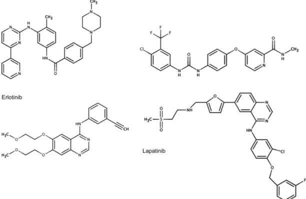
In an attempt to counter these disadvantages, there were developed small molecules inhibitors (SMIs). SMIs can specifically target tumor addresses in order to block cellular pathways or mutant proteins required for cancer cell growth and survival.3,25 Today, a lot of SMIs are under pre-clinical and clinical trials, being tyrosine kinase inhibitors group the most approved for tumor therapy, such as imatinib, sorafenib, erlotinib and lapatinib (Figure 3).3,25,26
Figure 3: Chemical structures of SMIs examples. Adapted.27
1.3. Peptides in Cancer Therapy
Peptides consist in small proteins, constituted by 50-100 amino acids (Table 1). Like proteins, peptides can organize structurally in a primary structure composed by an amino acids sequence and in a secondary structure with a special arrangement, like α helixes and β sheets.28
19
Table 1: Amino acids and their abbreviations. Taken from 29
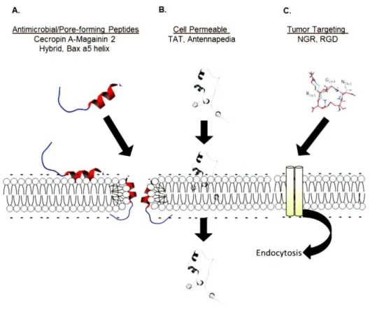
In cancer therapy, peptides are functional domains of proteins with specific bioactivities, including binding to receptors, structural sensitivity to chemical conditions (like acidity and temperature), penetration of the plasma membrane and activation or inhibition of cellular pathways.2,5 Taking into account anti-tumor therapy, there are three main groups of peptides with increased value: antimicrobial peptides or pore-forming peptides (AMPs), cell penetrating peptides (CCPs) and tumor targeting peptides (TTPs) (Figure 4).15
Figure 4: Classes of peptides with advantageous features in cancer therapy and respective examples. A)
20
AMPs (also called host defense peptides or cationic antimicrobial peptides) are cytotoxic agents based in naturally occurring peptides of protective innate immune response to microbes in many species. This peptides has a cationic amphipathic structure that allows them to interact with anionic lipid membranes and induce pores formation or membranes disruption, causing cellular necrosis or apoptosis. Whereas necrosis is the result of AMPs targeting lipids membrane, leading to cells lysis, apoptosis is triggered by mitochondrial membrane disruption. AMPs constitute alternative anti-tumor drugs since their cytotoxicity occurs within minutes, decreasing drug resistance.2
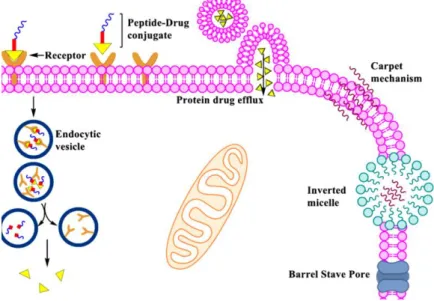
On the other hand, CPPs have the ability to deliver cargos into cells, from small molecules to large microparticles, without having tumor cell specificity. CPPs are able to penetrate cell membranes directly through the plasma membrane or through several internalization mechanisms, such as clathrin- or caveolin-mediated endocytic pathway, micropinocytosis or an endocytosis-independent mechanism, including the carpet, inverted micelle, barrel stave pore and toroidal models (Figure 5).2,15 CPPs can be organized into cationic, hydrophobic and amphipathic groups.15
Figure 5: Different mechanisms used by CCPs to cellular internalization. Adapted.11
1.3.1. Peptides in Tumor Targeting Therapy
Since their discovery about 30 years ago, TTPs, also known as tumor homing peptides, have become an useful tool for tumor therapy and diagnosis, due to their ability to mimic targeting properties of antibodies without macromolecular disadvantages.14 Thus, TTPs bind to specific addresses of tumor tissues with low to no affinity to normal cells. In comparison to antibodies, TTPs have a much smaller size
21
(less than 30 amino acids on average), which improves tissue penetration, low immunogenicity and a production process more simple and at a much lower cost.2,14 Relating to SMIs, peptides are more biocompatible and amenable to introduce structural modifications.15,30,31
Today, there are more than 740 TTPs identified, being RGD and NGR, discovered in 1980, the most studied.2,11,13–15 Besides TTPs with targeting tumor cells ability, TTPs able to target vascular and lymphatic tumor systems and tumor microenvironment cells were found.2,15 In the last 5 years, this vascular homing peptides were intensively investigated since they can destroy tumor microvasculature, promoting tumor cell death by direct accessibility from healthy vasculature system, not needing extravasation to reach the target that may be compromised by poor blood perfusion and high interstitial pressure.2,12,15,17 Furthermore, vasculature systems are more genetically stable, unlikely to acquire drug resistance.13 More recently, cancer therapy research integrated tumor microenvironment homing peptides, due to their important role in metastasis formation.2,14,15
Although “targeting” is a recent concept, there are already classic peptides with tumor targeting ability utilized in clinical practice. For example, goserelin, a gonadotropin releasing hormone (GnRH) receptor agonist, is utilized for breast and prostate cancer therapy, due to its capacity to suppress the expression of testosterone and estrogen. Another example is octreotide, a somatostatin receptors (SSTR) agonist, utilized for treatment of growth hormone producing tumors (Figure 6).32
Figure 6: Chemical structures of classic peptides with targeting ability. Adapted.33,34
1.4. TTPs Discovery Techniques
TTPs can be developed by molecular modeling when the X-ray structure of the receptor is available or by screening combinatorial peptide libraries with known molecular targets. These libraries can be divided into three main types: focused Abs
22
libraries (natural peptides or antibodies), one-bead one-compound (OBOC) libraries and phage display peptide libraries.14 In focused Abs libraries, peptides displaying specific antigen affinity are identified by amino acid sequences of complementarity determining regions (CDRs) of Abs, variable domains of Abs responsible for antigen recognition. However, recent findings show that not all CDRs regions are important for antigen binding and CDRs outside regions can contribute to its coupling.
OBOC library screen methods are based on a chemical technique composed of 90 μm-sized beads, each containing a different peptide ligand.14 In this technique, peptides are subject to a split mix strategy and screened against cell surface targets previously labeled. The advantage of this method is the possibility of incorporating non-natural components in peptides ligands, like D-amino acids, cyclization and branches.14,35
Phage display peptide libraries, mostly utilize to identify TTPs, are focused in a biological approach that creates a random combinatorial library, through genetic modifications of the DNA of phages, allowing the expression of surface ligands.14,36 These type of libraries are exposed to protein targets (for instance, through whole cells, tissue samples and live animals) and then phages with binding capacity are analyzed by DNA sequencing, immunohistochemistry, in vivo imaging and mass spectrometry to identify target-binding peptides.13,14,37 In this method, there are multiple repetitions of selection and amplification steps (panning) to enrich the number of surface targets with the highest binding affinity, allowing the screening of a large number of peptide sequences without the need to have previous knowledge of the molecular composition of the binding site (Figure 7).14,35
Figure 7: Steps of screening and identification of TTPs using the in vivo phage display technology. Taken
23
1.5. Targeted Drug Delivery Systems
Nowadays, both in academia and industry, the development of site-specific drug delivery systems (DDS) and efficient drug targeting approaches are being investigated to surpass the systemic “off-target” effects shown by antitumor therapies and to maximize their therapeutic efficacy.11,13,17,38
Generally, the starting point of a drug targeting approach is a prodrug construction by a covalent modification of the drug with a carrier unit, a small moiety that inactivates the pharmacological effect of the drug during delivery and provides adequate pharmacokinetic properties.38 The carrier unit and the drug are attached by a chemical linker that controls the intracellular release of the drug (for example, acid-cleavable and reduction-sensitivity spacers, where environment changes trigger their intracellular cleavage).11
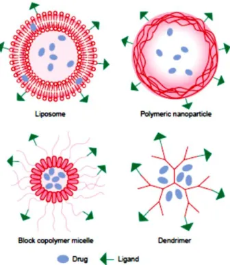
Alternatively, delivery vehicles (also known as particulate drug carriers) may be used to encapsulate the drug and control its biodistribution (Figure 8).38 Delivery vehicles are molecular assemblies with high loading capacity that confine the drug in a loading space (commonly the core of the particulate), providing protection against enzymatic inactivation. Due to the absence of covalent conjugation to entrap the drug molecule, delivery vehicles allow the use of a single carrier unit to formulate several drugs. Furthermore, the composition and size of vehicles can be adjusted to determine particulates with ideals physicochemical properties, which diversifies their stability and, per consequence, the rate of drug release. In the field of cancer therapy, the ideal size is in the 50-150 nm range (nanocarriers) to avoid extravasation into normal tissues and, at the same time, enabling the extravasation from most tumor blood vessels into tumor interstitium.11
24
Figure 8: Delivery vehicles used in targeted DDS to cancer therapy application. Adapted.17
The most common nanocarriers are liposomes, polymeric nanoparticles and block copolymer micelles. Whereas liposomes are lipid molecules orderly in a lamellar disposition, generally used as unilamellar vesicles, polymeric nanoparticles are biocompatible polymers where the drug is entrapped to a vesicle surrounded by a polymer membrane or dispersed in a matrix, named nanocapsules or nanospheres, respectively. Moreover, block copolymer micelles are characteristically spherical amphiphilic copolymers, whose loading space accomodates hydrophobic drugs, and the hydrophilic outer layer promotes dispersal of the micelles in water.17 As drug delivery nanocarriers or imaging probes, nanoparticles have been shown more advantages in comparison with others nanocarriers, including the better tumor penetrating ability, higher ability to carry cargos and quality of imagine information.12 However, the prodrug approach tends to prevent premature drug liberation and decreases inert materials quantity.38
It is common to conjugate hydrophilic polymers, like poly(ethylene glycol) (PEG), onto carrier units’ surface to inhibit their uptake by the reticuloendothelial system, therefore prolonging the in vivo half-life and, consequently, the targeting potential. PEG forms a hydrophilic barrier around the particulate that blocks proteins and other macromolecules to reach the particulate surface, including antibodies raised against the particulate. In this case, the ligand must be conjugated to the terminal of
25
the stabilizing component to prevent the shielding effect that leads to a severe reduction of ligand-receptor interaction.17
For the majority of targeted DDS, the cargo needs to be internalized into cells to exercise its pharmacological activity. After the peptide-receptor interaction, conjugates can be internalized through receptor-mediated endocytosis, where they may move through the early and late endosomes and finally, into lysosomes. Furthermore, to exert pharmacological activity, the cargo has to exit these organelles to reach the cytosol or the nucleus (the two sites of action of intracellularly active drugs). In lysosomes, due to enzymes and/ or pH dropping, the linker between the cargo and the carrier is broken, enabling the cargo escape to cytosol while, in the meantime, receptors are recycled (Figure 9).17,39 If the microenvironmental conditions do not allow the rapid disintegration of the particle, the diffusion of the drug out of the endosomes is necessary, through lytic peptides, pH-sensitive polymers and swellable dendritic polymers.17,32 In the case of nanocarriers, internalization of the drug can occur without concomitant internalization of the particulate, due to atypical conditions of the tumor microenvironment (like acidic pH, presence of lipases, enzymes and oxidizing agents), which results in the accelerated release of the drug and its diffusion into tumor cell, through passive diffusion or active transport.17
Figure 9: Schematic cell internalization process for targeted DDS. Adapted.39
1.5.1. TTPs as Carrier Units in DDS
During the past two decades, more efficient functional peptides were developed.15 In particular, TTPs have been studied as carrier units of peptide-based drug conjugates (PDCs),an emerging class of prodrugs, formed through the covalent
26
attachment of a specific peptide sequence to a drug via a cleavable linker (Figure
10).11,38
Figure 10: Schematic representation of PDCs. A) Schematic representation of tumor targeting approaches
using PDCs; B) Necessary key-properties of PDCs components. Adapted.11
To ensure drug delivery to tumor cells, is necessary to improve stability in vivo and evaluate pharmacokinetic properties of PDCs, that will improve selectivity for cancer cells.2 Generally, peptides have relatively large size, hydrophilicity and hydrogen-binding potential, which makes them unsuitable to be orally administered, due to the difficult passage by intestinal mucosal barriers. Thus, the parenteral route is the most chosen to delivery TTPs conjugates.39 Moreover, it is possible to conjugate multiple TTPs to a single delivery vehicle to increase cargo delivery and prevent peptides degradation by proteolysis, by blocking N- and C-terminally, through incorporation of D-amino acids (unnatural amino acids), cyclization and insertion of reduced peptide bonds.14,31 The most commonly utilized cyclization method is disulfide bridge formation from two Cys residues.40 Other important property of PDCs that promotes efficient delivery of drugs is the internalizing ability of some TTPs into cell membranes.17 Since some TTPs cannot internalize their cargos, CPPs-TPPs
27
conjugates have been developed, which contribute for a better intracellular drug delivery mechanism (Figure 11).11
Figure 11: Principal of cell-selective peptide targeting and delivery. A) TTPs without internalization ability;
B) TTPs-CPPs conjugates; C) TTPs with cell penetration ability. CPHP – Cell-penetrating homing peptides. Adapted.19
TTPs conjugates with cytotoxic drugs, in addition to being exhaustively studied in several preclinical and clinical studies, were also investigated as carriers of radioisotopes.12,30 In this case, there is an attachment of a radio-ligand to the peptide carrier through the aid of a chelator, such as diethylenetriaminepentaacetic acid (DTPA) and 1,4,7,10-tetraazacyclododecane-1,4,7,10-tetraacetic acid (DOTA), but there is no release of the cargo, since it is an unnecessary process. Radionuclides like 99mTc, 111In, 68Ga, 123I, 64Cu and 18F can be use in cancer radiotherapy, due to their longer half-life.31
28
2. Objectives
Nowadays, cytotoxic drugs used in antitumor therapies are not very effective and cause serious adverse effects due to their poor ability to distinguish tumor cells from the normal cells. To solve this problem, molecular systems based on conjugation of a cytotoxic drug and an antibody responsible for the tumor targeting ability are available on the market. However, these therapeutic conjugates have a high economic cost and structural disadvantages. On the other hand, since their discovery about 30 years ago, tumor targeting peptides (TTPs) are able to recognize specifically tumor targets, such as Abs but without their structural disadvantages. Thus, the use of these peptides in targeted therapeutic delivery systems to cure cancer patients is very promising. However, many of the features of these peptides are not yet fully understood.
The aim of this work is to review the literature regarding tumor targeting approaches and to organize and summarize the TTPs that contributed in a significant way to the advances of this field, including the development of therapeutic conjugates. This way, it is intended to focus their structural and functional characteristics, addresses, structure-activity relationship (SAR) studies, internalizing capacity and application in the development of drug conjugates.
29
3. Materials and Methods
3.1. Materials
In this work journal articles were used, mainly review articles published in several international scientific journals, as sources of information. These articles were collected and selected from the National Library of Medicine’s search service (PubMed database), that provides access to millions of references in United States National Library of Medicine and other related databases.
3.2. Methods
Firstly, the literature review started with the collection and reading of articles (especially review articles) regarding tumor targeting-based approaches and peptides with tumor targeting abilities. Then, the most representative peptides of the scientific advances made in this field were selected based on these articles. The selection criteria were based on the number of articles that referred the respective peptides, the quantity of information available, the actuality of the articles and the tumor addresses. Finally, others articles of each selected peptide were collected to summarize information of each, in order to highlight structural and functional features, addresses, structure-activity relationship (SAR) studies, internalization capacity and applications in the development of therapeutic conjugates (Table 2).
30
Table 2: Key-words and expressions utilized to obtain the selected articles. Key words and expressions References
Tumor targeting internalizing peptides review 17 Tumor targeting peptides review 7,11,12,14–16,30–32,37,41
Tumor homing peptides review 2,13,18,19,42 Peptides targeting cancer review 5,35
Peptide drug conjugates review 38,39 Tumor therapy review 1,8,10,26 Cancer therapies review 3,25 Novel cancer treatment review 4,43 F3 peptide nucleolin 44–47
RGD 48–63
NGR 36,64–66
NGR structure activity relationship 67 NGR structure activity 68
NGR pharmacophore 69
Lyp 70,71
Lyp peptide binding 72
Bombesin 73
Bombesin receptor cancer review 74 Bombesin receptor interaction structure 75–77
CREKA 78–82
SSTR mediated endocytosis 83–86
Angiopep 2 87–91
31
4. Results
Following the literature review, nine peptides were selected: RGD, NGR, F3, Octreotide, LyP-1, Bombesin, Angiopep 2, CREKA and M2pep. These peptides were organized according to the targeting localization type: tumor vasculature, tumor lymphatic vessels, tumor cells and tumor microenvironment. Relevant and recent information concerning others TTPs was collected and organized (Annexes 1 and 2).
4.1. Peptides Targeting Tumor Vasculature
4.1.1. RGD
The RGD tripeptide (Arg-Gly-Asp) is the most widely investigated among all TTPs (Figure 12A).14 This tumor targeting peptide was discovered by phage display techniques, as an essential cell recognition site of several blood, extracellular matrix (ECM) and cell surface proteins like fibronectin, vitronectin and fibrinogen.2,7,15,35,42,48,49 The RGD peptide can selectively target endothelial cells of tumor blood vessels expressing αvβ3 and αvβ5 integrins receptors.13,14
Integrins are cell adhesion receptors for ECM proteins, immunoglobulins, growth factors, cytokines and matrix-degrading proteases. These divalent heterodimeric membrane glycoproteins, composed by non-covalently associated α- and β-subunits are expressed at the surface of normal tissue and blood vessels.7,14,42,55 Despite 24 integrins subtypes being known, endothelial cells of angiogenic vessels express a different set of integrins, being αvβ3 and αvβ5 integrins specifically upregulated in tumor vasculature.5,14,35 Furthermore, αvβ3, αvβ5, α5β1, α6β4, α4β1 and αvβ6 integrins are also overexpressed in tumor cells, being the most studied integrins in cancer.51 However, RGD can be recognize by 8-12 of the 24 known integrins.35 The activation of this receptors triggers cellular pathways involved in tumor angiogenesis and metastasis promotion.11,37 In particular, αvβ3 integrin regulates intracellular signaling that protects tumor cells from the anti-proliferative action of anti-tumor drugs.37
There are several RGD recognition sites in α and β subunits of integrins. In all cases, these binding sites are localized at or near a binding site for divalent cations, being the β3 subunit of αvβ3 integrins the primary site for RGD binding.48,57,58 Adjacent binding sites of divalent cations helps to keep a favorable binding conformation. By itself, a simple linear RGD ligand presents low affinity for integrins receptors. This is
32
related to the conformation freedom of the RGD peptide that determines its selectivity.5,60 For example, the RGD-TNFα conjugate is promising, although the
presence of four Cys residues in the peptide structure makes it difficult to fold in a homogeneous manner, compromising its effectiveness.14,42 This way, to obtain a biologically active conformation, several cyclic RGD analogues (cRGD) were developed via “head-to-tail” modification, to create a more rigid structure, more stable at neutral pH and better at resisting proteolysis.5,52,54,60,62
In cyclic peptides, the RGD motif is flanked by other amino acids to build a ring system, which may induce receptor affinity or selectivity and other biological properties.55 cRGD peptides include RGD-4C (ACDCRGDCFCG) and Cilengitide (Figure 12).5,54 Cilengitide, the salt of the cyclic pentapeptide with the sequence Arg-Gly-Asp-DPhe-(NMeVal) - c(RGDf[NMe]V) - demonstrated encouraging results in patients with glioblastoma, which implies penetration of the blood brain barrier (BBB), although clinical trials have not fully proved its effectiveness.2,7,11,19,35,37,59 Cilengitide can act as a specific antagonist of αvβ3 and αvβ5 integrins.9,35 Others structural modifications in Cilengitide, including the introduction of the unnatural D-conformation of Phe amino acid and N-methylation, improve its receptor affinity and pharmacokinetic properties.60 More recently, three PDCs were developed based on this pentapeptide, in which the mutilated Val was mutated to Lys or Ser amino acids for the creation of a primary amine or hidroxyl group, potential sites for drug conjugation.35
33
On the other hand, others RGD peptides were designed to improve tumor penetration.2 Internalizing RGD (iRGD) is a 9-amino acid, disulfide-bridged cyclic peptide with the ability to facilitate the internalization into endothelial cells, in addition to target αvβ3 integrins.2,13,14,97 This extra capacity allows the intracellular uptake of the conjugated cargos, contributing to the internalizing process of antitumor drugs. The penetration capacity is due to a specific sequence contained in the peptide, the sequence R/KXXR/K (X: any amino acid) at C-terminal, named C-end Rule (CendR), that binds to neuropilin-1 (NRP-1), a transmembrane protein expressed on membranes of endothelial cells that, when activated, triggers downstream signal pathways to increase cell permeability.2,13,14,63 After iRGD connection to an integrin, a protease cleaves the peptide to produce CRGDK/R and exposes the CendR motif that can interact with NRP-1 receptors (Figure 13).55,61 The CendR penetration is receptor-mediated, energy-dependent, effective with small molecules and causes extravasation of the peptide (or their cargos) within minutes.61
Figure 13: Penetration mechanism of iRGD. Taken from 55
The current clinical trials involving RGD motif (Table 7, Annex 3) are testing its application not only in tumor therapy, but also in diagnostic imaging, through conjugation with cytotoxic drugs, peptides or proteins, nucleic acids, radionuclides and contrast agents. For instance, the first RGD-modified positron emission tomography (PET) tracer in clinical trials was Galacto-RGD, an RGD analog obtain by cyclization of a RGD pentapeptide and modification of Phe to the unnatural configuration – c(RGDfV).5,52
More specifically, in clinical trials, the improvement of the delivery and therapy efficacy of tumor therapy agents when conjugated with RGD peptide or analogues,
34
namely cytotoxic drugs like paclitaxel (PTX) and doxorubicin (DOX), therapeutic peptides such as KLAKLAKKLAKLAK (KLA), cytokines like tumor necrosis factor α (TNFα), interferon-γ (IFN-γ) and interleukin-12 (IL-12), Abs and their fragments such as the Fc fragment of immunoglobulin G (Ig G) was proven. 12,13 Furthermore, RGD-targeted nanocarriers benefit of the possibility to enhance the internalization process via integrin-mediated endocytosis, instead of standard uptake mechanisms after drug release, such as the RGD-modified PEGylated liposome-encapsulates DOX.5,55
4.1.2. NGR
The NGR tripeptide (Asn-Gly-Arg), discovered by the in vivo phage display method, specifically binds to tumor endothelial cells expressing the aminopeptidade N (APN) receptor, also called CD13.13,37,64 Like RGD, this sequence are encountered in natural proteins, such as fibronectine.36
CD13 is a membrane-bound and highly glycosylated metalloproteinase, with a significantly role in protein degradation and regulation of cytokines, antigen presentation, cell proliferation and migration and angiogenesis.13,42,66 Although it is overexpressed in tumor blood vessels, CD13 can be find in tumor cells, fibroblasts and pericytes, and in normal tissue like mast cells, keratinocytes, proximal renal tubules, myeloid cells and epithelial cells.13,36,42 There are several CD13 isoforms, which could explain the binding of NGR peptides to tumor vasculature but not to other CD13-rich tissues.14,42,68 Furthermore, in endothelial cells, CD13 interacts with galectin-3 (a
proangiogenic protein) in a carbohydrate recognition-dependent manner, forming a complex that, together with the abnormal architecture of tumor blood vessels, may be related to differential glycosylation or conformational changes and, therefore, with the selectivity of NGR motif to endothelial CD13.36
Recent studies suggest that NGR can interact with integrins by a spontaneous and unusual mechanism that consists in the rapid deamidation of the Asn residue, through the formation of a succinimide ring, followed by hydrolysis, forming isoaspartate and transforming NGR into the isoaspartate-glycine-arginine (isoDGR) derivate, a cell adhesion motif with high affinity for αvβ3 and αvβ5 integrins (Figure
14A).36,65,68,69,98 Thus, isoDGR can promote endothelial cell adhesion, having a binding site located within the RGD binding pocket, being as well an antagonist of ανβ3 integrin.36,65 However, mostly in injured tissues and wound healing, cells produce protein-L-isoAsp-O-methyltransferase (PIMT), an enzyme that converts isoaspartate to aspartate, causing the deactivation of isoDGR function (Figure 14B).36
35
Figure 14: Formation of isoDGR (A) and schematic representation of effects on receptor interactions (B).
Taken from 36
Several therapeutic molecules have been conjugated to NGR peptides, namely cytotoxic drugs, therapeutic proteins, proapoptotic peptides, viral particles, imaging agents and DNA complexes. Specific examples include conjugation of NGR with DOX, the proapoptotic peptide D and TNFα.13,64 This conjugates may penetrate cell membrane via receptor mediated endocytosis.68
36
Generaly, small cyclic NGR peptides are more selective than linear forms, due to conformational constraining that improves binding affinity. The most effective cyclic NGR peptides developed are c(CNGRC) and (KNGRE)-NH2 (Figure 15A and B). The c(CNGRC) was designed with the disulfide bonding of the two Cys residues, increasing the targeting efficiency due to stabilization of the bent conformation.14,64,65,68 Particularly, cCNGRC-TNFα (currently tested in Phase III clinical trials) (Table 8, Annex 3) have shown a promising potent anticancer activity due to its capacity to facilitate drug penetration and infiltration of immune cells, since TNFα increases intracellular adhesion molecules on endothelial cells, expression of pro-inflammatory cytokines and recruitment of tumor-specific cytotoxic T cells.13,64 Moreover, to surpasses the disulfide bond disadvantages (susceptibility to biodegradation and chemical modification), the “head-to-side-chain” amide bond cyclized peptide - c(KNGRE)-NH2 - was developed.65,68 Other examples of NGR peptides are GNGRG, NGRAHA and CVLNGRMEC.64 SAR studies of GNGRG (Figure 15C) suggest that the linear NGR peptide without disulfide constraints is more thermodynamically favorable when its configuration is based in a β-turn in Gly3 and Arg4and hydrogen bonding interactions between Asn2 and Cys5. Thus, like the RGD receptor binding, there is necessary a folded structure for receptor binding, with intramolecular stabilizing interactions (like hydrogen bonds) that stabilize the folded state.67
37
Figure 15: Structure of common NGR peptides. Adapted.65
Similar to the iRGD, internalizing NGR (iNGR) possesses the CendR motif and, therefore, the ability to bind to NRP-1 and penetrate tumor endothelial cells.13
4.1.3. F3
F3 peptide (KDEPQRRSARLSAKPAPPKPEPKPKKAPAKK) is a 34–amino acid fragment of the human high mobility group protein 2 (HMG2N), a nucleosomal protein that participates in unfolding of chromatin structure, facilitating the transcriptional activation of genes.44,46,47,99 This tumor targeting peptide, discovered by the phage display libraries technique, binds to the nucleolin (NCL) receptor overexpressed in tumor cells, tumor endothelial cells and in a bone marrow subpopulation, precursor of endothelial cells.44,46,99
NCL is a non-ribosomal protein important to polymerase I transcription, being found in high quantity in the cell nucleolus, where ribosome assembly and ribosomal ribonucleic acid (rRNA) transcription occurs. Other nucleoplasmic NCL receptors are responsible for the regulation of gene expression, mostly of oncogenes, and genome
38
stability, due to its interaction with RNA polymerase II. Moreover, in cytoplasm, nucleolin interacts with messenger ribonucleic acid (mRNAs) coding proteins related with cell proliferation and apoptosis. NCL structure can be divided in three domains: the negatively charged N-terminal domain (with acidic regions due to the abundance in aspartic acid and glutamic acid) disunited by basic stretches, the central domain containing fours RNA binding domains and the C-terminal domain, predominantly with Gly, Arg and Phe residues.45,99
In tumor cells and tumor endothelial cells, overexpressed NCL promotes the cell internalization of several ligands related with angiogenesis, proliferation and apoptosis.45 F3 peptide can be internalized by NCL due to its NH2-terminal domain (via receptor-mediated endocytosis) and carried into the nucleus after cell penetration.14,99 SAR studies show that the D-amino acid form of F3 has equal internalization ability, although it is not efficiently transported into the nucleus. This internalizing mechanism is not defined, but studies have shown energy dependence for the mechanism occurrence and the possibility of F3 interaction with NRP-1.14,46
F3 peptide was conjugated with radiotherapeutic agents for tumor therapy, such as 225Ac, 213Bi and 111In.13 Nonetheless, there are no studies about the conjugation between this tumor targeting peptide and cancer drugs.19
4.1.4. Octreotide
SST is an endogenous acid popypetide with the sequence Ala-Gly-c(Cys-Lys-Asn-Phe-Phe-Trp-LysThr-Phe-Thr-Ser-Cys), with high-affinity for SSTRs. This peptide acts as an endogenous inhibitory regulator and has various biological functions, including inhibition of many hormone secretions, cell survival and cell proliferation.41,83 However, SST possesses an extremely short half-life (about 2-3 minute), due to its rapid inactivation by peptidases, being limited its clinical utility.41,86 Thus, several synthetic SST analogues were developed to treat endocrine tumors, through shortening of its sequence and introducing D-amino acids to increase the half-life time. SAR studies demonstrate that the key sequence for binding and biological activity was the β-turn fragment Phe-Trp-Lys-Thr sequence, the residues 7-10 of the SST, in addition to the existing disulfide bridge.83,86 After interacting with the receptor, SST
analogues are rapidly internalized into tumor cells (via SSTR-mediated endocytosis), being able to translocate to the nucleus.83,84 Furthermore, these analogues have been
used to developed conjugates with application in targeting therapy, radiotherapy and tumor imaging (Figure 16).83
39
Figure 16: Schematic chemical structures of SSTs and analogues. Taken from 83
SSTRs are transmembrane G-protein coupled receptors expressed in the central nervous and immune systems, endocrine tissue, gastrointestinal tract and skin, playing different physiological roles.35,41,85 There are five distinct SSTR subtypes, being the SST2R and SST5R receptors overexpressed in various types of tumors, not only in tumor cells, but also in tumor endothelial cells.32,83
Octreotide, a cyclic octapeptide with the sequence D-Phe1-c(Cys2-Phe3-D-Trp4-Lys5-Thr6-Cys7)-Thr8-ol), is an agonist of SSTRs, namely SST2R subtype, that mimics the natural SST hormone, although it is a more potent inhibitor of growth hormone, insulin and glucagon.32,41,86 Nowadays, octreotide is utilized successfully against growth hormone-producing tumors in many countries.32,41 The remarkable feature of octreotide is its stability towards degradative enzymes.84 The β-turn is a stable conformation due to the N- and C-terminal of the D-Phe1 and Thr(ol)8 or Trp8-NH2 amino acids by intramolecular hydrogen bonds that stabilize the conformation of the peptide.86 Besides, SAR studies also showed that both C- and N-terminal residues of octreotide are important for binding affinity.39
Potent chemotherapeutics agents including PTX, camptothecin (CPT), DOX and methotrexate (MTX) have been used for the development of octreotide conjugates.32,83 For example, two molecules of PTX were coupled to the N-terminal of octreotide by an ester bond and utilizing a succinic acid spacer (Figure 17).32 More
40
recently, the redox-sensitive prodrug octreotide(Phe)-polyethylene glycol-disulfide bond-paclitaxel - OCT(Phe)-PEG-ss-PTX - was design.41 On the other hand, the first radiolabeled SST analog clinically applied was the (111In-DTPA0)-octreotide.5,84
Figure 17: Chemical structure of ester bond-linked PTX-octreotide. Adapted.31
4.2. Peptides Targeting Tumor Lymphatic Vessels
4.2.1. LyP-1
LyP-1 is a cyclic nonapeptide (CGNKRTRGC) with the capability to target tumor lymphatic vessels and tumor cells in hypoxic areas, by interaction with p32, a mitochondrial/ cell surface protein receptor (Figure 18).14,16 LyP-1 can penetrate the plasma membrane into the cytoplasm and nucleus.72 By itself, the linear form of LyP-1 peptide has cytotoxic activity, since it accumulates in tumor hypoxia areas, decreasing the number of lymphatic vessels and promoting apoptosis of tumor cells undergoing stress.14,70,72 For instance, LyP-1 was used to improve the delivery of DOX-loaded PEGylated liposomes.15
41
Figure 18: Cyclic structure of LyP-1. Adapted.72
The p32 receptor, also called as hyaluronic acid binding protein 1, is overexpressed on tumor lymphatics, tumor cells and TAMs.15,16 However, the molecular mechanism of LyP-1 recognition by the receptor is poorly understood.72 On the other hand, LyP-1 internalization is similar to iRGD penetrating mechanism. The binding of LyP-1 to the p32 receptor triggers its proteolytic cleavage into tLyP-1 (CGNKRTR), allowing the exposure of CendR motif and the binding to NRP receptor.71
4.3. Peptides Targeting Tumor Cells
4.3.1. Bombesin
Bombesin is a 14-amino acid neuropeptide (QQRLGNQWAVGHLM), containing high affinity to the gastrin realizing peptide receptor (GRPR), a G protein-coupled receptor overexpressed in tumor cells, also named as bombesin receptor type 2 (BB2).11,39,41,73,75
Mammalian bombesin receptors (BBRs) can be divided into four receptor subtypes: neuromedin B receptor (NMBR or BB1), GRPR and bombesin receptor subtypes 3 and 4 (BB3 or BRS-3 and BB4, respectively).35,73 These receptors can naturally occur in central nervous system (CNS) and in peripheral tissues, having a wide spectrum of actions in physiological processes.74 The activation of BBRs promotes the release of others peptide hormones like insulin, glucagon, prolactin and gastrin and cytokine activity, being able to act as a growth factor.75 Although there are four subtypes of BBRs, the natural ligand of BB3 is unknown and it has low affinity for bombesin peptides.74 As tumors can secrete bombesin to stimulate tumor growth, tumor targeting approaches based on this peptide may interrupt tumor autocrine-growth.35,74
42
BBRs antagonists can block the tumor growth stimulated by bombesin peptides. While BBRs agonists are internalized via receptor-mediated endocytosis, their antagonists have low to no internalization ability.74 To enhance bombesin antagonist
activity, it is possible to cleave the peptide bond in its active site (positions one to five or Tyr4-Lys3, and Tyr4-D-Phe12), introduce a nonpeptide between C-terminal and adjacent amino acid residues or replace them, incorporate D-amino acids, ester modifications, disulfide bridges and non-peptide bonds.75 The selectivity of GRPR antagonists depends mostly on interactions with amino acids Thr297, Phe302, and Ser305 in the fourth extracellular domain of the receptor. All of these amino acid faces forward in the binding pocket, demonstrating that cation and hydrogen bonding interactions are essential to occur between antagonists and the three respective amino acids.76,77 Examples of GRPR antagonist are JMV594 [(D-Phe6, Stat13)Bn(6–14)], and JMV641 [D-Phe-Gln-Trp-Ala-Val-GlyHis-Leu(CHOH-CH2)-(CH2)2-CH3].76
The bombesin sequence D-Tyr6-β-Ala11-Phe13-Nle14 is responsible for the rapid cell internalization in all three types of BBRs, being able to be used to develop targeting conjugates based in bombesin.32,41 For instance, bombesin conjugates through the coupling with MTX via a Lys spacer were developed, as well as DOX via a glutaric acid spacer and taxol using a PEG linker.11,39 Others conjugates of bombesin were prepared by loading them with CPT, PTX, mitochondria-disruptive peptides, marine toxins and siRNA.35 Moreover, a large number of bombesin analogues were conjugated with radioisotopes like 99mTc, 111In and 125I and other contrast agents, and bivalent probes with 64Cu as the cargo of the conjugate were prepared.74
4.3.2. Angiopep 2
Angiopep 2 is a 19-amino acid peptide with the ability to cross the BBB, targeting specifically the low-density lipoprotein receptor-related protein 1 (LRP-1).14
The BBB is responsible for brain protection from potentially harmful substances circulating in the bloodstream and for maintaining the homeostatic environment of the central nervous system. This neurovascular unit is constituted by endothelial cells with extensive tight junctions, neurons, astrocytes and a contractile apparatus of smooth muscle cells and pericytes.89,91
LRP-1 is a member of the large receptor family of low density lipoprotein (LDL). Like other receptors of this family, the modular structure of LRP-1 includes Cys-rich complement-type repeats, a cytoplasmic domain and a transmembrane domain. This receptor is highly expressed in astrocytes, smooth muscle cells, neurons and perycites,
43
but is not as expressed in the endothelium.89,91 LRP-1 mediates the BBB transcytosis process for several ligands such as lactoferrin, thyroglobulin, α2 macroglobulin and tissue-type plasminogen activator. In addition to being expressed on the surface of the BBB and controlling its permeability, LRP-1 is also present on a variety of tumors, mainly in high malignant glioma cells, participating in the cytoskeleton organization and in the adhesive complex turnover by modulating integrins functions in malignant cells.88,89,91 Thus, although BBB has low permeability for small-molecule drugs,
angiopep 2 is capable to penetrate the BBB and target brain tumor cells, using the same receptor-mediated transporter.14,32,87,90
The most promising drug conjugate based on angiopep 2 consists in a 19-amino-acid linear angiopeptin-2-PTX conjugate (GRN1005). This peptide drug conjugate is composed by three PTX molecules linked to the two Lys residues (positions 5 and 9) of angiopep 2 and to the N-terminal Thr, by cleavable ester linkers.32,35 GRN1005 crosses the BBB by LRP-1 receptor-mediated transcytosis and is distributed broadly throughout brain parenchyma (Figure 19).32,88 Howerver, the exact molecular mechanism of angiopep transcytosis remains unknown.90 Further studies showed that GRN1005 is not a substrate for P-glycoprotein-mediated drug efflux, improving anti-tumor efficacy.88
Other angiopep 2 drug conjugates were created, including angiopep-2– doxorubicin, angiopep-2–dimethylglycine etoposide and angipep 2–trastuzumab (efficient against HER2-positive intracranial tumors).35
44
Figure 19: Internalization of GNR1005 through BBB (A) and tumor cell surface (B). Taken from 88
4.4. Peptides Targeting the Tumor Microenvironment
4.4.1. CREKA
CREKA is a linear pentapeptide (Cys-Arg-Glu-Lys-Ala) identified by phage display libraries that targets fibrin-fibronectin complexes deposited in tumor vessels walls and tumor interstitial spaces, forming a meshwork of clotted plasma proteins detectable only in tumor tissues.14,15,78,81 Moreover, CREKA can induce tumor clotting, creating new binding sites in a self-amplifying effect. The exact binding site of the CREKA peptide is still unknown.
The bioactive conformation of CREKA is based on a turn-like structure where the charged groups of Glu, Lys and Arg form stable intermolecular interactions.82 Moreover, the sulfhydryl group of the Cys residue of CREKA is not required for binding interactions with its receptor, being the preferred site to conjugate the peptide with
45
other moieties.11,78 This knowledge promoted the design of CREKA-based peptides with enhanced features. For example, the CR(NMe)EKA has a N-metil derivate that increase tumor-homing response by protecting the peptide against proteolytic degradation (Figure 20).82 Moreover, in order to block the action of tumor-associated platelets on tumor metastatic progression, created liposomal nanoparticles bearing CREKA and ticagrelor (a platelet inhibitor) were created.80
Figure 20: Chemical structures of CREKA and CR(N-Me)EKA. Taken from 82
4.4.2. M2pep
The sequence YEQDPWGVKWWY, termed M2pep, identified using a phage display strategy, is a unique M2-selective peptide that specifically recognizes and internalizes M2 cells, including TAMs, having low affinity to others leukocytes.14,15,94
Macrophages are phagocytic cells originated from circulating blood monocytes that extravasate into tissues and differentiate into macrophages with several functional phenotypes. Whereas M1 macrophages phenotype is stimulated by mediators, like interferon γ or lipopolysaccharide, resulting in a pro-inflammatory and microbial functional phenotype, M2 macrophages are stimulated by IL-4 and IL-13 in tissue remodeling and inflammation resolution cases.94 TAMs are originated from circulating
monocytes, differentiating within the tumor microenvironment, in M1 or M2 activated macrophages.93,95 Most of TAMs are M2-like macrophages and have the ability to produce immunosuppressive cytokines and have low antigen-presenting and co-stimulating capacity, facilitating tumor progression by suppressing the adaptive immune


