Braz. Arch. Biol. Technol. v.54 n.1: pp. 99-106, Jan/Feb2011 Vol.54, n. 1: pp. 99-106, January-February 2011
ISSN 1516-8913 Printed in Brazil BRAZILIAN ARCHIVES OF
BIOLOGY AND TECHNOLOGY
A N I N T E R N A T I O N A L J O U R N A L
A Nonspecific Primer Anchored PCR Technique for
Chromosome Walking
Yan Zhang, Daqun Liu
*, Wenxiang Yang, Yaning Li and Hongfei Yan
College of Plant Protection; Agricultural University of Hebei; Biological Control Center for Plant Diseases and Plant Pests of Hebei Province; Baoding 071001; China
ABSTRACT
A chromosome walking method was improved in this work. The new method was named as nonspecific primer anchored PCR (NPA-PCR). Nested gene specific primers were designed based on the known region and long random primer using degeneracy oligonucleotides for nonspecific anchoring. Annealing temperatures were varied to control the priming. Target sequences were obtained by PCR with random primer and gene-specific primer. Nonspecific sequence with long random primers at both ends formed stem loop structure due to inverted terminal repeats. The method was employed to isolate a gene with newly-isolated actinomycin producing strain Streptomyces setonii Z-L-22. A 0.77 kb fragment of actinomycin synthetase gene cluster was isolated from the strain. The fragments of 1474bp and 701bp were obtained, respectively, at the up and down streams of known fragment through the this method. NCBI Blast analysis showed that the walking sequence and the known sequence were located conjointly in the same cluster gene. It demonstrated that the result was correct and this technique could be useful and efficient for chromosome walking or isolating the gene.
Key words: nonspecific primer anchored PCR; chromosome walking; random primer
*Author for correspondence: [email protected]
INTRODUCTION
The hunt for missing sequence data is often required in the experiments. Several polymerase chain reactions (PCR)-based methodologies are available for walking from a known region to cloned or uncloned genomic DNA, including inverse PCR (IPCR)(Triglia et al., 1988; Melanie et al.,2004, Ochman et al.,1988), adaptor ligation (LM-PCR) (Espelund and Jakobsen, 1992; Kilstrupand Kristiansen, 2000; Yan et al.,2003; Chen et al., 2004) and randomly primed PCR (RP-PCR). The first two methods have high specificity
but require enzyme digestion and ligation, making them relatively expensive and time-consuming. Thermal asymmetric interlaced PCR (TAIL-PCR) (Liu et al., 1995) is a representative method of the third one. This method has its specific advantage but the amplified products are generally limited to a length of <1 kb.
MATERIALS AND METHODS
Template DNA
Streptomyces setonii Z-L-22 was used in this
work. Its genomic DNA was extracted according to the method described by Kieser et al., (2000).
Walking primers
Two sets of nested specific primers were designed
based on the known sequence. GSP1 and GSP2 started respectively from 216bp and 89bp in the upstream of the known region. GSP3 and GSP4 started respectively from 502bp and 701bp in the downstream of the known region. The sequence of anchoring primers SAP1, SAP2 used in NPA-PCR and SSP1, SSP2 used in NASS-PCR together with gene specific primers are shown as follows.
PCR procedure
Four steps were included in NPA-PCR. In the first step, PCR mixture (15 l final volume) including 50 ng of template DNA, 1 U of long and accurate
Taq DNA polymerase (TaKaRa Biotechnology Co., Ltd), 0.2 M dNTP mixture, 10 l 2 × GC buffer I, 0.1 M SAP1. 5 l GSP1 (with the final concentration of 0.5 M) was added, and the reaction cycles were run (Table 1) then the second step started. The third step was carried out in 25 l volume of reaction mixture composed of the reaction products above and 2.5 l SAP2 with the final concentration of 0.5 M and 2.5 l 2 × GC buffer I and run the program in table 1. For the fourth step, 1 l of the third step PCR products diluted 50 times with Milli-Q water was used as the PCR template. The reaction mixture was composed of 0.5 M GSP2, 0.5 M SAP2, 0.2 M
dNTPs, l × GC buffer I and 1 U Taq DNA polymerase. After the PCR, the reaction mixture was resolved by electrophoresis on a 1.2% agarose gel and DNA fragments were visualized by staining in ethidium bromide. Reactions of NPA-PCR and NASS-NPA-PCR are shown in Table 1.
Braz. Arch. Biol. Technol. v.54 n.1: pp. 99-106, Jan/Feb2011 Table 1 - Reactions of NPA-PCR and NASS-PCR.
NPA-PCR NASS-PCR
steps Primers adding cycles procedure steps Primers adding cycles procedure
1 SAP1
1 94°C 5min 30°C 3min
ramp to 65°C at the rate of 0.25°C/min 65°C 5min
2 GSP1
(GSP3)
10 94°C 30s 65°C 3min 1 72°C 5min
1
SSP1 GSP1 (GSP3)
1 94°C 5min
10 94°C 30s 60°C 1min 1 72°C 3min
94°C 30s 35°C 3 min ramp to 65°C at the rate of 0.25°C/min
72°C 5min
3
SAP2 1 94°C 2min 20 94°C 30s
65°C 3min 1 72°C 5min
2 SSP2
1 94°C 2min 20 94°C 30s
60°C 1min 1 72°C 7min
4
GSP2 (GSP4)
SAP2
1 94°C 2min 30 94°C 30s
65°C 3min 1 72°C 5min
3
GSP2 (GSP4)
SSP2
1 94°C 2min 30 94°C 30s
60°C 1min 1 72°C 7min
RESULTS
The principle of NPA-PCR
The principle of NPA-PCR is illustrated in Figure 1. Four primers were used in NPA-PCR. Two sequence anchoring primer (SAP), SAP1 and SAP2 (a part of SAP1), were as the nonspecific anchoring. Two gene specific primers (GSP), GSP1 and GSP2, were nested primers (Fig. 1a). Four steps were included in NPA-PCR. First, a single cycle of PCR was carried out at a low annealing temperature with only primer SAP1. At this temperature, SAP1 could prime at target or non-target positions on the DNA template (Fig. 1b). Then the annealing temperature was increased to the point corresponding to the annealing temperature of GSP1. GSP1 was added at this time the amplification products were produced when SAP1 hybridized with target position by SAP1 and GSP1, meanwhile the non-target gene amplification was suppressed by the stem-loop structure of the DNA (Fig. 1c). SAP2 was added in the third step. The amplification was carried out by GSP1 and SAP2 to generate the template (Fig. 1d). The products of third step were diluted and used as template in the last step. Target DNA was exponentially amplified by the nested PCR with
GSP2 and SAP2 (Fig. 1e).
NPA-PCR and NASS-PCR were carried out according the protocol described above. The PCR products were separated on 1.2% agarose gel and
examined (Fig. 2). The amplification results were the same for the upstream walking by NPA-PCR and NASS-PCR. Bands with the length of approximately 1500bp were obtained. Different results were obtained at downstream walking: a band of approximately 700bp by NPA-PCR while 600bp by NASS-PCR. The products were purified and sequenced.
Sequence analysis
Upstreamed 1474bp-fragments were obtained both by NPA-PCR and NASS-PCR. The sequences were the same except for the primer sequences of SAP1 and SSP1. The walking sequences had 86bp overlap with known sequence. A fragment of 701bp was obtained by NPA-PCR at the downstream with 70bp overlap to the known one and was consistently with design. A 2716bp-fragment obtained by assembling the walking sequence and the original sequence was shown below. The sequence had 79% identity with actinomycin synthetase III (acmC) gene cluster of
Streptomyces chrysomallus, which was consistent
Figure 1 - Schematic outline of NPA-PCR method for chromosome walking Known and unknown sequences are depicted with thick and thin lines, respectively. DNA sequences with thin white arrows in them represent the same sequences as the primers. SAP1 is composed of grey, blue and black segments. Grey segments show the expected specific hybridized sites, black segments show the same part of SAP1 and SAP2, blue segments show the nonspecific hybridized parts particular in SAP1.
Figure 2 - Amplification results of genomic DNA of Streptomyces setonii Z-L-22 by NPA-PCR and NASS-PCR M, DNA ladder: DL2000. 1: upstream walking by NPA-PCR; 2: upstream walking by NASS-PCR; 3: downstream walking by NPA-PCR; 4: downstream walking by NPA-PCR.
b. Add SAP1, annealing at 30℃
c. Add GSP1, PCR with GSP1 and SAP1
GSP1 GSP2
SAP1 SAP1
GSP1
d. Add SAP2, PCR with SAP2 and GSP1
SAP2
GSP1
SAP2
GSP2
e. Add GSP2, PCR with GSP2 and SAP2
GSP1 GSP2 GDGGDG
a. Primer design target sequence Non target sequence
SAP1
SAP1
SAP1
SAP1
SAP1
Braz. Arch. Biol. Technol. v.54 n.1: pp. 99-106, Jan/Feb2011
CCTGAGACCTATTACCGCCCAGAGGATCTCCGTGTCCTCTGCATAACGAACTCCTCGGCGA
CCAGAACGACCCCCACAGCCTCTTCACCAACCAGATCACCCACTGGAAAAACACCCTCGAA
AACCTCCCCGACCACATCACCATCCCCACCGACCGACCCCGCCCCACCATCACCACCTACCA
AGGCGACTACACCACCATCACCATCAACCCCACCCTCCACCAAAAAATCACCCACCTCGCCC
ACACCACCAACACCACCGTCTTCATGGTCCTCCAAGCAGGACTCACCACCCTCCTCACCAAG
CTCGGCGCCGGAAACGACATCCCCATCGGCACCCCCATCGCCGGACGCACCGACCACAACC
TCGACAACCTCATCGGCTTCTTCGTCAACACCCTCGTCCTCCGCACCGACACCACCAACAAC
CCCACCTTCACCCAACTCATCAACCGCATCCGCGAAACCAACCTCACCGCCTACGCCCACCA
AGACGTCCCCTTCGAATACCTCGTCGAAATCCTCAACCCCACCCGCACCCTCAACCACCACC
CCCTCTTCCAAATAATGCTCGCCCTCCAAAACGCACCCGAAGGAGCATTCCAACTCCCCGGC
CTCACCATCGACATCGCCCCCGGACGCACCGGAACCGCCAAATTCGATCTCTTCTTCAGCCT
CGTCGAGAAACGCGGACCCCACGGCGAACCCGAAGGCATCACCGGCGCCATCGAATACTCC
AGCGACCTCTACGACGAAACCACCGTCCACACCCTCTTCGAACGCCTCACCCGCATCCTCGA
AACCGCCACCGACCACCCCGACCAACCCCTCAGCCACATCGACGCCCTCAGCCCGCACGAG
AAGCACCAGGTGCTGGACTCCTGGCTGGAGACGGCGACGGAGGTAGGGGCCGGTCTGCTGC
CTGCCCGCTTCTCAGAGCAAGCGTCCGCGACACCGGACGCCGTGTCCCTGGTCGCCGGCGAC
ACCGTCCTGACCTATGCCGAACTGGACCGGCGGGCCAACCGGCTGGCCCGAGTCCTGCTCGA
CCGAGGTGCGGGAGCCGGCCGTGTGACCGCCATCGCGCTGCCGCGCTCGGCCGACCTGGTG
GTGGCACTGCTGGCAGTGCTCAAGTCCGGCGGCGCCTACCTGCCGCTCGACCCCGACCACCC
GGCGGCACGCCTTACGCACGTCCTGGAGGATGCCCGCCCCTCACTGCTGCTCACGACCACGG
CGACGGACTGGATGATCCCGGAGGTGGGCGCCGCACAGCGGCTGGTCCTCGATTCCGACCC
CGTACGGGAGGCATCGGACGCCGCCCTCGACAGCGATCCGGCCGACGACGGCGGGGTCGCC
CCCCTCCGGCCGGAGGACGCCGCGTACGTCATCTACACCTCGGGCTCGCACCGGCCGGCCGA
AGGGCGTGGTGGTCCCGCACGGCGCCTTGCTCAACTTCCTCGTGGGCATGCGGGAGAAGGCT
CCGATCCGGCCGGAGGACCGGCTGCTC
GCGGTCACCACCGTCGCCTTCGACATCGCCGCCCTCGAGCTCTACCACCCCCTGCTCTCCGGC
GCCGCCGTCGTCATCGCACCCAAGGAAGCCGTTCCGCAGCCCTCGGCCGTGCTGGACCTGAT
CGAACGGCACAGCGTCACGGTCGTGCAGGGCACCCCGTCCCTGTGGCAACTGCTCGTGGCGC
ACGACGCGGAGGCGCTTCGAGGGCTACGGATACTGGTCGGCGGCGAAGCGCTGCCGGTCTC
CCTCGCCGAGGCCCTGCGCGGGCTCACCGACGACCTGGTCAACCTGTACGGCCCGACCGAG
ACCACCATCTGGTCGACCGCTGCGGATCTGGCCGGCCACACCGGAGGGCCGGCACCGATCG
GGCGGCCGATCGCAAACACCCGGGTCTACGTGCTGGACTCCGCTCTGCAGCCGGCCGCTCCC
GGTGTGGTGGGTGAGCTGTACGTCGCGGGCGAGGGCCTGGCCCGCGGCTACGCCAACCAGC
CCGGGCTCACCGCCGAACGCTTCGTCGCAGATCCGTTCCACCCCGAACCCGGGCAACGGATG SAP1
TACCGCACCGGCGACCTCGCCCGCTGGAACACCCACGGCGAACTCGAATACCTCGGCCGTAC
CGA
CCACCAGGTCAAGATGCGGGGCTACCGCATCGAACCCGGCGAGATCGAGAAGACCCTCACC
GACCACCCCGACATCGCCCAGGCCGCCGTGATCGTACGGGAGGACAGGCCGGGAGACCTCC
GCCTGGTCGGGTACGTCGTGGCCGACAGCGGGGGCGGAGTCCGTGACGAGCATGTCGAGCG
GGACCAGCTGAGCGAGTGGCAGGACCTCTACGACTCCGTCTACACGTCCGCCGGCGAAACT
GACATCGGCGAGAACTTCGCGAGCTGGAACAGCAGTTACGACGGTCAGCCCATCCCACTGG
AAGACATGCGGGAATGGCGCGACAGCACCGTCGACCGCATCAAAGCACTCCACCCACGACG
CGTCCTGGAAATCGGCGTCGGCACCGGCCTCCTGCTCGCCAAAATCGCACCGGACTGCGACG
AATACTGGGGAACCGACTTCTCCCCGACCGTCATCGAAGACCTACGACACCACATAGAAGC
CGACCCCCACCTCACCGAGAAAGTCACCCTCCGCACCCAAGCCGCACACGAACACGGCGAC
CTCAAGAAGAACCACTTCGACACCATCATCCTCAACTCCGTCGTGCAGTACTTCCCCAACGC
CGGATGCATGGAGCTGCCTCTAGGAGACCCGCCATTATCCAGAGTCC
Amplification of the assembling sequence from genome DNA
The PCR were carried out using the gene specific primers designed from the assembling sequence with genome DNA to verify the walking results.
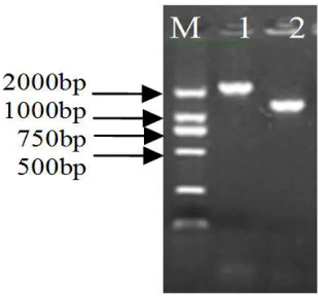
The amplification results are shown in Figure 3. Expected fragments were 2100bp and 1300bp. The amplification fragments shown in the gel were consistent with the expected. Sequencing results were same as the expected.
Figure 3 - Validation the walking results by amplification M, DNA ladder marker, DL2000. 1: Validation the assembling sequence at 5’ by amplification; 2: Validation the assembling sequence at 5’ by amplification.
GSP4
Braz. Arch. Biol. Technol. v.54 n.1: pp. 99-106, Jan/Feb2011
DISCUSSION
Anchored PCR is often used for the amplification of cDNA ends due to the same homo- oligonucleotide at 3’ends of mRNA (Ohara et al., 1989; Frohman et al., 1988) and generally could get good results. However, the DNA structures are not same, anchored PCR can’t be used in DNA without any manipulation. Nonspecific anchoring primer, equal to adding the same structure to DNA, was used in this work, which could accomplish the amplification of unknown region. Chromosome walking methods based on the random primer is well accepted due to simplicity and specialty (Jiang et al., 2007; Tan et al., 2005). The NPA-PCR method described in this work was designed on the basis of NASS-PCR. Random primers were redesigned and reactions were improved. NPA-PCR showed a higher specialty when employed in chromosome walking in Streptomyces setonii. The method was based upon the principle that a primer may initiate PCR at any sequences that had only partial homology to the 3' end. Chromosome walking was accomplished by using two gene specific primers (GSP) which annealed to the known sequence, and a nonspecific anchoring primer (SAP1) which had sequences to be complement to a particular region that were not known near the target sequence. SAP1 hybridized to multiple sites on both the coding and complementary strands of template DNA. Targeted PCR products would only be produced when SAP1 primer annealed to a DNA strand that was contiguous with and complementary to the strand of DNA to which gene specific primer had hybridized.
SAP1, a partially degenerate primer, plays a key role in the process. It bears the effect of priming, anchoring by nonspecific hybridization and suppressing amplification of non-target sequence. The oligonucleotides 5’-NNNNNTGCAT-3’ at the 3’end of SAP1 were utilized to find the TGCAT sites on the template molecules at low temperature with the help of random nucleotides NNNNN (Tan et al., 2005). Non-target molecule, amplified by single primer SAP1, contained SAP1 sequences at both the ends. The ends of the single non-target strands formed a stem-loop structure when annealing during every cycle in the second step. Stem-loop structures, stable than the primer-template hybrid, suppressed the amplification. The length of SAP1 (44bp) facilitated the formation of PCR suppression effect.
For SAP1, as random primer, the length and kind of oligonucleotide could be varied. Different number and kinds of specific anchoring oligonucleotides at 3’ ends of SAP1 would lead to different amplification results. Five specific oligonucleotides were chosen in this work and a sequence of 1474bp was obtained in upstream chromosome walking in S. setonii Z-L-22. Analyzing the sequence between SAP2 and GSP2, TGCAT sites appeared once thus creating one clear band in the gel electrophoresis. There would be more TGCAT sites of contiguous DNA far from the target sequence, which wasn’t be amplified due to the length. To be addressed, two aspects increasing the specificity should be noticed in NPA-PCR in the second step. First, the concentration of GSP1 should be low while the concentration of SAP1 should be high (Wang et al., 2007). Second, GSP1 should be added at its annealing temperature.
ACKNOWLEDGEMENTS
This work was supported by doctor’s foundation of Hebei province (05547005D-1)
REFERENCES
Chen, B.J., Sun, C., Wang, Y., Hu, Y. L., Lin, Z. P. (2004), Anchored PCR (A-PCR): a new method for chromosome walking. Chin Sci Bull, 49, 1772-1774 Espelund, M., Jakobsen, K. S. (1988), Cloning and
direct sequencing of plant promoters using primer-adapter mediated PCR on DNA coupled to a magnetic solid phase. BioTechniques, 13, 74-81 Frohman, M. A., Dush, M. K., Martin, G. R. (1988),
Rapid production of full-length cDNAs from rare transcripts: Amplification using a single gene-specific oligonucleotide primer. Proc. Natl. Acad. Sci. USA, 85, 8998-9002
Jiang, Y, Pei, J. J., Song, X., Shao, W.L. (2007), Restriction Site-dependent PCR: An Efficient Technique for Fast Cloning of New Genes of Microorganisms. DNA Research, 73, 1-8
Kieser, T., Bibb, M. J., Buttner, M. J., Chater, K. F., Hopwood, D. A. (2000), Preparation and analysis of genomic and plasmid DNA. Practical Streptomyces Genetics. John Innes Foundation Norwich, England, pp. 169-171
Liu, Y. G., Whittier, R. F. (1995), Thermal asymmetric interlaced PCR: automatable amplification and sequencing of insert end fragments from P1 and YAC clones for chromosome walking. Genomics, 1995, 25, 674–681
Melanie,K., Williams, R. S., Harwood, A. J. (2004), An inverse PCR technique to rapidly isolate the flanking DNA of dictyostelium insertion mutants.
Molecular Biotechnology, 26, 221-224
Ochman, H., Gerber, A. S., Hartl, D. L. (1988), Genetic applications of an inverse polymerase chain reaction.
Genetics, 120, 621–623
Ohara, O., Dorit, R. L., Gilbert, W. (1989), One-sided polymerase chain reaction: the amplification of cDNA. Proc. Natl. Acad. Sci. USA, 86, 5673–5677. Peng, H. Z. (2005), Studies on the characteristic of
meristem development and cloning and functional analysis of related genes in bamboo. PhD Thesis, Zhejiang University, Zhejiang, China
Tan, G.H., Gao, Y., Shi, M., Zhang, X.Y., He, S.P., Chen, Z.L., An, C.C. (2005), Site finding-PCR: a simple and efficient PCR method for chromosome walking. Nucleic Acids Res, 33, e122
Triglia, T., Peterson, M.G., Kemp, D.J. (1988), A procedure for in vitro amplification of DNA segments that lie outside the boundaries of known Sequences.
Nucleic Acids Res, 16, 8186
Wang, S.M., He, J., Cui, Z.L., Li, S.P. (2007), Self-formed adaptor PCR: a Simple and Efficient Method for Chromosome Walking. Applied and Environmental Microbiology, 73, 5048–5051
Yan, Y. X., An, C. C., Li, L., Gu, J.Y., Tan, G. H., Chen, Z. L. (2003), T-linker-specific ligation PCR (T-linker PCR): an advanced PCR technique for chromosome walking or for isolation of tagged DNA ends. Nucleic Acids Res, 31, e68